Clinical and Radiologic Outcomes of Acute Acromioclavicular Joint Dislocation: Comparison of Kirschner’s Wire Transfixation and Locking Hook Plate Fixation
Yong Girl Rhee , Jung Gwan Park, Nam Su Cho, Wook Jae Song
Shoulder and Elbow Clinic, Department of Orthopaedic Surgery, Kyung Hee University School of Medicine, Seoul, Korea
Background: Kirschner’s wire (K-wire) transfixation and locking hook plate fixation techniques are widely used in the treatment of acute acromioclavicular joint (ACJ) dislocation. The purpose of this study was to compare the clinical and radiologic outcomes between K-wires transfixation and a locking hook plate fixation technique.
Methods: Seventy-seven patients with acute ACJ dislocation managed with K-wire (56 shoulders) and locking hook plate (21 shoulders) were enrolled for this study. The mean follow-up period was 61 months.
Results: At the last follow-up, the shoulder rating scale of the University of California at Los Angeles (UCLA) was higher in patients treated with locking hook plate than with K-wires (33.2 ± 2.7 vs. 31.3 ± 3.4, p=0.009). In radiologic assessments, coracoclavicular dis- tance (CCD) (7.9 mm vs. 7.7 mm, p=0.269) and acromioclavicular distance (ACD) (3.0 mm vs. 1.9 mm, p=0.082) were not statistically different from contralateral unaffected shoulder in locking hook plate fixation group, but acromioclavicular interval (ACI) was significant difference. However, there were significant differences in ACI, CCD, and ACD in K-wire fixation group (p<0.001). Eleven complications (20%) occurred in K-wire transfixation group and 2 subacromial erosions on computed tomography scan occurred in locking hook plate fixation group.
Conclusions: ACJ stabilization was achieved in acute ACJ dislocations treated with K-wires or locking hook plates. Locking hook plate can provide higher UCLA shoulder score than K-wire and maintain CCD, and ACD without ligament reconstruction. K-wire transfixation technique resulted in a higher complication rate than locking hook plate.
(Clin Shoulder Elbow 2014;17(4):159-165)
Key Words: Shoulder; Acromioclavicular; Dislocation; Kirschner wires
Introduction
Injuries to the acromioclavicular joint (ACJ) are common, representing about 9% of all shoulder injuries.1) Despite the high frequency of ACJ dislocations, there continues to be substan- tial controversy about their management. Numerous surgical techniques have been described for the surgical treatment for ACJ dislocations and controversy remains over which method is the gold standard.2) Beitzel et al.3) reviewed 120 studies and reported 151 techniques for operative reconstruction of the
ACJ dislocations. Phemister or modified Phemister technique, Bosworth technique, Weaver-Dunn technique, tightrope using technique, Wolter plate, conventional hook plate, locking hook plate fixation have been reported. Recently, arthroscopic reduc- tion technique is being utilized.3)
Among these numerous methods, the relatively simple Phe- mister technique with Kirschner’s wire (K-wire) is a commonly used surgical technique for last decades in ACJ dislocation and many literatures reported clinical results of this technique.4,5) Nowadays hook plate ACJ stabilization technique has been
Clinics in Shoulder and Elbow Clinics in Shoulder and Elbow Vol. 17, No. 4, December, 2014
http://dx.doi.org/10.5397/cise.2014.17.4.159
Received July 4, 2014. Revised October 22, 2014. Accepted October 24, 2014.
Correspondence to: Yong Girl Rhee
Department of Orthopaedic Surgery, Kyung Hee University School of Medicine, 23 Kyungheedae-ro, Dongdaemun-gu, Seoul 130-872, Korea Tel: +82-2-958-8370, Fax: +82-2-964-3865, E-mail: [email protected]
Financial support: None. Conflict of interests: None.
widely used.6-8) Unlike the old Wolter plate, hook plate has been designed with an extension hook under the acromion to pro- vide more stable fixation.9) Its use has increased since the advent of locking plate with locking screws and good results are being reported.10,11) But there are few reports comparing the clinical outcomes of these two techniques, and there is no report com- paring two techniques in Korea.
The purpose of this study is to use K-wire transfixation and locking hook plate fixation technique for ACJ stabilization in acute ACJ dislocation patients and retrospectively compare the clinical and radiologic outcomes. We hypothesized that K-wire transfixation and locking hook plate fixation technique both can achieve ACJ rigid fixation, and that anatomical reduction is dif- ficult without coracoclavicular ligament fixation.
Methods
This study was retrospective in nature, and final approval of exemption by the Kyung Hee University Medical Center Institu- tional Review Board was obtained (KMC IRB 1431-06).
Patient Selection
Among patients who had surgical treatment for acute ACJ dislocation from January 2002 to December 2012, a consecu- tive series of 77 patients receiving ACJ stabilization with K-wires or locking hook plates in Kyung Hee University Medical Center were studied retrospectively. Inclusion criteria of patients were as follows: (1) radiographically confirmed, closed Rockwood type III or higher acromioclavicular dislocation patient between the ages of 17 and 70 years, (2) underwent surgery within 2 weeks after injury, and (3) more than 1 year of radiological follow-up.
Exclusion criteria included concomitant fractures of the proximal part of the contralateral humerus, a previous proximal humeral fracture on either side, cases underwent coracoclavicular liga- ment reconstruction, and an interval between the injury and surgery of more than 2 weeks. We excluded patients who had polytrauma; those with associated nerve and/or vessel injury; pa- tients who had received radiation and/or chemotherapy prior to, during, or within the last year; patients with an active malignant lesion; and those with existing neuromuscular and/or rheumatic disease or psychiatric and/or metabolic disorders that would pre- clude accurate assessment; patients undergoing regular systemic therapy with corticosteroids due to chronic disease. Finally, 74 males and 3 females with a mean age of 36.6 years were in- cluded in the study. Mean duration of follow-ups was 61 months (range, 15 to 148 months) from surgery; 64 months (range, 20 to 148 months) in K-wire fixation group and 55 months (range, 15 to 67 months) in locking hook plate group.
Diagnosis of ACJ dislocation was made on the basis of clinical and radiological assessments. The radiological examination was performed for all patients, including anteroposterior, lordotic
and stress radiographic views and computed tomography (CT) scan before surgery. According to Rockwood’s classification,12) there were 8 in type III lesions (10%), 15 in type IV lesions (19%) and 52 in type V lesions (70%) (Table 1). In overall series there were coracoclavicular ligament injuries. Patients with grade IV and V lesions were immediately addressed to surgical treatment.
In patients with grade III lesion, indication for surgery was given on the basis of the patient’s functional demand. The included participants received either K-wire transfixation or locking hook plate fixation. All patients underwent surgery within a mean of 4 days (range, 1 to 12 days). All data related to complications and reoperations were recorded.
Operative Techniques
All patients were placed in the beach-chair position, and the lateral end of the clavicle and ACJ were exposed through a longi- tudinal skin incision through a Langer’s line. After the open reduc- tion of dislocation, K-wires were trans-articularly inserted through the ACJ under the fluoroscopic control and all K-wires were insert- ed bicortically for more stable fixation and bent to prevent forward migration in K-wire fixation group. In locking hook plate fixation group, a tunnel was made in the subacromial space behind the ACJ. The 3.5-mm locking compression plate Clavicle Hook Plate manufactured by Synthes (Paoli, PA, USA) was then inserted into this tunnel, and the plate was fixed with regular 3.5 mm cortex screws and locking screws. The medial side was fixated with corti- cal screws, and the lateral side was fixated with locking screws.
During plate implantation, there was an underlying assumption that hooked portion of the plate is inserted sufficiently posterior to the ACJ to avoid subacromial impingement with range of motion (ROM). Also, the hook was bent parallel to the acromion to avoid subacromial erosion. Deltoid and trapezius muscle suture was done for supraclavicular reinforcement. Postoperatively the oper- ated arm was supported with a sling. Patients did not receive spe- cific physiotherapy. K-wires were removed at 8 weeks and hook plates were removed at 12 to 16 weeks after ACJ stabilization.
The computed tomography (CT) scan was performed routinely for locking hook plate fixation group before implant removal.
Clinical Assessment
Postoperative clinical evaluations were performed regu- Table 1. Distribution of Cases according to Rockwood’s Classification12)
Rockwood’s
classification K-wire (n=56) Locking hook
plate (n=21) Overall series (n=77)
Type III 5 (9) 3 (14) 8 (10)
Type IV 11 (20) 4 (19) 15 (19)
Type V 40 (71) 14 (67) 54 (70)
Values are presented as number (%).
K-wire: Kirschner’s wire.
larly on an outpatient basis (at 4 weeks, 6 weeks, 3 months, 6 months, 12 months, and at the last follow-up) and the results at the last follow-up were analyzed. At the time of follow-up, all the patients were evaluated using visual analogue score (VAS) for subjective pain scale and the shoulder rating scale of the Univer- sity of California at Los Angeles (UCLA) for clinical assessment.
Postoperative shoulder ROM including forward flexion, external rotation at the side, internal rotation to the back and abduction were assessed. ROM was compared to the contralateral unaf- fected shoulder to decide limitation in ROM.
Radiologic Assessment
Anteroposterior radiographs of the affected and contralateral unaffected ACJ in neutral rotation made with the patient in a standing position were obtained immediately postoperatively, each follow-ups and at the latest follow-up examination. Analysis of the immediate postoperative and latest follow-up radiographs included assessment of the following factors (Fig. 1):
1. Acromioclavicular interval (ACI)13) 2. Coracoclavicular distance (CCD)12) 3. Acromioclavicular distance (ACD)12)
ACI was defined as the perpendicular distance between clavicle distal end and acromion, CCD was defined as the per- pendicular distance between the upper border of the coracoid process and the inferior cortex of the clavicle, and ACD was de- fined as the perpendicular distance between the line passing the upper border of acromion and the line parallel to the upper bor- der of the lateral part of clavicle. All radiographs were analyzed by two authors who reached a consensus. Reduction loss was decided by comparing ACI, CCD, and ACD in the immediately postoperative and the last follow-up radiographs. Radiologic re- sult of contralateral unaffected side and affected side at the last follow-up was compared to decide whether anatomical reduc-
tion was appropriate.
Statistical Analysis
The metrics of both groups were evaluated for normality using Shapiro-Wilk test. Statistical differences of metrics with normal distributions were evaluated using the independent t-test and chi-square test for data that was found to be normal. Non- parametric analysis (Mann-Whitney U test and Wilcoxon signed rank test) was used to compare data found not to be normal.
Significance was set at a level of 0.05 with associated 95% con- fidence intervals. The IBM SPSS Software package version 20.0 (IBM Co., Armonk, NY, USA) was used for all statistical analysis.
Results
Clinical Outcomes
All patients were able to return to their pre-surgical occupa- tion after surgery. Immediate postoperative radiographic imaging reported successful ACJ reduction in all cases. There’s no signifi- cance in total operation time and VAS for pain at the last follow- up. The average time for plate removal was statistically shorter in the K-wire transfixation group. In the locking hook plate fixation group, the mean UCLA shoulder score at latest follow-up was significantly higher than that in the K-wire transfixation group (33.2 ± 2.7 points vs. 31.3 ± 3.4 points, p=0.009) (Table 2).
Fig. 1. Radiologic analysis. a: acromioclavicular interval, b: coracoclavicular distance, c: acromioclavicular distance.
a b
c
Table 2. Clinical Outcomes of K-wire Transfixation and Locking Hook Plate Fixation Groups
Variable K-wire
(n=56) Locking hook plate (n=21) p-value
Age (yr) 36.4 (17–69) 34.8 (20–54) 0.632
Sex (male/female) 53/3 21/0 0.558
Duration of follow-up (mo) 63.6 ± 23.5 54.7 ± 10.5 0.052 Total operation time (min)* 39.8 ± 10.1 39.3 ± 10.4 0.713
VAS (points) 1.1 ± 1.3 0.9±1.0 0.354
UCLA shoulder score (points) 31.3 ± 3.4 33.2±2.7 0.009§ Range of motion (deg)†
Active further flexion -7.9 ± 13.9 -6.9 ± 10.9 0.759 External rotation at side -3.3 ± 9.1 -5.0 ± 8.2 0.456 Internal rotation to back -2.0 ± 3.0 -2.6 ± 2.5 0.403
Abduction -8.3 ± 16.2 -9.5 ± 11.2 0.752
Implant removal (mo)‡ 2.0 ± 0.8 3.2 ± 0.8 <0.001§ Values are presented as median (range), number only, or mean ± standard deviation.
K-wire: Kirschner’s wire, VAS: visual analogue scale, UCLA: the University of California at Los Angeles.
*Time from skin incision to skin closure. †Difference between affected side and contralateral unaffected side. ‡Time of implant removal from acromiocla- vicular stabilization. §Statistically significant (p<0.05).
There’s a significant decrease of ROM comparing the results of contralateral unaffected side and affected side at the last follow- up in either K-wire transfixation group or locking hook plate fixa- tion group (Table 3). But there’s no difference in the degree of decrease between two groups (Table 2).
Radiological Outcomes
There’s no difference in ACI, CCD and ACD between two groups on radiographs of contralateral unaffected side. In com- parison of immediate postoperative and last follow-up radio- graphs, there was no case with more than 10 mm reduction loss. 5 to 9 mm radiologic change was present in 9 cases in the K-wire transfixation group, and 3 cases in the locking hook plate fixation group. However, no additional surgery was done in the cases with 5 to 9 mm radiologic change and clinical outcomes at the last follow-up were not affected. When comparison was made between the K-wire transfixation group and the locking hook plate group, the difference between unaffected shoul- der and affected shoulder was statistically not different in ACI and ACD but CCD showed less difference in the hook plate group than the K-wire group (0.4 ± 0.4 mm vs. 2.2 ± 2.1 mm, p<0.001) (Table 4). In comparison of ACI, CCD, and ACD val- ues of contralateral unaffected shoulder and affected shoulder at the last follow-up, K-wire transfixation group showed significant average increase of ACI by 2.2 mm, CCD by 2.0 mm, and ACD by 1.8 mm. However, while ACI was increased by 2 mm (5.3 ±
2.4 mm vs. 3.3 ± 1.7 mm, p<0.001) in the locking hook plate fixation group, the increase of CCD by 0.2 mm (7.9 ± 2.0 mm vs. 7.7 ± 2.2 mm, p=0.269) and ACD by 1.1 mm (3.0 ± 2.3 mm vs. 1.9 ± 1.9 mm, p=0.082) was not statistically different to the contralateral unaffected shoulder (Table 5).
Complications
Eleven complications (20%) occurred in K-wire transfixation group, including 4 backward migration of K-wire, 4 breakage of K-wire, 2 skin irritations of K-wire and 1 superficial infection.
Two complications (10%) occurred in locking hook plate fixation group, and both were subacromial erosion on CT scan. When K- wire migration or breakage occurred in the K-wire transfixation group, hardware removal was done after ACJ stabilization was confirmed. Two of the patients with K-wire migration or break- age were Rockwood classification type IV, and 6 were type V.
The mean VAS, UCLA shoulder score, and radiologic results of patients with K-wire migration or breakage at the last follow-up showed tendency of inferior outcomes compared to patients without such complications, but it was not significant (Table 6). There were two cases with skin irritation and one case with superficial wound infection in K-wire transfixation group that Table 3. Difference of Range of Motion in Affected Side and Contralateral
Unaffected Side
Variable Affected side Unaffected side p-value*
In overall series (n=77), deg
Active further flexion 154.4 ± 15.0 162.1 ± 7.3 <0.001 External rotation at side 53.9 ± 15.6 57.7 ± 9.6 <0.001 Internal rotation to back T9.6 ± 3.1 T7.4 ± 2.4 <0.001 Abduction 155.5 ± 23.9 164.1 ± 16.8 <0.001 K-wire transfixation group (n=56), deg
Active further flexion 154.6 ± 16.0 162.6 ± 8.0 <0.001 External rotation at side 51.4 ± 13.9 54.7 ± 13.5 <0.001 Internal rotation to back T9.7 ± 3.1 T7.7 ± 2.2 0.003 Abduction 156.3 ± 22.3 164.6 ± 12.9 <0.001 Locking hook plate fixation group (n=21), deg
Active further flexion 153.8 ± 12.1 160.7 ± 4.6 0.007 External rotation at side 60.5 ± 18.0 65.5 ± 15.3 0.016 Internal rotation to back T9.4 ± 3.2 T6.8 ± 2.7 0.001
Abduction 153.3 ± 28.2 162.9 ± 24.7 0.003
Values are presented as mean ± standard deviation.
K-wire: Kirschner’s wire.
*Statistically significant (p<0.05).
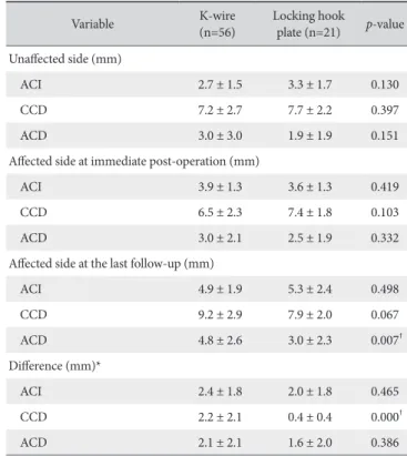
Table 4. Radiologic Assessments of K-wire Transfixation and Locking Hook Plate Fixation Groups
Variable K-wire
(n=56) Locking hook plate (n=21) p-value Unaffected side (mm)
ACI 2.7 ± 1.5 3.3 ± 1.7 0.130
CCD 7.2 ± 2.7 7.7 ± 2.2 0.397
ACD 3.0 ± 3.0 1.9 ± 1.9 0.151
Affected side at immediate post-operation (mm)
ACI 3.9 ± 1.3 3.6 ± 1.3 0.419
CCD 6.5 ± 2.3 7.4 ± 1.8 0.103
ACD 3.0 ± 2.1 2.5 ± 1.9 0.332
Affected side at the last follow-up (mm)
ACI 4.9 ± 1.9 5.3 ± 2.4 0.498
CCD 9.2 ± 2.9 7.9 ± 2.0 0.067
ACD 4.8 ± 2.6 3.0 ± 2.3 0.007†
Difference (mm)*
ACI 2.4 ± 1.8 2.0 ± 1.8 0.465
CCD 2.2 ± 2.1 0.4 ± 0.4 0.000†
ACD 2.1 ± 2.1 1.6 ± 2.0 0.386
Values are presented as mean ± standard deviation.
K-wire: Kirschner’s wire, ACI: acromioclavicular interval, CCD: coracocla- vicular distance, ACD: acromioclavicular distance.
*Difference between the value of unaffected side and affected side at the last follow-up. †Statistically significant (p<0.05).
did not lead to further surgical treatment after implant removal.
There was no ACJ arthritis case at the last follow-up. In locking hook plate fixation group, two cases with subacromial erosion on CT scan was type V according Rockwood classification. They did not complain of pain after hardware removal, with VAS 0 points and had no functional deficiency, with UCLA shoulder score 35 points at last follow-up.
Discussion
The 4 main surgical options for ACJ dislocations are (1) ACJ fixation with pins, screws, suture wires, plates and hook-plates, (2) coracoacromial ligament transfer, (3) coracoclavicular inter- val fixation, and (4) ligament reconstruction in the literatures.14) And each of these techniques has had numerous modifications with inherent potential complications.14) ACJ fixation technique is a relatively simple surgical option with good outcome re- ports.11,14-16) For ACJ fixation, various types of devices are used.
In the current study, ACJ stabilization was achieved in acute ACJ dislocation patients treated with K-wires or locking hook plates without ligament reconstruction.
The early surgical treatment techniques of K-wires or Stein- mann pins transarticulation for ACJ dislocations still remain a popular procedure.14) Many literatures reported that satisfactory outcomes may be achieved with the use of K-wires.4,5,16) The au- thors also did not find any problems with achieving ACJ stabiliza- tion by the K-wire transfixation technique. Functional outcomes were satisfactory, and the technique still seems to be a simple with low hardware’s costs. However there were several reports about complications of K-wire such as wire breakage, migration, and ACJ cartilage injury resulting in ACJ arthritis.17,18) Simovitch et al.19) reported that the wider range of better implants which
is now available, K-wires fell out of favor. In our study, the high rate of complications was a persistent problem. Especially, K- wire breakage or migration occurred in 8 cases (14%). Although statistically insignificant, the patients with K-wire breakage or migration had inferior clinical outcomes in VAS, UCLA shoulder score, ROM when compared to patients without breakage or migration. Despite attempts to prevent migration like bicortical insertion and distal wire bending during surgery and postopera- tive motion restriction after surgery, migration or breakage of K-wire seems to be unsolvable problems of K-wire transfixation technique. Although ACJ stabilization was achieved with con- servative treatment and no additional surgery in this study, other surgical options should be given priority considering potential complications such as K-wire migration or breakage.
The hook plate is one of the most commonly used implants for acute ACJ dislocations with good clinical and biomechanical results.20,21) Koukakis et al.9) reported that the use of hook plate results in excellent functional outcome for the treatment of ACJ dislocations and it is suitable for inexperienced surgeons. Unlike the previously used Wolter plate, hook plates do not require drilling into the acromion and is a relatively less challenging sur- gical technique.9,22) Recently, locking plates shaped as conven- tional hook plates but with locking screws are being commonly used, and good results are being reported.6-8) An advantage of the hook plate is the relatively easy implantation procedure and early postoperative mobilization.23) However, serious complica- tions from the hook plate including subacromial impingement and pathologic acromial fractures require attention.24,25) In the current study, comparison of radiologic outcomes at last follow- Table 5. Radiologic Assessments of K-wire Transfixation and Locking Hook
Plate Fixation Groups
Variable Unaffected
side Affected
side* p-value K-wire fixation group (n=56), mm
ACI 2.7 ± 1.5 4.9 ± 1.9 <0.001†
CCD 7.2 ± 2.7 9.2 ± 2.9 <0.001†
ACD 3.0 ± 3.0 4.8 ± 2.6 <0.001†
Locking hook plate fixation group (n=21), mm
ACI 3.3 ± 1.7 5.3 ± 2.4 <0.001†
CCD 7.7 ± 2.2 7.9 ± 2.0 0.269
ACD 1.9 ± 1.9 3.0 ± 2.3 0.082
Values are presented as mean ± standard deviation.
K-wire: Kirschner’s wire, ACI: acromioclavicular interval, CCD: coracocla- vicular distance, ACD: acromioclavicular distance.
*The value at the last follow-up. †Statistically significant (p<0.05).
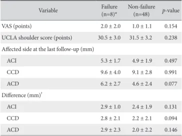
Table 6. Clinical and Radiologic Results of K-wire Failure and Non-failure Cases in K-wire Transfixation Group
Variable Failure
(n=8)* Non-failure (n=48) p-value
VAS (points) 2.0 ± 2.0 1.0 ± 1.1 0.154
UCLA shoulder score (points) 30.5 ± 3.0 31.5 ± 3.2 0.238 Affected side at the last follow-up (mm)
ACI 5.3 ± 1.7 4.9 ± 1.9 0.497
CCD 9.6 ± 4.0 9.1 ± 2.8 0.991
ACD 6.2 ± 2.7 4.6 ± 2.4 0.077
Difference (mm)†
ACI 2.9 ± 1.0 2.4 ± 1.9 0.131
CCD 2.8 ± 2.1 2.2 ± 2.1 0.094
ACD 2.9 ± 2.3 2.0 ± 2.2 0.146
Values are presented as mean ± standard deviation.
K-wire: Kirschner’s wire, ACI: acromioclavicular interval, CCD: coracocla- vicular distance, ACD: acromioclavicular distance.
*The cases with K-wire migration or breakage. †Difference between the value of unaffected side and affected side at the last follow-up.
up showed that there were significant differences in ACI, CCD, and ACD of contralateral unaffected shoulder and the shoulder with ACJ fixation in the K-wire transfixation group, while no sta- tistical difference of CCD and ACD to the unaffected side was seen in the locking hook plate group. Consequently, hook plate seems to be capable of anatomical reduction in superoinferior reduction even without coracoclavicular ligament reconstruc- tion. Modi et al.10) reported that the goal of fixation of ACJ is to restore the CCD in order to allow healing of the ruptured ligaments. Also, Luis et al.26) reported that coracoclavicular liga- ment repair is unnecessary with hook plates because anatomical reduction is done. Therefore, using hook plate in anatomical reduction of ACJ dislocation, which is advantageous for supero- inferior displacement reduction, will normalize CCD and ACD to achieve superoinferior reduction and yield coracoclavicular ligament healing effect. Due to the design characteristics of hook plates, anatomical reduction of superiorly displaced distal clavicle is relatively easy, but it is technically demanding to ana- tomically reduce the anterior-posterior displacement of clavicle and acromion. So the authors aimed to achieve anatomical re- duction and rigid fixation by anteriorly shifting the distal clavicle while using conventional screws at the medial holes of the hook plate, and locking screws for the rest. Known complications of hook plates include subacromial migration and rotator cuff impingement, infection, and reduction loss. Recently, erosions due to hook plates are being reported.7,20) But it has been re- ported in the literature that early plate removal can minimize complications.20) The authors also performed plate removal at postoperative average 3.2 months to avoid complications like erosion. Hook bending procedure was also done to make the hook parallel to the acromion and prevent subacromial impinge- ment. Since the morphology of acromion or clavicle differed individually, hook plates with various depths (12, 15, or 18 mm) were used to prevent overcorrection when the hook was placed in the subacromial space. The 2 patients in the current study subacromial bony erosion in CT scans did not report subjective pain and complaint, and there was no functional deficiency. Fa- vorable clinical and radiological outcomes were achieved in all cases with hook plates with no clinically significant complication.
It is widely accepted that Rockwood types I and II ACJ dis- locations can be treated conservatively and types IV to VI ACJ dislocations require surgical treatment.27) But ideal management of type III injuries is still controversial.7,16) Recently, many authors recommend surgical reconstruction exclusively for young pa- tients, in athletes or for heavy workers.2,28-30) In the current study, out of 77 cases, 8 cases (10%) were Rockwood classification type III. Although the authors chose conservative treatment for type III ACJ dislocations in most cases, operative treatment was indicated in patients with demands for greater functionality and early ROM. The type III patients achieved satisfactory clinical and radiological outcomes without complications.
Our study has a few limitations. First, being retrospective in nature, our study has limitations similar to other retrospec- tive studies. But we conducted a retrospective analysis of the prospectively collected patients’ data. Second, it was not ran- domized, which could produce selection bias. In addition, the number of reviewed patients was relative small, but this is single surgeon’s result at one institution. Finally, the interobserver reli- ability and intraobserver reliability were not evaluated. And our study was performed using manually drawn measurements.
However we believed that the authors participating in this study had ripe experience and it was easily reproducible.
Conclusion
ACJ stabilization was achieved in acute ACJ dislocations treat- ed with K-wires or locking hook plates. Locking hook plate can provide higher UCLA shoulder score than K-wire and maintain CCD and ACD without ligament reconstruction. K-wire transfix- ation technique resulted in a higher complication rate than lock- ing hook plate.
References
1. Mazzocca AD, Arciero RA, Bicos J. Evaluation and treat- ment of acromioclavicular joint injuries. Am J Sports Med.
2007;35(2):316-29.
2. Bishop JY, Kaeding C. Treatment of the acute traumatic acromioclavicular separation. Sports Med Arthrosc.
2006;14(4):237-45.
3. Beitzel K, Cote MP, Apostolakos J, et al. Current concepts in the treatment of acromioclavicular joint dislocations. Arthros- copy. 2013;29(2):387-97.
4. O’Carroll PF, Sheehan JM. Open reduction and percutaneous Kirschner wire fixation in complete disruption of the acromio- clavicular joint. Injury. 1982;13(4):299-301.
5. Leidel BA, Braunstein V, Kirchhoff C, Pilotto S, Mutschler W, Biberthaler P. Consistency of long-term outcome of acute Rockwood grade III acromioclavicular joint separations after K-wire transfixation. J Trauma. 2009;66(6):1666-71.
6. Eschler A, Gradl G, Gierer P, Mittlmeier T, Beck M. Hook plate fixation for acromioclavicular joint separations restores coraco- clavicular distance more accurately than PDS augmentation, however presents with a high rate of acromial osteolysis. Arch Orthop Trauma Surg. 2012;132(1):33-9.
7. Kienast B, Thietje R, Queitsch C, Gille J, Schulz AP, Meiners J.
Mid-term results after operative treatment of rockwood grade III-V acromioclavicular joint dislocations with an AC-hook- plate. Eur J Med Res. 2011;16(2):52-6.
8. Liu HH, Chou YJ, Chen CH, Chia WT, Wong CY. Surgical treat- ment of acute acromioclavicular joint injuries using a modified Weaver-Dunn procedure and clavicular hook plate. Orthope-
dics. 2010;33(8). doi: 10.3928/01477447-20100625-10.
9. Koukakis A, Manouras A, Apostolou CD, et al. Results using the AO hook plate for dislocations of the acromioclavicular joint. Expert Rev Med Device. 2008;5(5):567-72.
10. Modi CS, Beazley J, Zywiel MG, Lawrence TM, Veillette CJ.
Controversies relating to the management of acromioclavicular joint dislocations. Bone Joint J. 2013;95(12):1595-602.
11. von Heideken J, Boström Windhamre H, Une-Larsson V, Eke- lund A. Acute surgical treatment of acromioclavicular disloca- tion type V with a hook plate: superiority to late reconstruc- tion. J Shoulder Elbow Surg. 2013;22(1):9-17.
12. Rockwood C, Williams G, Young D. Disorders of the acromio- clavicular joint. In: Rockwood CA, ed. The shoulder. 3rd ed.
Philadelphia: WB Saunders; 2004. 521-95.
13. Moon SC, Lee CH, Baek JH, Cho NS, Rhee YG. Tension band wiring for distal clavicle fracture: radiologic analysis and clini- cal outcome. J Korean Fract Soc. 2014;27(2):127-35.
14. Johansen JA, Grutter PW, McFarland EG, Petersen SA. Ac- romioclavicular joint injuries: indications for treatment and treatment options. J Shoulder Elbow Surg. 2011;20(2 Suppl):S70-82.
15. Verdano MA, Pellegrini A, Zanelli M, Paterlini M, Ceccarelli F. Modified Phemister procedure for the surgical treatment of Rockwood types III, IV, V acute acromioclavicular joint disloca- tion. Musculoskelet Surg. 2012;96(3):213-22.
16. Lizaur A, Sanz-Reig J, Gonzalez-Parreño S. Long-term results of the surgical treatment of type III acromioclavicular disloca- tions: an update of a previous report. J Bone Joint Surg Br.
2011;93(8):1088-92.
17. Larsen E, Bjerg-Nielsen A, Christensen P. Conservative or surgical treatment of acromioclavicular dislocation. A prospec- tive, controlled, randomized study. J Bone Joint Surg Am.
1986;68(4):552-5.
18. Lyons FA, Rockwood CA Jr. Migration of pins used in opera- tions on the shoulder. J Bone Joint Surg Am. 1990;72(8):1262- 7.
19. Simovitch R, Sanders B, Ozbaydar M, Lavery K, Warner JJ.
Acromioclavicular joint injuries: diagnosis and management. J Am Acad Orthop Surg. 2009;17(4):207-19.
20. Sim E, Schwarz N, Höcker K, Berzlanovich A. Repair of com- plete acromioclavicular separations using the acromioclavicu- lar-hook plate. Clin Orthop Relat Res. 1995;(314):134-42.
21. De Baets T, Truijen J, Driesen R, Pittevils T. The treatment of acromioclavicular joint dislocation Tossy grade III with a clavi- cle hook plate. Acta Orthop Belg. 2004;70(6):515-9.
22. Gstettner C, Tauber M, Hitzl W, Resch H. Rockwood type III acromioclavicular dislocation: surgical versus conservative treatment. J Shoulder Elbow Surg. 2008;17(2):220-5.
23. Wagner M. General principles for the clinical use of the LCP.
Injury. 2003;34 Suppl 2:B31-42.
24. Lin HY, Wong PK, Ho WP, Chuang TY, Liao YS, Wong CC. Cla- vicular hook plate may induce subacromial shoulder impinge- ment and rotator cuff lesion: dynamic sonographic evaluation.
J Orthop Surg Res. 2014;9:6.
25. Nadarajah R, Mahaluxmivala J, Amin A, Goodier DW. Clavicu- lar hook-plate: complications of retaining the implant. Injury.
2005;36(5):681-3.
26. Luis GE, Yong CK, Singh DA, Sengupta S, Choon DS. Acromio- clavicular joint dislocation: a comparative biomechanical study of the palmaris-longus tendon graft reconstruction with other augmentative methods in cadaveric models. J Orthop Surg Res. 2007;2:22.
27. Babhulkar A, Pawaskar A. Acromioclavicular joint dislocations.
Curr Rev Musculoskelet Med. 2014;7(1):33-9.
28. Beim GM. Acromioclavicular joint injuries. J Athl Train.
2000;35(3):261-7.
29. Galpin RD, Hawkins RJ, Grainger RW. A comparative analy- sis of operative versus nonoperative treatment of grade III acromioclavicular separations. Clin Orthop Relat Res. 1985;
(193):150-5.
30. Mönig SP, Burger C, Helling HJ, Prokop A, Rehm KE. Treat- ment of complete acromioclavicular dislocation: present indi- cations and surgical technique with biodegradable cords. Int J Sports Med. 1999;20(8):560-2.