Displaced Scapula Fracture (Ideberg Type IIb) Combined with a Large Rotator Cuff Tear in Anterior Shoulder Dislocation:
A Case Report
Young-Min Noh , Chul-Hong Kim1, Seung-Hyun Lee, Chul-Soon Im
Department of Orthopedic Surgery, College of Medicine, Dong-A University, 1Orthopedic Surgery Center, Metro Hospital, Busan, Korea
Traumatic anterior shoulder dislocation combined with scapular fracture in elderly patients is relatively rare. In this case, a patient visited Emergency Room of Dong-A University Hospital for shoulder pain after falling off a ladder. Radiographs demonstrated anterior shoulder dislocation with displaced Ideberg type IIb scapula (glenoid fossa) fracture combined with a large rotator cuff tear on magnetic resonance imaging. We performed arthroscopic rotator cuff repair, but a large fragment in the inferior glenoid was left untreated. At the 1 year fol- low-up visit, the pain visual analogue scale of the patient was 2, the American Shoulder and Elbow Society score was 88 and the patient had gained nearly full range of motion without any apprehension.
(Clin Shoulder Elbow 2017;20(3):162-166)
Key Words: Scapula fracture; Shoulder fracture dislocation; Ideberg classification; Glenoid fossa Clinics in Shoulder and Elbow Vol. 20, No. 3, September, 2017
https://doi.org/10.5397/cise.2017.20.3.162
Received March 28, 2017. Revised June 27, 2017. Accepted July 30, 2017.
Correspondence to: Young-Min Noh
Department of Orthopedic Surgery, College of Medicine, Dong-A University, 32 Daesingongwon-ro, Seo-gu, Busan 49201, Korea Tel: +82-51-240-2867, Fax: +82-51-243-9764, E-mail: [email protected]
IRB approval (No. DAUHIRB-EXP-17-071).
Financial support: None. Conflict of interests: None.
Traumatic anterior shoulder dislocation is common in elderly patients, but is rarely accompanied by scapula fracture. In most cases, rotator cuff tear or greater trochanter (GT) fracture are combined in old age and only require surgical treatment if the rotator cuff tear is large or the GT fracture is displaced signifi- cantly because of shoulder instability. More typically, glenoid fractures are observed in elderly patients as the type of anterior avulsion or rim fracture in anterior shoulder dislocations, and approximately 10% are substantially displaced (>5 mm). Mini- mally displaced (<2 mm) scapular fractures that involve the gle- noid neck and fossa are known to respond well to conservative treatment, but surgical reduction is usually recommended for displaced articular fractures of the scapula, especially in young active patients.
Based on our literature review, no previous reports have ad- dressed severely displaced Ideberg type IIb scapular fracture combined with rotator cuff tear in anterior shoulder dislocations.
Therefore, this case study was written to report the clinical short
term results of an arthroscopic repair of a large rotator cuff tear without surgical fixation of the inferior glenoid fragment in an Ideberg type IIb scapular fracture associated with anterior shoul- der dislocation in an elderly patient.
Case Report
A 62-year-old male patient visited Emergency Room of Dong-A University Hospital for right shoulder pain after he fell off a ladder. The shoulder motion was unavailable because of severe pain; however, shoulder pain was absent before the acci- dent. He felt that his shoulder was dislocated after the fall on the outstretched arm, but could still move his elbow and fingers.
Passive range of motion (ROM) of the affected shoulder in- cluding other shoulder exams could not be conducted because of severe pain. His plain radiograph showed anterior shoulder dislocation and a large displaced fragment involving inferior glenoid fossa. We reduced his shoulder with gentle force by
the traction-countertraction reduction technique and checked that his shoulder was reduced concentrically by a plain radio- graph (Fig. 1). We also confirmed that any neurovascular deficits including the axillary nerve and radial artery were not found after reduction maneuver. Upon computed tomography (CT), a severely displaced Ideberg type IIb scapular fracture that ex- tended about 3 cm through the lateral border of the scapula was observed, but glenohumeral (GH) articulation was not disturbed and concentric reduction was maintained (Fig. 2). His shoulder magnetic resonance imaging (MRI) revealed full thickness of su- praspinatus, and an anterior portion of the infrasupinatus tendon with retraction about 3 cm in lateral to medial length, as well as mild muscle atrophy of the suprasipinatus was found with grade I fatty degeneration (Fig. 3). The pain visual analogue scale (pVAS) was 9.
After general anesthesia, we confirmed that the inferior dis- placed fragment did not block the passive motion of the shoul- der, concentric glenohumeral contact was achieved in the full ROM and there was no motion found in the inferior fragment under c-arm guided dynamic examination. Based on his age,
we planned to perform an arthroscopic rotator cuff repair while leaving the large fragment in the inferior glenoid alone.
Surgery was performed in the beach chair position under general anesthesia. In the glenohumeral joint, we could see that the large solitary unreducible fragment (about 1 cm displaced) in the inferior glenoid fossa and inferior glenohumeral ligament and capsule were disrupted in that fragment. Upon arthroscopic evaluation, the fractured portion was far below the level of gle- nohumeral articulation and the fragment did not impinge with the humeral head in any motions. The long head of the biceps was already torn and debridement was complete. We move to the subacromial space, debrided the bursal tissue, and indenti- fied retreacted large rotator cuff tear (approximately 2.5 cm at the anterioposterior diameter) involved supraspinatus and anterior portion of infraspinatus. The torn tendon edge was de- generated and delaminated, but a tendinous portion remained, which was possible to repair without defect. After adequate release, two medial row anchors were inserted in the articular margin of the humeral head and the medial row sutures were tied. Lateral row fixation was conducted using a knotless suture
A B
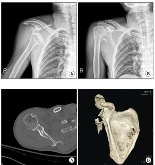
Fig. 1. (A) Plain radiograph (anteroposterior, AP) shows an anterior shoulder dislocation and combined scapular fracture at the lateral border of the scapula in a 62-year-old male patient. (B) Plain radiograph (AP) shows that the shoulder is reduced after manual reduction.
A B
Fig. 2. (A) Axial view of computed tomog- raphy shows concentric reduction of the glenohumeral joint of the right shoulder. (B) 3-dimensional-reconstruction image shows displaced Ideberg type IIb scapular fracture, which was extended by about 3 cm through the lateral border of the scapula.
anchor by suture bridge fashion (Fig. 4). The shoulder was kept in a sling with a small pillow for 6 weeks for protection of repair.
Pendulum exercises were started on day 2 after operation, but no passive motion exercises were initiated until 6 weeks after surgery. Passive shoulder motion was started after 4 weeks, and strengthening exercises were started after 3 months. Follow-up MRI was conducted three months after surgery, which showed good contact of the tendon at the repair site (Fig. 5). Upon 1 year follow-up, shoulder ROM was as follows: forward flexion 160°, abduction 150°, external rotation at side 15° and internal rotation at waist (L3). The pVAS was 2, American Shoulder and Elbow Society score was 88, and constant score was 68 (Fig. 6).
One year follow-up CT show showed no progression of arthritis in the glenohumeral joint.
Discussion
Fractures of the scapula constitute only 3% to 5% of injuries of the shoulder girdle, but this level rises to 5% to 10% in mul-
A B C
Fig. 3. Magnetic resonance imaging T2-weight coronal image (A) and sagittal image (B) show large rotator cuff tear, mild muscle atrophy and fatty degeneration on the T1-weght sagittal image (C).
A B C
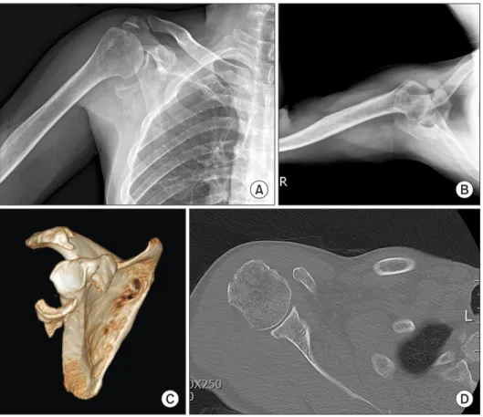
Fig. 4. Arthroscopic finding shows displaced inf glenoid fragment (A), large rotator cuff tear (B), and final view of repaired rotator cuff with suture bridge tech- nique (C).
Fig. 5. Postoperative 3 months magnetic resonance imaging follow-up reveals good continuity of the tendon at the greater tuberosity with good coverage.
tiple injuries, whereas there is no clear comparative evidence re- garding outcomes for surgical versus nonsurgical management.1-5) Ideberg classification has been widely accepted in subdivision of glenoid fractures of the scapula, and it has been reported that the anterior chip fragment fracture is the most common intraarticular glenoid fracture type, occurring in about two-thirds of cases and being associated with shoulder dislocation.6) Goss7) reported that 10% of scapula fractures involve the glenoid cavity, and Jaeger et al.8) analyzed and classified the fracture patterns by CT scan, demonstrating that f articular segment fractures were involved in approximately 38% of the 120 patients. Inferior glenoid fractures most often occur after a fall as a result of land- ing on the elbow of the abducted limb, with the humeral head impacted directly at the inferior half of the glenoid. There is ongoing debate regarding the indication of operative treatment for scapular fractures, but in general, intra-articular step-off (>3 mm) and medial displacement of the lateral border (>2.5 cm) is commonly accepted as surgical indication in Ideberg type II fractures to prevent subtle instability or posttraumatic arthritis.9)
In the current case, the fracture type of the scapula was Ide- berg type IIb (transverse, infra-equatorial in CT scan), which was a displaced (>5 mm) oblique fracture through the glenoid fossa existing inferiorly, combined with an anterior shoulder disloca- tion and large rotator cuff tear. Based on existing literature, open reduction and internal fixation with or without rotator cuff repair may have been a treatment option in this case for preventing
ongoing arthritis; however, the authors decided not to fix scapu- lar fracture based on several reasons. The most important factor was the size of fragment and the fracture pattern. Joint congru- ency of the articular contact surface of the GH joint, which for the inferior fragment was far below at the equator, and no im- pingement and subtle instability was found in c-arm guided mo- tion. Moreover, the inferiorly dominant fragment in the patient required extensive soft tissue dissection with accurate fixation, which posed the risk of brachial plexus damage, especially to the axillary nerve. The second factor was the patient’s age. The re- current instability in elderly is mostly a result of a combined rota- tor cuff tear or displaced unhealed GT fracture rather than a re- sult of capsulolabral disruption. Itoi et al.10) reported that glenoid bone loss of more than 20% can cause glenohumeral instability, and that bone loss mainly occurs at the anterior or anteroinferior quadrant in GH instability. Even though defect of the antero- inferior quadrant glenoid might cause re-dislocation in young and active patients, the fragment in our case primarily involved the inferior portion of the glenoid, which showed relatively less extension of the anterior glenoid; therefore, the authors were not concerned about instability of the GH joint in this patient. Fi- nally, the authors were concerned about postoperative shoulder stiffness after extensive dissection of soft tissue considering his age as well as the risk of neurovascular damage, such as axillary nerve injury during reduction procedure with inevitable massive dissection. We were also concerned about the chronicity of the
A B
C D
Fig. 6. Postoperative 1-year follow-up x-ray anteroposterior (A), axial (B) and 3-dimen- sional computed tomography image (C), axial scan (D) shows no interval change of inferior fragment without arthritis.
rotator cuff tear and how to manage the tear in this patient. In the T1 sagittal series of MRI, mild to moderate muscle atrophy was found with mild fatty infiltration, and the torn tendon edge was smoothly thinned; therefore, these findings indicate the pos- sibility of chronic or acute on chronic rotator cuff tear. However, even if concomitant rotator cuff tear was not acute, the originally existing large rotator cuff tear was considered to be one cause of shoulder dislocation; therefore, we decided to repair the tear.
Based on these considerations, we deliberated on surgical fixa- tion of the fracture and decided to repair the rotator cuff tear and leave the inferior fragment.
The fragment did not achieve bony union, and the 1 year follow-up x-ray revealed that the position and size of the frag- ment had not changed. Moreover, no arthritis was observed on the 1 year follow-up x-ray and CT scan, and the patient reported minimal pain with good shoulder function and ROM an no ap- prehension on short-term follow-up (Fig. 6).
References
1. Rowe CR. Fractures of the scapula. Surg Clin North Am.
1963;43:1565-71.
2. Mayo KA, Benirschke SK, Mast JW. Displaced fractures of the glenoid fossa. Results of open reduction and internal fixation.
Clin Orthop Relat Res. 1998;(347):122-30.
3. Jones CB, Cornelius JP, Sietsema DL, Ringler JR, Endres TJ.
Modified judet approach and minifragment fixation of scapu- lar body and glenoid neck fractures. J Orthop Trauma. 2009;
23(8):558-64.
4. Schandelmaier P, Blauth M, Schneider C, Krettek C. Fractures of the glenoid treated by operation. A 5- to 23-year follow-up of 22 cases. J Bone Joint Surg Br. 2002;84(2):173-7.
5. Kim KC, Rhee KJ, Shin HD, Yang JY. Can the glenopolar angle be used to predict outcome and treatment of the floating shoulder? J Trauma. 2008;64(1):174-8.
6. Ideberg R. Unusual glenoid fractures: a report on 92 cases.
Acta Orthop Scand. 1987;58:191-2.
7. Goss TP. Double disruptions of the superior shoulder suspen- sory complex. J Orthop Trauma. 1993;7(2):99-106.
8. Jaeger M, Lambert S, Südkamp NP, et al. The AO Foundation and Orthopaedic Trauma Association (AO/OTA) scapula frac- ture classification system: focus on glenoid fossa involvement. J Shoulder Elbow Surg. 2013;22(4):512-20.
9. Hardegger FH, Simpson LA, Weber BG. The operative treat- ment of scapular fractures. J Bone Joint Surg Br. 1984;66(5):
725-31.
10. Itoi E, Lee SB, Berglund LJ, Berge LL, An KN. The effect of a glenoid defect on anteroinferior stability of the shoulder af- ter Bankart repair: a cadaveric study. J Bone Joint Surg Am.
2000;82(1):35-46.