www.e-arms.org 101 Reconstruction of soft tissue defects of the knee has always
been a challenging task for plastic surgeons. Local or free flaps have been used to repair defects in this region with varying success rates.1-3 There are several characteristic features of knee joint defects. The use of a free flap is preferred for reconstructions involving obliteration of large-cavity defects, but recipient pedicle isolation can be difficult because of the extent of the injury zone. Moreover, the true defect during knee joint flexion is larger than during knee joint extension and a durable flap is necessary for joint movement. The use of fasciocutaneous flaps is advantageous as it does not require the sacrifice of precious regional muscles but provides durable coverage and is easily re-elevated later if further work is needed on the underlying bone. There is, however, a paucity of local fasciocutaneous flaps of significant size that can be reliably used
around the knee.
The pedicled perforator flap is currently a very popular soft tissue flap. Its use in the knee region as a distally based flap is appealing for its low donor site morbidity and the availability of a large amount of tissue. Although use of the distally based freestyle perforator flap has been reported,2 concerns remain about the viability of this option in view of the significant anatomic variation with regards to the vascular supply of the flap as well as the locations of the dominant skin perforators.
This article reports our experience with the use of the distally based pedicled perforator flap for coverage of knee defects and highlights the relevant anatomic variations of the flap that impact its use in the knee as well as our approach for circumventing the anatomy in these situations.
Pedicled Perforator Flaps for Reconstruction of Bilateral Knee Defects: A Case Report
Joo Seok Park, Joon Pio Hong, Tae Suk Oh*
Department of Plastic Surgery, Asan Medical Center, University of Ulsan College of Medicine, Seoul, Korea
CC This is an open-access article distributed under the terms of the Creative Commons Attribution Non-Commercial License (http://creativecommons.org/licenses/by-nc/3.0) which permits unrestricted noncommercial use, distribution, and reproduction in any medium, provided the original work is properly cited.
Copyright © 2014 by the Korean Society for Microsurgery. All Rights Reserved.
Received July 7, 2014 Revised August 12, 2014 Accepted August 14, 2014
*Correspondence to: Tae Suk Oh Department of Plastic Surgery, Asan Medical Center, University of Ulsan, 88 Olympic-ro 43-gil, Songpa-gu, Seoul 138- 736, Korea
Tel: +82-2-3010-3600 Fax: +82-2-476-7471 E-mail: [email protected]
Financial support: None.
Conflict of interest: None.
Reconstruction of soft tissue defects of the knee has always been a challenging task for plastic surgeons. Various reconstructive choices are available depending on the location, size, and depth of the defect relative to the knee joint. Defects on the knee joint have several characteristic features. The use of a free flap is preferred for reconstructions involving obliteration of large-cavity defects, but recipient pedicle isolation can be difficult because of the extent of the injury zone. Furthermore, the true defect during knee joint flexion is larger than during knee joint extension, and a durable flap is necessary for joint movement. We report for the first time on the use of pedicled perforator flaps for reconstruction of bilateral knee defects in a 76-year-old woman. The operative procedure required skeletonizing the perforators of an antero-lateral thigh flap and antero-medial thigh flap and rotating the flap in the defect. The patient returned to normal daily activity and had a full range of motion two months after the accident. The shorter operating time with decreased donor site morbidity and its durability make this flap a valuable alternative for soft tissue reconstruction of the knee.
Key Words: Knee, Pedicled flap, Perforator flap
ARMS
Archives of Reconstructive MicrosurgeryCase Report
pISSN 2383-5257 eISSN 2288-6184 Arch Reconstr Microsurg 2014;23(2):101-104 http://dx.doi.org/10.15596/ARMS.2014.23.2.101
Arch Reconstr Microsurg Vol. 23. No. 2. November 2014
102
CASE REPORT
A 76-year-old woman found lying unconscious in a sauna presented with a deep thermal wound of both knees in the anterior aspect of the patella (Fig. 1). A 15×8-cm soft tissue defect on the right knee and a 16×7-cm soft tissue defect on the left knee with bony exposure remained after debridement.
We designed a distally based pedicled perforator flap for reconstruction of the defect on the right knee. Preoperatively, the cutaneous perforators were mapped with Doppler flowmetry and computed tomography (CT) angiography around the wound. A 15×8-cm flap was marked adjacent to the defect and centered over the perforators. The flap margins were incised and the dissection was performed in the subfascial plane. The perforators were easily identified due to the previous mapping with Doppler flowmetry and CT angiography.
The motor branches of the femoral nerve were preserved to minimize donor site morbidity.
After skeletonizing all of the perforators, flap mobility was assessed. Perfusion of the flap via the selected perforators was confirmed. Finally, three perforators hindering the advancement of the flap were ligated and only one was preserved, providing adequate perfusion and flap mobility (Fig. 2). Then, the flap was rotated by 180° and insetted without undue tension and the donor site was closed primarily. Vacuum-assisted closure (VAC) therapy was applied to the left knee. The right knee was splinted for 2 weeks postoperatively.
Two weeks after placement of the pedicled perforator flap on the right knee, the other pedicled perforator flap on the left
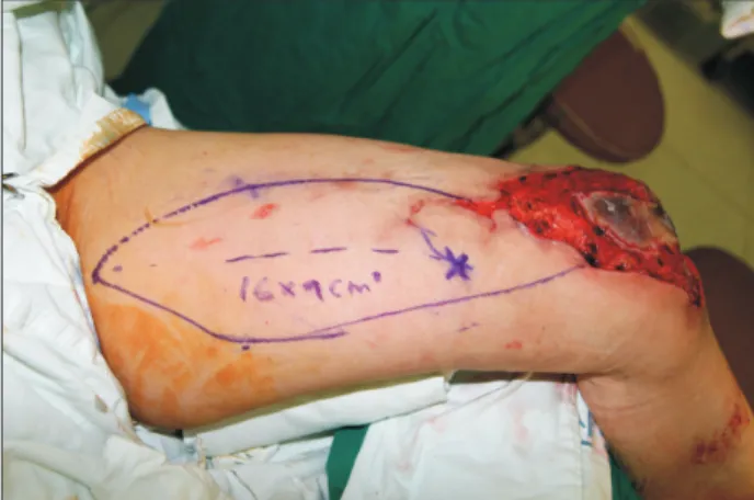
knee was planned. Preoperative Doppler tracing, subfascial plane dissection, 180o rotation, and insetting were the same as for the first pedicled perforator flap. However, during the insetting, there was mild tension at the distal tip (Fig. 3).
Accordingly, necrosis began at the distal tip of the flap on the left knee 10 days after the operation. After debridement of the necrotic portion of the flap, another pedicled perforator flap was placed on the left knee. However, due to the anatomical location of the perforator, we were unable to cover the knee without tension. Thus, distal tip necrosis on left knee occurred again and secondary healing with VAC therapy was performed.
Fig. 2. A pedicled perforator flap 15×8 cm in size was elevated with one perforator on the right knee. Three perforators hindering the advancement of the flap were ligated and only one was preserved, providing adequate mobility and perfusion to the flap.
Fig. 3. A pedicled perforator flap 16×7 cm in size was elevated with one perforator on the left knee.
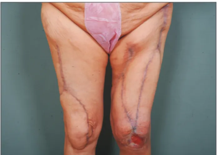
Fig. 1. A 76-year-old woman was found lying unconscious in a sauna and presented with a deep thermal wound of both knees in the anterior aspect of the patella.
Joo Seok Park, et al. Pedicled Perforator Flaps for Bilateral Knee Defects
www.e-arms.org 103
Stretching exercises had been performed before the first pedicled perforator flap was placed and we encouraged the patient to ambulate before healing of both knees was completed. Three weeks after the placement of the first pedicled perforator flap on the right knee, wheelchair ambulation was allowed with a splint on the left knee. The patient demonstrated slight decreases in range of motion at the hip and knee in the early postoperative period because of knee immobilization. Even though the soft tissue defect on the left knee was not healed completely, the patient was encouraged to ambulate with VAC therapy on the left knee. Two months after the trauma, the patient resumed normal daily activity with a full range of motion of both knees and after physiotherapy. Five months after the first pedicled perforator flap on the right knee, only a 3.5×2.5-cm dermal depth wound remained (Fig. 4).
DISCUSSION
Small-size soft tissue defects of the knee are treated with local muscle, musculocutaneous, and fasciocutaneous flaps.1-3 Because of their short pedicle length and small flap size, these flaps provide limited coverage. For large defects, the use of a free flap is preferred. Anterolateral thigh (ALT) flaps have recently become the workhorse for free flap reconstruction of soft tissue defects4 due to the following advantages: large defect coverage, moderate thickness, a remarkably long pedicle, reliable vas- cularity, design versatility, and minimal donor site morbidity.
Free flaps require a longer operating time and have variable success rates, particularly in technically demanding situations (e.g., deep recipient vessels). Nonetheless, the isolation of a recipient pedicle can be difficult when there is an extensive injury zone.5 Accordingly, in our case, we did not expect to find available recipient vessels.
Zhang6 was the first to describe the use of a distally based ALT flap for knee defect coverage. Since then, many others have reported its use.7,8 The flap is supplied by reverse blood flow from the lateral superior genicular artery and the pivot point is 3 to 10 cm above the lateral superior angle of the patella, usually at the distal portion of the vastus lateralis muscle.7 The flap’s long pedicle with a wide arc of rotation allows coverage of the entire knee region with a sufficient amount of tissue. However, arterial or venous insufficiency may easily develop due to pedicle compression or twisting during flap advancement.
Elevation of Type II and IV distally based ALT flaps, which are based on the perforators derived from the transverse branch of the lateral circumflex femoral artery (LCFA), can be technically demanding.8
Preservation of the blood supply from the superior genicular artery is essential for flap survival. In our case, we decided to use pedicled perforator flaps based on Doppler flowmetry and CT angiography. There are several advantages to this procedure:
a tailored design is possible according to a preoperative study such as Doppler flowmetry or CT angiography, lateral superior genicular artery integrity is not critical, a shorter operating time (no microsurgery needed), and the LCFA is intact.
Skeletonizing the perforators maximizes flap mobilization. Out of all of the perforators, a single dominant perforator should be enough to support the vascularity of the entire pedicled perforator flap.9 If the flap cannot be advanced to fill the defect, some of the remaining perforators restricting mobility of the flap can be ligated to permit more advancement. This technique can be applied in reconstruction of soft tissue defects at the patellar region. However, the positions and directions of the perforators vary. If sufficient advancement cannot be obtained, a propeller ALT perforator flap10 can be considered. Flap rotation by 180o over the pivot point (the distal perforator) can locate the proximal part of the ALT flap over the defect, thus enabling further distal coverage. This flap does not require ligation of the LCFA either. When these options fail to achieve coverage, a free flap is indicated. In our procedure, some parts of the vastus
Fig. 4. Five months after placement of the pedicled perforator flap on the left knee. Only absorbent soft silicone foam dressing was needed for the left knee. The patient could walk without discomfort and take a shower.
Arch Reconstr Microsurg Vol. 23. No. 2. November 2014
104
lateralis muscle and vastus medialis muscle were dissected to skeletonize all of the perforators. However, the motor branches of the femoral nerve were all preserved intraoperatively.
A pedicled anteromedial thigh (AMT) flap was planned for the soft tissue defect of the left knee in the absence of an available perforator for the ALT flap. The AMT flap also preserved the LFCA. However, necrosis at the distal tip of AMT flap on the left knee occurred due to tension during insetting. Improper perforator position might result in partial flap loss and could prolong the healing period. Thus, preoperative evaluation that includes Doppler flowmetry and CT angiography should be carefully done. Before complete healing, the patient was allowed to walk with VAC therapy on the left knee. Early ambulation may have helped her to do daily activities without limitations. The patient did not show any donor site complications. In conclusion, the pedicled perforator flap is a valuable alternative for soft tissue reconstruction of the knee with careful preoperative evaluations.
REFERENCES
1. McCraw JB, Fishman JH, Sharzer LA. The versatile gastro- cnemius myocutaneous flap. Plast Reconstr Surg 1978;62:15-23.
2. Wallace CG, Kao HK, Jeng SF, Wei FC. Free-style flaps: a further
step forward for perforator flap surgery. Plast Reconstr Surg 2009;124(6 Suppl):e419-26.
3. Yuen JC, Zhou AT. Free flap coverage for knee salvage. Ann Plast Surg 1996;37:158-66.
4. Wei FC, Jain V, Celik N, Chen HC, Chuang DC, Lin CH. Have we found an ideal soft-tissue flap? An experience with 672 anterolateral thigh flaps. Plast Reconstr Surg 2002;109:2219-26.
5. Hong JP, Lee HB, Chung YK, Kim SW, Tark KC. Coverage of difficult wounds around the knee joint with prefabricated, distally based sartorius muscle flaps. Ann Plast Surg 2003;50:484-90.
6. Zhang G. Reversed anterolateral thigh island flap and myocutaneous flap transplantation. Zhonghua Yi Xue Za Zhi 1990;70:676-8, 46.
7. Pan SC, Yu JC, Shieh SJ, Lee JW, Huang BM, Chiu HY. Distally based anterolateral thigh flap: an anatomic and clinical study.
Plast Reconstr Surg 2004;114:1768-75.
8. Lin RY, Chien WH. Experiences in harvesting type II distally based anterolateral thigh flaps. Plast Reconstr Surg 2006;118:
282-4.
9. Saint-Cyr M, Schaverien M, Wong C, Nagarkar P, Arbique G, Brown S, et al. The extended anterolateral thigh flap: anatomical basis and clinical experience. Plast Reconstr Surg 2009;123:1245- 55.
10. Chen CY, Hsieh CH, Kuo YR, Jeng SF. An anterolateral thigh perforator flap from the ipsilateral thigh for soft-tissue recon- struction around the knee. Plast Reconstr Surg 2007;120:470-3.