INTRODUCTION
Despite the many improvements made in the field of lower extremity reconstruction in recent decades, amputation is still recommended for patients with extensive lower extremity wounds that require coverage.1 Although the feet contribute relatively little to total body surface area, they are essential organ for ambulation, and the mortality rate after amputation has been reported to be high.2 Furthermore, cosmetic and
psychiatric aspects cannot be ignored.
A mangled foot is a mutilated and disfigured foot, probably caused by trauma. In terms of the treatment strategy, the pros and cons of salvage versus amputation remain controversial.
Foot reconstruction is not straightforward because of the scarcity of local tissues. Based on considerations of functional and cosmetic aspects, especially in patients with a large affected area or a contaminated wound, free tissue transfer is viewed as a first (and often the last) reconstructive option. Among
Reconstruction of a Mangled Foot with an Anterolateral Thigh Free Flap
Kyung Hoon Cook, Myong Chul Park, Dong Ha Park, Il Jae Lee*, Hyung Keun Song1, Young Uk Park1
Departments of Plastic and Reconstructive Surgery, 1Orthopedic Surgery, Ajou University Hospital, Suwon, Korea
CC This is an open-access article distributed under the terms of the Creative Commons Attribution Non-Commercial License (http://creativecommons.org/licenses/by-nc/4.0) which permits unrestricted noncommercial use, distribution, and reproduction in any medium, provided the original work is properly cited.
Copyright © 2016 by the Korean Society for Microsurgery. All Rights Reserved.
Received April 24, 2016 Revised May 17, 2016 Accepted May 19, 2016
*Correspondence to: Il Jae Lee
Department of Plastic and Reconstructive Surgery, Ajou University Hospital, 164 WorldCup-ro, Yeongtong-gu, Suwon 16499, Korea
Tel: +82-31-219-5614 Fax: +82-31-219-5610 E-mail: [email protected]
Financial support: None.
Conflict of interest: None.
Purpose: In recent decades, amputation is still recommended for patients with extensive lower extremity wounds requiring coverage. Although the feet contribute relatively little to total body surface area, they are essential organ for ambulation, and a high mortality rate after amputation has been reported. We report on 10 challenging cases of a mangled foot which was reconstructed using an anterolateral thigh (ALT) free flap, and analyze the advantages and disadvantages of this technique.
Materials and Methods: This retrospective study was conducted on 10 patients who underwent reconstructive surgery on a foot. Patients’ charts were reviewed for age, sex, causes, defect size and site, flap size and type, flap type, and complications. Cases with a defect size of >100 cm2 were included.
Results: Seven of the 10 patients were male, and overall mean age was 38.5 years (range, 22 to 61 years). Mean defect size was 179.6 cm2 (range, 104 to 330 cm2), and mean flap size was 193 cm2 (range, 120 to 408 cm2). Three cases were reconstructed with a musculocutaneous free flap and seven cases were reconstructed with a fasciocutaneous free flap. There were two occurrences of local wound complication. All ten flaps survived well, however five patients underwent a debulking procedure to reduce flap volume.
Conclusion: Reconstruction of a near completely degloved soft tissue defect or a wide defect containing two or more surfaces of extremity with an ALT free flap was performed.
The purpose of this case study is to report on free tissue transfer using the ALT flap for salvage of the lower extremity.
Key Words: Soft tissue injuries, Foot injuries, Free flaps
ARMS
Archieves of Reconstructive Microsurgery http://dx.doi.org/10.15596/ARMS.2016.25.1.7the many types of free tissue transfers, the anterolateral thigh (ALT) flap is one of the most popular for the reconstruction of defects following trauma or cancer ablative surgery. The ALT free flap has many reported advantages, and the availability of soft tissue is its greatest advantage for the reconstruction of a mangled foot. We have experienced many types of mangled feet, and have used an ALT free flap as a first choice for flap coverage. We present 10 challenging cases of mangled foot which were reconstructed using an ALT free flap, and analyze the advantages and disadvantages of this technique.
MATERIALS AND AND METHODS
This retrospective study was conducted on 10 patients who underwent reconstructive surgery on a foot between March 2012 and December 2013 at the Department of Plastic and Reconstruction Surgery at Ajou University Hospital. Patients’
charts were reviewed for age, sex, causes, defect size and site, flap size and type, flap type, and complications (Table 1).
Cases with a defect size of >100 cm2 were included. Seven of the 10 patients were male, and overall mean age was 38.5 years (range, 22 to 61 years). Mean defect size was 179.6 cm2 (range, 104 to 330 cm2), and mean flap size was 193 cm2 (range, 120 to 408 cm2).
Defects were limited to a foot and all were reconstructed using an ALT free flap in a fasciocutaneous manner or using vastus lateralis muscle depending on the recipient site.
RESULTS
Five cases were due to traffic accidents, two cases were due to crush injuries in an occupational setting, two were caused by accidents to military personnel, and it was caused by a mine explosion. We performed free tissue transfer using two types of flap elevation, that is, either fasciocutaneous or musculocutaneous. Three cases were reconstructed using a musculocutaneous free flap with vastus lateralis and seven cases were reconstructed using a fasciocutaneous free flap.
Two local wound complications occurred. One was due to arterial rupture at the anastomosis site at 11 days postoperatively requiring an emergency operation, at which the arterial leakage site was repaired. The arterial rupture was caused by detachment of dressing material. The other was due to venous congestion at the flap site. The congestive condition progressed over 5 days and over this period the flap skin blackened. Debridement of skin and of the subcutaneous layer revealed viable tissue. Daily dressing and close monitoring after debridement resulted in flap survival within 1 month.
No events postoperative complications occurred. All ten flaps survived well, though five patients underwent a debulking procedure to reduce flap volume.
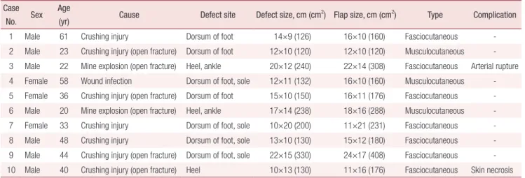
Case I (case No. 6)
A 20-year-old man had a 17×14 cm (238 cm2) soft tissue defect on heel of his left foot caused by a mine explosion (Fig.
1). Calcaneal bone was exposed and an open fracture was noted. Following debridement orthopedic surgeons performed
Table 1. Patients data Case
No. Sex Age
(yr) Cause Defect site Defect size, cm (cm2) Flap size, cm (cm2) Type Complication
1 Male 61 Crushing injury Dorsum of foot 14×9 (126) 16×10 (160) Fasciocutaneous -
2 Male 23 Crushing injury (open fracture) Dorsum of foot 12×10 (120) 12×10 (120) Musculocutaneous - 3 Male 22 Mine explosion (open fracture) Heel, ankle 20×12 (240) 22×14 (308) Fasciocutaneous Arterial rupture 4 Female 58 Wound infection Dorsum of foot, sole 12×11 (132) 16×10 (160) Musculocutaneous - 5 Female 36 Crushing injury (open fracture) Dorsum of foot 15×10 (150) 16×11 (176) Fasciocutaneous - 6 Male 20 Mine explosion (open fracture) Heel, ankle 17×14 (238) 18×16 (288) Musculocutaneous - 7 Female 33 Crushing injury Dorsum of foot, sole 10×20 (200) 11×21 (231) Fasciocutaneous -
8 Male 48 Crushing injury Dorsum of foot, sole 13×10 (130) 15×12 (180) Fasciocutaneous -
9 Male 44 Crushing injury (open fracture) Dorsum of foot, sole 22×15 (330) 24×17 (408) Fasciocutaneous - 10 Male 40 Crushing injury (open fracture) Heel 10×13 (130) 11×16 (176) Fasciocutaneous Skin necrosis
external fixation with K-wire and an Ilizarov system to preserve the bony structure of the injured foot. After serial debridement with antibiotic therapy, a left ALT musculocutaneous flap was elevated and applied to the recipient site. In this patient, end- to-end anastomosis was performed between the posterior tibial artery and the descending branch of lateral circumflex femoral artery.
Case II (case No. 9)
A 44-year-old man had a 22×15 cm (330 cm2) soft tissue defect on the dorsum of right foot caused by a crush injury sustained during a traffic accident, and concurrently had bone exposure at the metatarsal level of the 1st to 5th toes (Fig. 2).
After arthrodesis of mid foot bones, amputation from the 1st to 5th metatarsal bone level, open reduction, and internal fixation,
we elevated a right ALT musculocutaneous flap. In this patient, the anterior tibial artery was anastomosed end-to-side to the descending branch of the lateral circumflex femoral artery.
DISCUSSION
In patients with a large foot defect, treatment can be undertaken in two ways, that is, by amputation at the non- salvageable tissue level or by covering the defect and preserving the ankle joint and foot structure. Although amputation has some advantages, foot and ankle salvage are obviously the better option in terms of quality of life.3
Basically, three methods can be used to cover soft tissue defects, namely, skin grafting, local flap transfer, and free tissue transfer. Skin grafting has advantages for the coverage of large
Fig. 1. A 20-year-old man had injury on heel of his left foot caused by a mine explosion.
Fig. 2. A 44-year-old man had a soft tissue defect on the dorsum of right foot caused by a traffic accident.
defects and is relatively straightforward, but grafted skin is unsuitable for the weight-bearing sole area, and uptake on extensive wounds on a muscle tendon or bone bed.
Local flap transfer provides good coverage of small defects because of the similarity between flaps and adjacent tissues, but it cannot be used to cover large defects and has disadvantages in the context of infection control because of the low vascularity of utilized tissues.4
Therefore, the free muscle flap is more appropriate for extensive wounds, regardless of the defect site, and the latissimus dorsi muscle, rectus abdominis muscle, and gracilis muscle are primarily used.5 The latissimus dorsi muscle is broad and relatively flat and has a long vascular pedicle, and latissimus dorsi flap is appropriate for the treatment of large defects.6 However, a position change is required while performing the operation, and it is prone to donor site seroma. The rectus abdominis muscle free flap provides a large volume of soft tissue but is narrower than the latissimus dorsi flap. On the other hand, the gracilis flap is a good surgical option for smaller defects and its donor-site morbidity is minimal.7
Some unique features of foot surfaces required consideration prior to defect reconstruction. On the dorsum of the foot the skin is thin and flexible and devoid of subcutaneous adipose tissue. Thus, to ensure that a patient can wear shoes and obtain a satisfactory cosmetic outcome, a thin flap should be elevated.8 On the other hand, the weight-bearing plantar area is characterized by thick skin and a large amount of fat, and must perform a sensation protective function to enable weight bearing while walking.9
The thigh contains abundant soft tissue, and thus, defects of the plantar area can be reconstructed with an ALT free flap.
Actually, all ten of our patients were eventually able to ambulate unaided.
We reconstructed defects with two types of ALT free flap, one is fasciocutaneous free flap and the other type is musculocutaneous free flap. Musculocutaneous flap has advantages of bacterial infection rates reduction and high blood flow. In a series of animal studies, Calderón and Leniz10 compared the hemodynamic features, histological details, and bacterial changes between the musculocutaneous and fasciocutaneous flaps. Decreased bacterial count reveals 30 to 60 times better than fasciocutaneous flaps.11
In recent years, however, it has been reported not only that
there is no significant difference in surgical outcomes between the muscle flap and the fasciocutaneous flap, but also that there is no need to sacrifice muscle tissue at donor sites.12
Notably, the shearing force applied to the plantar area is considerable, and thus, usually a medial plantar artery based flap is used in weight bearing areas. Furthermore, should ulceration later develop, we have a plan to make coverage by medial plantar artery flap focused on ulcerated area if it is required, but we also have to consider vascular status of injured vessel by trauma itself.
However, problematic cases of lower limb wounds that cannot be simply reconstructed are also encountered. When an area of injury is too wide, possible recipient vessels may be located too far from the wound, and when a defect is large and complex one flap may be insufficient to achieve coverage.
CONCLUSION
Reconstruction of a near totally degloved soft tissue defect or a wide and contains two or more surface of extremity with ALT free flap was performed. The purpose of this case report is to present free tissue transfer using the ALT flap for salvage of the lower extremity.
REFERENCES
1. Kim TG, Kim IK, Kim YH, Lee JH. Reconstruction of lower extremity complex wounds with combined free tissue transfer using the anterolateral thigh flap as a link. Microsurgery 2012;32:575-9.
2. Hong JP, Oh TS. An algorithm for limb salvage for diabetic foot ulcers. Clin Plast Surg 2012;39:341-52.
3. Gil J, Schiff AP, Pinzur MS. Cost comparison: limb salvage versus amputation in diabetic patients with charcot foot. Foot Ankle Int 2013;34:1097-9.
4. Kang MJ, Chung CH, Chang YJ, Kim KH. Reconstruction of the lower extremity using free flaps. Arch Plast Surg 2013;40:575-83.
5. Jeon BJ, Lee KT, Lim SY, Pyon JK, Bang SI, Oh KS, et al. Plantar reconstruction with free thoracodorsal artery perforator flaps. J Plast Reconstr Aesthet Surg 2013;66:406-13.
6. Gomez MM, Casal D. Reconstruction of large defect of foot with extensive bone loss exclusively using a latissimus dorsi muscle free flap: a potential new indication for this flap. J Foot Ankle Surg 2012;51:215-7.
7. Reddy V, Stevenson TR. MOC-PS(SM) CME article: lower extremity reconstruction. Plast Reconstr Surg 2008;121(4
Suppl):1-7.
8. Redett RJ, Robertson BC, Chang B, Girotto J, Vaughan T. Limb salvage of lower-extremity wounds using free gracilis muscle reconstruction. Plast Reconstr Surg 2000;106:1507-13.
9. Hong JP. Reconstruction of the diabetic foot using the anterolateral thigh perforator flap. Plast Reconstr Surg 2006;117:
1599-608.
10. Calderón WL, Leniz P. Comparison of the vascularity of fasciocutaneous tissue and muscle for coverage of open tibial
fractures. Plast Reconstr Surg 2010;125:1582.
11. Kuo PJ, Chew KY, Kuo YR, Lin PY. Comparison of outcomes of pressure sore reconstructions among perforator flaps, perforator- based rotation fasciocutaneous flaps, and musculocutaneous flaps. Microsurgery 2014;34:547-53.
12. Sofiadellis F, Liu DS, Webb A, Macgill K, Rozen WM, Ashton MW. Fasciocutaneous free flaps are more reliable than muscle free flaps in lower limb trauma reconstruction: experience in a single trauma center. J Reconstr Microsurg 2012;28:333-40.