INTRODUCTION
Solving the issues related to the knee soft tissue defects after total knee arthroplasty (TKA) has been a challenge from the past to the present. There are a number of causes; however it is because the soft tissue defects by wound infection are required to be approached by simultaneously solving two tasks-infection control and wound coverage. Thus, the soft tissue defects, unless resolved properly, might lead to a variety of results:
the removal of prosthesis, arthrodesis, and even a loss of the
limbs in the worst case. The wound infection, once it occurs, is required to be treated carefully and surely, considering the importance and characteristics of knee joints. The coverage of the soft tissue defects by the infection after TKA has been approached in various manners and continuously; yet no consensus on the management has been made until now.
Authors accomplished the successful coverage of the soft tissue defects by the infection after TKA by using various perforator flaps around knees.
Soft Tissue Reconstruction Using Perforator Flap in Patients with Infected Knee Prosthesis
Jin Won Lee, Sung Hoon Kim, Jun Ho Yoo, Si Gyun Roh*, Nae Ho Lee, Kyoung Moo Yang
Department of Plastic and Reconstructive Surgery, Chonbuk National University Medical School, Jeonju, Korea
CC This is an open-access article distributed under the terms of the Creative Commons Attribution Non-Commercial License (http://creativecommons.org/licenses/by-nc/3.0) which permits unrestricted noncommercial use, distribution, and reproduction in any medium, provided the original work is properly cited.
Copyright © 2014 by the Korean Society for Microsurgery. All Rights Reserved.
Received November 3, 2014 Revised November 14, 2014 Accepted November 18, 2014
*Correspondence to: Si Gyun Roh Department of Plastic and Reconstructive Surgery, Chonbuk National University Hospital, 20 Geonji-ro, Deokjin-gu, Jeonju 561-712, Korea
Tel: +82-63-250-1860 Fax: +82-63-250-1866 E-mail: [email protected]
Financial support: None.
Conflict of interest: None.
Purpose: Soft-tissue reconstruction in the knee area requires thin, pliable, and tough skin. The range of motion of the knee also acts as a limitation in using only local flaps for coverage. The author has successfully used various perforator flaps for soft tissue reconstruction around the knee while preserving its functional and cosmetic characteristics.
Materials and Methods: Out of the twenty patients assessed from April 2009 to March 2011, seven received anterolateral thigh perforator flaps, four received medial sural perforator island flaps, four received lateral supragenicular perforaor perforator flaps, and five received medial genicular artery flaps. The age of the patients ranged from 44 to 79 and the size of the defects ranged from 4×5 cm to 17×11 cm. Fifteen of the twenty patients had histories of total knee replacement (TKR) surgery.
Results: There were no flap losses in any of the twenty patients assessed. Two patients showed partial losses in the distal area of the flap, but were treated through careful wound care. One patient presented with pedicle adhesion at the drainage site from a past TKR, but it did not hinder the flap survival. Primary closure at the donor site was possible in nine patients, while split skin graft was necessary for the other 13.
Conclusion: In soft tissue reconstruction of the knee, various perforator flaps can be used depending on the condition of the preoperation scar, wound site, and size. It also proved to provide better functional and cosmetic results than in primary wound closure or skin grafts.
Key Words: Reconstructive surgical procedures, Perforator flap, Knee prosthesis
ARMS
Archives of Reconstructive Microsurgery http://dx.doi.org/10.15596/ARMS.2014.23.2.70MATERIALS AND METHODS
This study has been conducted from April 2009 to February 2012, targeting the patients with the soft tissue defects after TKA (Table 1). The patients-related data sheet demonstrates that the subjects included 14 female patients (68%) and 6 male patients (32%), and their average age was 66 years. The main cause of primary TKA was osteoarthritis (OA) (13 patients, 65%). Age, gender, etiology, pathogen, defect size, exposed
tissue, time from the primary TKA to infection, kind of flap, complication, and donor site coverage of a total of 20 patients were recorded and analyzed.
Depending on the result of appropriate the bacterial culture test after the detection of infection, the most susceptible antibiotics to the bacteria was intravenously administered and appropriate debridement and incision and drainage were performed in accordance with the wound condition. After the administration of antibiotics for 2 weeks on an average basis, the
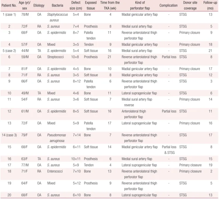
Table 1. Summary of cases Patient No. Age (yr)/
sex Etiology Bacteria Defect size (cm)
Exposed tissue
Time from the TKA (wk)
Kind of
perforator flap Complication Donor site coverage
Follow-up (mo) 1 (case 1) 78/M OA Staphylococcus
aureus
5×4 Bone 4 Madial genicular artery flap - STSG 13
2 72/F RA S. aureus 7×4 Prosthesis 8 Medial sural artery flap - STSG 5
3 68/F OA S. epidermidis 8×7 Patella
tendon
11 Reverse anterolateral thigh perforator flap
- Primary closure 9
4 57/F OA Mixed 3×5 Tendon 9 Madial genicular artery flap - Primary closure 18
5 (case 2) 44/M TA S. epidermidis 5×4 Soft tissue 16 Medial sural artery flap - STSG 21
6 59/M OA Streptococci 10×8 Prosthesis 21 Reverse anterolateral thigh perforator flap
Partial loss STSG 8
7 81/F OA S. epidermidis 4×5 Bone 10 Madial genicular artery flap - Primary closure 17
8 71/F RA S. aureus 3×5 Soft tissue 8 Madial genicular artery flap - STSG 12
9 66/F OA S. aureus 8×12 Patella
tendon
6 Reverse anterolateral thigh perforator flap
- STSG 3
10 49/M TA Mixed 4×6 Bone 11 Lateral supragenicular flap - STSG 6
11 54/F RA S. aureus 3×6 Soft tissue 7 Medial sural artery flap
reverse
- Primary closure 14
12 61/M OA S. epidermidis 8×5 Soft tissue 16 Anterolateral thigh perforator flap
Partial loss STSG 11
13 72/F OA Mixed 5×9 Patella
tendon
17 Lateral supragenicular flap - Primary closure 16
14 (case 3) 79/F OA Pseudomonas aeruginosa
7×14 Bone 7 Reverse anterolateral thigh perforator flap
- STSG 17
15 68/F OA S. epidermidis 6×11 Soft tissue 14 Madial genicular artery flap Partial loss
& STSG
STSG 8
16 63/F TA S. aureus 10×11 Prosthesis 6 Medial sural artery flap - STSG 15
17 77/M OA S. aureus 5×9 Tendon 4 Lateral supragenicular flap - Primary closeure 19
18 71/F RA Enterococci 7×10 Bone 13 Reverse anterolateral thigh
perforator flap
- Primary closeure 2
19 64/F OA Mixed 5×12 Prosthesis 9 Reverse anterolateral thigh
perforator flap
- STSG 5
20 68/F OA S. aureus 6×10 Bone 8 Lateral supragenicular flap - STSG 13
M: male, F: female, OA: osteoarthritis, RA: rheumatoid arthritis, TA: traumatic arthritis, STSG: split-thickness skin graft.
removal of the infected TKA and antibiotic impregnated spacer insertion were performed. Regarding the soft tissue defects around knees, an appropriate flap was selected by considering operative scars in the past, perforator condition, defect size, location, the degree of the exposed prosthesis or bone, and the condition of the surrounding soft tissues.
Especially we focused on defect location when selected the type of perforator flap.
RESULTS
Out of a total of 20 patients, the reversed anterolateral thigh flap was used for 7 patients; medial sural artery perforator flap was used for 4 patients; lateral supragenicular perforator flap was used for 4 patients; and medial genicular artery perforator flap was used for 5 patients. There was no flap loss. Partial loss of flap was observed in 3 patients; 2 patients were healed spontaneously; and split-thickness skin graft (STSG) was performed on the flap with a partial loss for 1 patient. In 7 patients, donor site primary closure was possible. The main pathogen of infection was Staphylococcus aureus (8 patients, 40%), followed by S. epidermidis (5 patients, 25%) and mixed bacteria (4 patients, 20%). Mean defect size was 49.3 cm2 (range, 3×5 cm to 10×11 cm) and the infect occurred 10 weeks after primary TKA on an average basis. Furthermore, bone and soft tissue were exposed in 6 patients respectively and prosthesis was exposed in 4 patients after the soft tissue defect by infection and the average follow-up period for each patient was about 12 months on an average basis.
One patient showed a sign of perforator adhesion, which made lap dissection difficult; yet there was no effect on flap
survival. Staged revision TKA was able to be performed 4 months after flap coverage on an average basis and there was no patient who was infected again. Furthermore, the patients showed no functional discomfort when they ambulated.
And cosmetically, there was no more secondary debulking procedure after the perforator flap operation.
Case 1
There was one male patient who underwent right knee TKA because of OA. Wound opening accompanied by infective discharge occurred 4 weeks after the primary TKA. S. aureus was cultured and thus wound irrigation and debridement were performed by using susceptible antibiotics. Prosthesis removal was performed followed by antibiotic impregnated spacer insertion. The 5×4 cm defect occurred after debridement was covered by using medial genicular artery perforator flap. There was no problem with flap survival; donor site was covered with STSG; the spacer was removed 3 months after flap operation;
staged revision TKA was performed; and no sign of infection has been observed until now (Fig. 1).
Case 2
There was a 44-year-old male patient who underwent TKA because of arthritis resulted from the car accident. The infection wound occurred in the center of right knee incision knee 16 weeks after the surgery and S. epidermidis was cultured and thus antibiotics was used for the patient. Since then, the unstable scar accompanied by soft tissue exposure was formed. Thus, debridement was performed and 5×4 cm defect was covered by using medial sural artery perforator flap. Donor site was covered with STSG. No sign of reinfection has been observed
Fig. 1. Soft tissue reconstruction by medial genicular artery perforator island flap of a 78-year-old male. (A) The 5×4 cm sized soft tissue defect is seen in the right knee caused by Staphylococcus aureus infection. (B) Immediate postoperative view. (C) Postoperative 13 months view.
A B C
during 21-month follow-up treatment and the patient showed excellent knee joint mobility (Fig. 2).
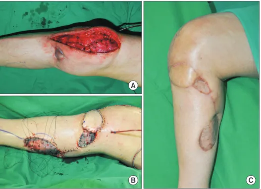
Case 3
There was a 79-year-old female patient diagnosed as degener- ative OA. The wound infection occurred in incision line in the distal area due to Pseudomonas aeruginosa 7 weeks after right knee TKA. Prosthesis removal was performed after conducting the administration of antibiotics; wound irrigation and debridement were performed; and then antibiotic impregnated spacer insertion was performed; and then, 7×14 cm soft tissue defect was covered by using anterolateral thigh perforator flap. Spacer removal from right knee and staged revision TKA
were performed simultaneously 6 months after. The patient did not show any specific sign of either wound complication or reinfection and did not have any problem with knee joint mobility (Fig. 3).
DISCUSSION
It has been reported that about 20% of patients who underwent TKA had the wound complication and from 1.1%
to 12.4% wound complication was resulted from infection.1,2 In particular, the wound complication resulted from infection might lead to the complicated issues such as the removal of TKA resulted from infection, the likelihood of arthrodesis
Fig. 3. Soft tissue reconstruction by reverse anterolateral thigh perforator flap of a 79-year-old female. (A) Soft tissue defect is seen caused by Pseudomonas aeruginosa infection. After debridement, defect size was 7×14 cm. (B) Immediate postoperative view. (C) Postoperative 17 months view.
A B C
Fig. 2. Soft tissue reconstruction by medial sural artery perforator island flap of a 44-year-old male. (A) Chronic infective wound is seen on the medial side of total knee arthroplasty incision wound. (B) Immediate postoperative view. (C) Postoperative 21 months view.
A
B C
or amputation, and the death resulted from sepsis. Thus, to reduce the likelihood of deep infection, prosthesis removal, and amputation, it is required to detect the infection in the early stage and start the infection control by using appropriate antibiotics.2-8
Knees are the biggest and important joints that tolerate all kinds of loads and absorb the shock.4,9,10 Thus, the protection of knee joint and the role as a major movement joint are important.2 The approaches for soft tissue defect coverage after TKA include tissue expander, local flap, skin graft, muscle flap, fasciocutaneous flap, and free flap and so on. However, the outcome of tissue expander or local flap is undesirable considering knee joint mobility; tissue expander or local flap is not appropriate approach in that it does not resolve the dead space; skin graft also is undesirable when deep structure is exposed.1,6 Free flap is useful and produces a good result; yet surgery requires a lot of time for the surgery itself, high skills and a lot of recovery time.10 Gastrocnemius muscle flap needs a short surgery time and is rather easy to handle or inset the flap and thus is the most widely used until now. However, there is a risk of donor site morbidity resulted from the sacrifice of muscle; defect size is likely to be large, or the site is more proximal than patella; and the use of the surgery is limited in case of quadriceps tendons.1,2,5,9-11
The development of perforator brought innovative changes to the wound coverage by using perforator flap. Since the introduction of a concept of cutaneous vasculature flap by Harvey in the 1600s and Tomas, Spalteholz in the 1800s, Carl Manchot in the late 1880s (quoted from reference 12) has conducted in-depth studies on vascular territories in human bodies. Having conducted anatomic dissection by using original lead oxide injection, Michel Salmon has visualized ‘vascular supply to the skins’. Since then, the introduction of angiosome reinforced the concept of perforator and thus the perforator flap using this concept has been actively used.12
The collateral circulation network is well developed around the knees and thus a wide range of perforators exist accordingly.13 Thus, there is a wide range of choices for flaps for the defect coverage depending on defect site, size, location of scar, and the condition of surrounding soft tissues.
At the location of the defect mainly, the flap can be selected as one of the three techniques. When the defect exists the front knees, the defect of lateral side, and especially the defect of the
lower third in thigh, distally based anterolateral thigh flap is able to be used as the perforator. And the defect locates the below or inside the patella, medial sural artery perforator flap can be considered. When the defect placed on the thigh’s lower third and the front or the sides of knees, superior lateral genicular artery and superior medial genicular artery are able to be selected.
Distally based anterolateral thigh flap, using the descending branch of lateral circumflex femoral artery, has a number of advantages. This flap is able to cover the front knees, the defect of lateral side, and especially the defect of the lower third in thigh. It is possible to design flaps in many different sizes and control the flap thickness by using debulking in accordance with the depth of defect. Furthermore the very consistent anatomy is an advantage.10,12 It is rather easy to get thick and long pedicle and there is the low likelihood of donor site morbidity and thus this author chooses this flap most frequently.
Medial sural artery perforator flap is ideal when the defect is below or inside the patella.10,14 My papers proved that the anatomy of perforator is very consistent and this is, in fact, the most widely used. The perforator is able to be observed on the imaginary line between popliteal crease and the midpoint of medial malleolus. The first perforator is located in a circle of 2 cm radius in about 8 cm away from the proximal of the imaginary line. Very thin and pliable flap can be obtained; flap elevation is easy; and there is the low likelihood of donor site morbidity.
Furthermore, it is able to be used as the perforator of the genicular artery that have been developed well around knees.
Typically, superior lateral genicular artery and superior medial genicular artery are able to be selected. These two flaps are useful to cover the defects thigh’s lower third and the front or the sides of knees. Furthermore it is rather easy to elevate the flap after finding the perforator by using dropper. It is possible to design the flap of various sizes and there is the less likelihood of donor site functional impairment.10
As mentioned earlier, the soft tissue coverage after TKA by using fasciocutaneous perforator flap is a very useful surgery method with a number of advantages. Furthermore this flap does not include muscle, which means there is the low likelihood of donor site morbidity and it is possible to fill the dead space around the knees without including muscle.6,9,11
It is most unlikely that the antibiotics alone can control
infected TKA. The early flap coverage after early aggressive debridement and irrigation is a method to preserve prosthesis and prevent arthrodesis or amputation.3
For the soft tissue defect occurred after the infected TKA, the elevation including the bacterial culture in the early stage is required to be identified. Furthermore it is required to use appropriate antibiotics; perform early debridement and irrigation; choose appropriate perforators; and select the flap by considering the defect size, location, the location of preoperative scar and the condition of the surrounding soft tissues for early coverage.
REFERENCES
1. Salibian AH, Anzel SH. Salvage of an infected total knee prosthesis with medial and lateral gastrocnemius muscle flaps.
A case report. J Bone Joint Surg Am 1983;65:681-4.
2. Casey WJ 3rd, Rebecca AM, Krochmal DJ, Kim HY, Hemminger BJ, Clarke HD, et al. Prophylactic flap reconstruction of the knee prior to total knee arthroplasty in high-risk patients. Ann Plast Surg 2011;66:381-7.
3. Bengston S, Knutson K, Lidgren L. Treatment of infected knee arthroplasty. Clin Orthop Relat Res 1989;(245):173-8.
4. McPherson EJ, Patzakis MJ, Gross JE, Holtom PD, Song M, Dorr LD. Infected total knee arthroplasty. Two-stage reimplantation with a gastrocnemius rotational flap. Clin Orthop Relat Res 1997;(341):73-81.
5. Sanders R, O'Neill T. The gastrocnemius myocutaneous flap used as a over for the exposed knee prosthesis. J Bone Joint Surg
Br 1981;63:383-6.
6. Tiengo C, Macchi V, Vigato E, Porzionato A, Stecco C, Azzena B, et al. Reversed gracilis pedicle flap for coverage of a total knee prosthesis. J Bone Joint Surg Am 2010;92:1640-6.
7. Arnold PG, Prunes-Carrillo F. Vastus medialis muscle flap for functional closure of the exposed knee joint. Plast Reconstr Surg 1981;68:69-72.
8. Galat DD, McGovern SC, Larson DR, Harrington JR, Hanssen AD, Clarke HD. Surgical treatment of early wound complications following primary total knee arthroplasty. J Bone Joint Surg Am 2009;91:48-54.
9. Menderes A, Demirdover C, Yilmaz M, Vayvada H, Barutcu A. Reconstruction of soft tissue defects following total knee arthroplasty. Knee 2002;9:215-9.
10. Wiedner M, Koch H, Scharnagl E. The superior lateral genicular artery flap for soft-tissue reconstruction around the knee:
clinical experience and review of the literature. Ann Plast Surg 2011;66:388-92.
11. Ries MD, Bozic KJ. Medial gastrocnemius flap coverage for treatment of skin necrosis after total knee arthroplasty. Clin Orthop Relat Res 2006;446:186-92.
12. Chen CY, Hsieh CH, Kuo YR, Jeng SF. An anterolateral thigh perforator flap from the ipsilateral thigh for soft-tissue reconstruction around the knee. Plast Reconstr Surg 2007;120:
470-3.
13. Hallock GG. Salvage of total knee arthroplasty with local fasciocutaneous flaps. J Bone Joint Surg Am 1990;72:1236-9.
14. Kim HH, Jeong JH, Seul JH, Cho BC. New design and identification of the medial sural perforator flap: an anatomical study and its clinical applications. Plast Reconstr Surg 2006;117:1609-18.