천공지 피판을 이용한 두경부 재건
Perforator Flaps in Head and Neck Reconstruction
김연환·김정태
한양대학교 의과대학 성형외과학교실
Youn Hwan Kim, M.D.
Jeong Tae Kim, M.D., Ph.D.
Department of Plastic and Reconstructive Surgery, School of Medicine, Hanyang University, Seoul, Korea
책임저자 주소: 김정태, 138-788, 서울시 성동구 행당동 17번지 한양대학교병원 성형외과
Tel: 02-2290-8563, Fax: 02-2295-7671 E-mail: [email protected]
투고일자: 2009년 5월 25일, 심사일자: 2009년 7월 5일, 게재확정일자: 2009년 8월 10일
Abstract
Basic requirements of head and neck reconstruc- tions are thin resurfacing, a long vascular pedicle, 3-dimensional and well customized reconstruction with a team approach.
Ideal reconstruction methods were thought to be free tissue transfer including radial forearm flap, latissmus dorsi or rectus abdominis myocutaneous flap. But recently, there has been concerns about sacrifice of donor structures in these conventional flaps. For minimal sacrifice of donor structures, there has been much evolution in flap concepts, which lead to the introduction of perforator flaps.
They are popular in every region for reconstruction.
Anterolatral thigh, latissmus dorsi or deep inferior epigastric artery perforator flaps are commonly used.
Perforator flaps could also be applied to head and neck reconstructions, because they could be used
for the controlled resurfacing of scalp, cheek, neck, oropharynx, and for customized 3-dimensional re- constructions, including diverse components ac- cording to each perforator, which may result in more comfortable handling and less restricted access to the defect. The perforator flaps also have long vas- cular pedicles compared to conventional myocuta- neous flaps, which can lead to less restriction in choosing recipient vessels.
Perforator flaps have known to have many advan- tages as described and they give one more option in head and neck reconstruction.
Key Words: Perforator flaps, Head and neck re- construction
서 론
두경부의 종양 적출술 후에 발생하는 결손을 재건하기 위해서는 여러가지 사항이 요구된다.1-4 1) 얇은 피복이 가 능하여야 하며, 2) 3차원적인 재건이 필요할 수 있고, 3) 긴 혈관경으로 문합이 자유로워야 하며, 4)혈관경의 혈류가 안정적이고, 5) 두께를 자유로이 조절할 수 있어 결손에 적 합하게 재단할 수 있는 피판(well customized flap)이어야 하며, 6) 재건팀과 종양 적출하는 팀이 함께 수술이 가능해 야 하고. 7) 공여부의 이환율이 적어야 한다.
이런 여러 가지 요건을 모두 갖춘 피판으로 전완부 요골 동맥 피판(radial forearm flap)을 두경부 재건에 있어 가 장 많이 사용하여 왔다. 하지만, 주요동맥인 요골 동맥을 희생해야 하는 문제점과 함께 공여부에 넓은 피부 이식으 로 인해 이환율이 크며, 얇은 피복은 가능하나, 두께 조절 의 자유로움이 떨어져 현재에는 그 사용이 줄어드는 추세 이다.5-10 이런 문제점을 극복하기 위해 광배근 근피판(latis- simus dorsi myocutaneous flap)11-13이나 복직근피판
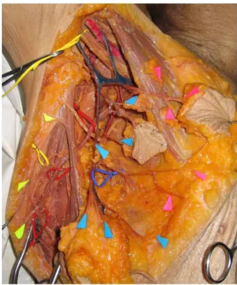
Fig. 1. Cadaver dissection shows three groups of perforator in axilla area. First rows(yellow) indicate musculotaneous perforator group, mid rows(blue) indicate septocutaneous perforator group and the other anterior rows(pink) indicate direct cutaneous group.
(rectus abdominis myocutaneous flap)14-16 등을 두경부 재건에 많이 적용하여 왔다. 하지만 이런 근피판들은 근육 을 포함하고 있어, 광범위 결손부를 채우는 데는 적합하지 만 얇게 피복을 필요로 하는 구강내 재건이나 경부 재건에 매우 어려움이 있으며, 근육의 결손으로 인해 공여부의 이 환율이 크다. 피판에 대한 개념이 새로이 적립되고 미세수 술의 발전이 거듭되면서, 1989년 Isao Koshima 등에 의해 천공지 피판(perforator flap)이 소개되었다. 천공지 피판 은 천공지에 의해 혈류를 공급받는 근육을 포함하고 있지 않는 피부 및 피하 조직의 일부를 포함하는 얇은 피판을 의 미하는 것으로, 배꼽 주변의 천공지에 의해 횡복직근을 포 함하지 않는 피판에 대해 보고하였으며, 이는 심하복벽 동 맥(deep inferior epigastic artery)을 혈관경으로 하며 혈 류 역시 기존의 심하복벽 근피판과 같이 안정적인 것으로 보고되었다.17
이 후 천공지 피판에 대한 많은 보고와 발전이 거듭되었 으며, 현재 유방재건, 상하지 재건, 안면 재건 등 다양한 분 야에서 여러가지 천공지 피판이 유용하게 이용되고 있
다.18-30 대표적인 천공지 피판으로 외측 대퇴 회선 동맥
(lateral circumflex femoral artery)의 분지를 혈관경으로 하는 전외측 대퇴 천공지 피판(anterolateral thigh perfo- rator flap, 이하 ALT perforator flap), 흉배 동맥(thora- codorsal artery)를 혈관경으로 하는 흉배 동맥 천공지 피 판(thoracodorsal artery perforator flap, 이하 TDP) 혹은 광배근 천공지 피판(latissimus dorsi perforator flap,이하 LDP), 심하복벽 동맥(Deep inferior epigastric artery)을 혈관경으로 하는 심하복벽 천공지 동맥 피판(deep inferior epigastric artery perforator flap, 이하 DIEP) 이 있다. 이 런 천공지 피판들이 공통적으로 추구하는 바로는 1) 근육 을 포함하지 않고 공여부의 희생을 최소화할 수 있으며, 2) 재피복시 두께를 자유롭게 조절할 수 있고, 3) 감각 피판이 가능하고, 4) 결손부에 맞추어 재단할 수 있는 작도의 자유 로움이 있으며, 5) 주요 혈관들(source vessel)을 보존할수 있으며, 6) 혈관경을 좀 더 길게 확보할 수 있고, 7) 피판의 선택에 있어 자유로움이 있다는 것이다. 최근 이런 천공지 피판이 가진 다양한 장점들은 두경부 재건에 있어서 갖추 어야 할 여러 가지 요건에 잘 부합하기에, 두경부 재건에 있어 기존의 피판을 대체할 수 있는 또 다른 유용한 피판으 로 생각되어 많은 적용이 이루어지고 있다. 저자들이 주로 사용하는 흉배 동맥 천공지 피판혹은 광배근 천공지 피판
과 전외측 대퇴부 천공지 피판에 대해 소개하고 이를 통해 천공지 피판의 두경부 적용에 대해 소개하고자 한다.
본 론
1. 천공지 해부와 위치(Anatomy and Location of Perforator)
액와부의 천공지는 대개 일정한 간격으로 열을 이루어 광배근을 따라 피부에 혈류를 공급하게 되는데, 광배근의 후방에 있는 근피천공지(musculocutaneous group)와 광 배근의 근막을 뚫고 올라오는 격막천공지(septocutaneous group)와 광배근의 전방에 바로 피부를 혈류를 공급하게 되는 직접천공지(direct cutaneous group)의 3가지로 크 게 분류할 수 있다(Fig. 1).23, 24 천공지 피판의 명명에 대해 서는 여러 저자들의 의견이 분분하며 아직 통일되지 못하 였으나, 저자들의 새로운 명명법에 따르면,31 근육을 뚫고 올라오는 천공지(musculocutaneous perforator)의 경우
는 근육의 이름을 이용하여 광배근 천공지 피판(latissimus dorsi perforator)로 명명할 수 있으며, 근육을 가로 지르지 않는 격막천공지나 직접천공지에서는 천공지의 혈관경이 되는 흉배 동맥(throacodorsal artery)을 기저로 하여, 흉 배 동맥 천공지 피판(thoracodorsal artery perforator)으 로 쉽게 구별하여 명명할 수 있다. 전외측 대퇴 천공지 피 판(ALT perforator flap)의 경우 역시 액와부의 경우와 비 슷하다. 역시 이곳에서도 격막천공지와 근피천공지로 구분 된다. 따라서 격막천공지의 경우는 그 혈관경의 이름을 기 저로 하여, 외측 대퇴 회선 동맥 천공지 피판(lateral cir- cumflex femoral artery perforator flap)으로 명명할 수 있으며, 근피천공지의 경우 뚫고 올라오는 외측 광근 천공 지 피판(vastus lateralis muscle perforator flap)이라고 명 명할 수 있다.31 이처럼 액와부와 전외측 대퇴부 하지에는 다양한 천공지들이 존재하며, 이를 이용하여 chimeric pat- tern으로 근육 및 피하 지방조직을 자유롭게 포함하여, 두 경부의 복합 결손에 대해 자유롭게 재건할 수 있는 이점을 제공한다.
2. 천공지의 확인(Perforator Identification) 광배근 천공지(LDP)의 경우, 광배근의 앞쪽 경계를 따라 Audible Doppler를 이용하여, 천공지를 확인할 수 있다.
전외측 대퇴 천공지 피판의 경우 전상 장골극(anterior superior iliac supine)에서 슬개골의 가측 경계를 연결한 중간 지점의 반경 3 cm 이내에서 천공지를 확인할 수 있 다.1, 19, 20 대게 혈류가 안정적인 천공지(reliable perfora- tor)는 액와부에서 10 cm 이내에서 확인할 수 있는데, Audible Doppler 의 경우 그 하방에 존재하는 흉배동맥이 나 대퇴동맥의 맥박에 의해서도 나타날 수 있으므로, 전적 으로 의존해서는 안된다.23, 24 최근에는 3차원 단면 영상을 이용하여 천공지의 위치를 정확하게 확인하고 수술에 임하 는 경우도 늘고 있다.32-34 혈류가 안정적이며, 믿을만한 천 공지는 그 직경으로 판단하기 보다는 피판을 거상하는 과 정에서 처음 발견된 천공지의 맥박이 눈으로 확인된다면 (visible pulsation), 피판의 혈관경으로 사용하기에 적합하 다고 할 수 있다.
3. 수술방법(Operative Techniques)
전외측 대퇴부 천공지 피판과 심하복벽 천공지 피판의 경우 두경부 종양 적출부위와 떨어져 있어, 두개의 팀이 동
시에 접근이 가능한 가장 큰 장점을 가지고 있으며, 술 중 에 자세를 바꾸지 않고, 앙와위에서 수술을 진행할 수 있어 시간을 절약할 수 있다. 광배근 천공지 피판의 경우, 자세 를 바꾸어야 하는 불편한 점과 종양 적출팀과 위치가 가까 워서 동시에 수술이 어렵다는 단점이 제기되었다. 하지만, 저자들의 경우 앙와위에서도 피판의 거상이 가능하였으며, 종양 적출팀과 동시에 수술적 접근이 가능하여 광배근 천 공지 피판 또한 두경부 재건에 유용하게 이용할 수 있다.
앙와위에서 팔을 약간 상승시키면, 광배근의 앞쪽 경계 를 쉽게 확인할 수 있으며, 이를 통해 먼저 천공지를 확인 한다. 물론 천공지의 위치가 정확하게 확인되었다면, 이를 바탕으로 작도하고, 절개선을 예정할 수 있다. 하지만, 두 경부 재건의 경우 대개 이비인후과 팀에서 종양을 적출한 후에 정확한 결손의 크기를 알 수 있으므로, Mardini 등이 전외측 대퇴 천공지 피판에서 기술한 바와 같이 절개를 한 후 믿을만한 천공지를 먼저 확인하고 이의 위치에 따라 결 손부에 맞게 피판을 작도하는 "free style perforator flap"
이 두경부 재건에는 적합하다.35 절개선은 광배근의 앞쪽 경계를 따라 시행하고, 대게 액와부에서 10 cm 이내에 눈 으로 확인되는 맥박을 가진 믿을만한 천공지(reliable per- forator)를 확인할 수 있는데, 앞에서 기술한 바와 같이 직 접천공지나 격막천공지가 있을 수 있으므로, 조심스럽게 지혈겸자(sharp hemostat)를 이용하여 박리를 해 들어가 야 한다. 전외측 천공지 피판의 경우 역시 마찬가지이다.
전상장골극과 슬개골을 잇는 가상의 선을 따라 전외측 하 지의 중심선을 따라 절개선을 넣고 조심스럽게 천공지를 찾은 후, 천공지가 확인되면 vessel loop을 이용하여 조심 스럽게 다루면서 작은 혈관 가지들은 bipolar cautery를 이 용하여 지혈을 한다. 근육속으로 주행하는 천공지의 경우 특히 세밀한 박리를 요하며, 술 중 천공지의 연축(spasm) 을 막기 위해 혈관확장제를 지속적으로 뿌려준다.24 피판 거상시 술중 천공지의 맥박은 빠른 시간내에 사라지므로, 다른 경우에서보다 혈관확장제를 더욱 많이 사용하여야 한 다. 근육 속으로 주행하는 천공지를 박리 시에는 천공지 주 변을 완전히 박리하기보다는 근육이나 연부조직을 조금 주 변에 붙여서 천공지가 손상을 받는 것을 방지하고, 외부의 압박으로부터 보호(cushion effect) 받을 수 있도록 한다.
감각피판이 필요한 경우 흉배 동맥 천공지 나 광배근 천공 지 피판의 경우 늑간 신경(intercostals nerve)을 함께 피판 에 포함시키고, 전외측 대퇴 천공지 피판의 경우 외측 대퇴
Fig. 3. (Above, Left) Recurrent squamous cell carcinoma in scalp. (Above, Right) 28x16 cm thoracodorsal artery perforator flap was used for resurfacing. (Below, Left) Preoperative view of skin defect and MRSA infection after cranioplasty. (Below, Right) A thoracodorsal artery perforator flap was used for the resurfacing of scalp defects.
Fig. 2. (Left) preoperative view of tongue cancer. (Right) posto- perative view of tongue resurfacing using perforator flap.
피신경(lateral femoral cutaneous nerve)을 포함시킨다.
근육내 박리가 끝나고 주혈관경이 확보되면 혈관경의 길이 를 확인하고 피판을 거상하게 되는데, 적출된 종양에 맞게 재건하고자 하는 결손부위를 작도한다. 천공지의 위치는 혈관경의 길이가 충분하다면 피판의 중심에 위치하는 것이 가장 이상적이지만, 혈관경의 길이가 짧거나 작도에 어려 움이 있는 경우에는 가장자리에 위치시켜 혈관경을 조금 더 길게 확보할 수 있다. 이처럼 천공지 피판의 경우 작도 의 자유로움이 두경부 재건에 적용할 수 있는 큰 장점 중에 하나가 된다. 천공지 중심부보다는 주변부에서부터 거상을 시작하며, 지방층의 두 층을 피판을 견인하면서 분리하면 얇은 피판의 거상이 가능하다. 다만 천공지가 피부로 들어 가는 중심부 반경 1~2 cm에서는 얇게 거상하기 보다는 주 변에 지방층을 충분히 붙여서 천공지의 손상을 막고 술 후 압박되는 것을 방지할 수 있는 것이 안전하다.23, 24 피판의 거상이 끝난 후에는 종양의 적출이 완료될 때까지 skin sta- pler를 이용하여 그 자리에 고정시키고, 혈관확장제를 충분 히 뿌려 천공지의 연축을 방지한다. 일시적으로 정맥 울혈 이 발생할 수 있으나 믿을만한 천공지를 택한 경우 시간이 지나면서 자연적으로 회복하게 된다.
4. 두경부 재건에의 적용(Application in the Head and Neck Reconstruction)
1) 두께 조절을 통한 재피복(Controlled Resurfacing) 두경부의 경우 매우 얇은 피판에서부터 어느 정도의 두 께를 필요로 하는 두피 재건에 이르기까지 다양한 두께를 필요로 한다. 특히 설기저(tongue base), 편도암(tonsil), 인두벽(pharyngeal wall)이나 연구개(soft palate)등의 종 양 적출 후에 얇은 두께의 피판으로 재건이 필요한데, 피판 이 두꺼운 경우 음식물을 삼키기가 곤란하고, 발음에 있어 서도 문제가 될 수 있다. 과거에 사용하던 전완부 요골 동 맥 피판이 매우 얇은 피판으로서 이런 목적으로 많이 사용 되었으나, 최근 천공지 피판으로도 매우 얇은 피판의 거상 이 가능하게 되어, 이를 대체할 수 있다. 특히 설암(tong cancer)으로 혀 재건이 필요한 경우는 부위별로 어느 정도 의 부피조절이 필요한데, 천공지 피판의 경우 결손부에 맞 게 부피 및 두께가 자유로이 조절이 된다(Fig. 2). 또 두피 재건의 경우 기존의 피판들은 너무 두꺼워 외형적으로 좋 은 결과를 얻지 못해서 이차적 수술이 필요한 경우가 많으
나, 천공지 피판을 이용할 경우 주변의 두께에 맞게 자유스 럽게 두께를 조절할 수 있으므로, 미적으로 만족할 만한 결 과를 얻을 수 있다(Fig. 3). 특히 뇌경막(dura)의 결손이 있 는 경우 전외측 대퇴부 천공지 피판 거상시 근막을 함께 포 함하여 두피의 재피복과 함께 경막의 재건에도 유용하게 이용할 수 있다(Fig. 4). 경부의 재피복의 경우에도 적절하 게 두께를 조절함으로서 구축 없이 기능적으로, 미용적으 로 만족할 만한 결과를 얻을 수 있다. 이처럼 천공지 피판 은 필요한 부위에 얇은 두께의 피판에서부터 결손부위의 두께에 맞게 부피를 자유롭게 조절하면서 재피복이 가능하 다.
Fig. 4. (Above, Left) Recurrent MRSA infection of artificial dura and cerebritis (Above, Right) Intraoperative view of cranium (Below, Left) Vastus Lateralis perforator flap which is known as anterolateral thigh perforator flap was elevated. (Below, Right) postoperative view of scalp resurfacing.
Fig. 5. (Above, Left) Old male patient had suffered pharyngeal cancer with fistula formation and huge chyle leakage. (Above, Right) A septocutaneous perforator flap for pharyngeal lining and a musculocutaneous perforator skin flap for outer lining were elevated in anterolateral thigh. (Below, Left) Flap insetting for customized reconstruction. (Below, Right) Postoperative view shows successful reconstruction of pharyngeal lining and outer resurfacing.
2) 결손부에 맞게 재단된 재건(Customized Reconstruc- tion)
기존 피판의 경우 근육 및 피하조직, 피부판들이 하나로 연결되어 있어, 결손부에 맞게 배치시키는데 어려움이 많 다. 하지만, 천공지 피판의 경우 천공지를 기저로 근육 및 피하조직 및 피판을 각각 분리시킬 수 있어 조작이 매우 용 이하며, 결손부의 접근이나 배치 또한 기존의 피판들보다 유리하다. 코의 재건에 적용하여 3차원적으로 재건이 가능 하며, 치조골 재건(alveolar reconstruction)의 경우 천공 지의 위치에 맞게 피판을 자유롭게 분리가 가능하여 구강 내에서 자유롭게 피판을 조작할 수 있어 유리하다. 특히 상 악골 재건(maxilla reconstruction)의 경우 3차원적인 재 건을 필요로 하는데, 피부 바깥쪽의 결손 재건, 비내벽의 재건(nasal lining), 구강내벽의 재건(oral lining) 과 함께 골재건(bony reconstruction)을 함께 필요로 하는 경우에 도 각각의 천공지에 따라 피판을 한 혈관경으로부터 각각 분리하며, 입체적으로 재건이 가능한 장점이 있다. 또한 갈 비뼈나 전거근(serratus anterior muscle) 및 피하 지방조 직과 같은 다양한 구성요소를 포함시켜서, 마치 재단하듯
이 원하는 대로 피판 제작이 가능하다. 전외측 대퇴 천공지 피판의 경우에서도 근육을 포함하여 사강 충전에 사용이 가능하며, 근피천공지와 격막천공지를 각각 분리하여 두개 의 피판을 통해 인두암(pharyngeal cancer)환자의 재건에 사용할 수 있다(Fig. 5).
3) 긴 혈관경의 확보(Elongation of Pedicle)
긴 혈관경은 두경부 재건에 있어 자유스러운 재건과 접 근이 가능할 수 있으므로 되도록 긴 혈관경을 확보하는 것 이 유리하다. 특히 방사선 치료 후에 적절한 혈관이 없는 경우 반대측 혈관을 사용해야 하는 경우도 발생하므로 더 욱 중요하다. 기존의 근피판은 근육내 박리가 없이 근육을 포함하므로 천공지 피판에 비해 혈관경이 짧다. 이에 반해 천공지 피판은 대부분 근육내 박리가 필요하며 이를 통해 3-5 cm 정도로 더 긴 혈관경을 확보할 수 있다. 이처럼 천 공지 피판은 기존의 피판보다 긴 혈관경을 통해 수혜부 혈 관 선택에 제한이 없어 재건에 자유로움을 제공한다.
4) 식도 재건(Esophageal Reconstrcution)
유리 공장 이식술(free jejunal flap)과 전완부 요골 동맥 피판은 하인두 및 식도 재건에서 유용하게 이용되어 왔다.
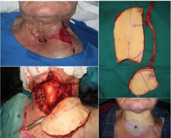
Fig. 6. (Above, Left) total pharyngolaryngectomy state. (Above, Right) tubed latissimus dorsi perforator flap was elevated for cervical esophageal reconstruction. (Below, Left) Flap insetting for neoesophagus. (Below, Right) Postoperative endoscopic view shows successful reconstruction of esophagus.
Fig. 7. (Above and Below) Generally latissimus dorsi or thraco- dorsal artery perforator flap is located more anterior to conven- tional muscle flap therefore you can harvest these flaps on supine position for two team approach.
유리 공장 이식술은 점막과 같은 성상으로 자연스러운 재 건이 가능하다는 점등의 장점을 지녔지만, 개복 수술로 인 한 탈장이나 장유착, 분비물로 인해 음성의 질이 떨어지거 나 방사선 치료 후 누공이나 협착 등 다른 문제점들이 제기
되었다.36-39 이에 반해 전완부 요골 동맥 피판은 근막 피판
이 가진 장점 즉, 음성의 질이 우수하며 감각 회복이 좋다 는 점을 지니고 있다. 하지만 공여부의 이환율이 너무 높아 사용에 제한이 있고 경부 식도 재건에 부족한 점이 있다.5, 6 최근 들어 전외측 천공지 피판을 관상 형태로 말아서 하인 두 및 식도 재건에 적용하여 좋은 결과를 보였다.40 저자들 역시 흉배 동맥 혹은 광배근 천공지 피판을 관상 형태로 말 아서 재건에 적용하여 좋은 결과를 얻었으며, 기존의 유리 공장 이식술이 가진 낮은 누공율과 전완부 요골 동맥 피판 이 가진 음성의 질이 우수한 장점을 천공지 피판을 통해 얻 을 수 있었다(Fig. 6).
전외측 대퇴 천공지 피판의 경우 큰 피판으로 거상할 경 우 역시 일차 봉합이 힘들어 피부 이식이 필요한 경우가 있
고, 또한 노출 부위에 흉이 남는다는 문제점을 지니고 있다.
흉배 동맥 혹은 광배근 천공지 피판의 경우 역시 전외측 천 공지 피판보다 좀 더 큰 피판의 거상이 가능하나, 술 후 젖 꼭지 등이 바깥쪽으로 변형될 수 있고, 흉부의 조임 등의 불편이 있을 수 있다. 하지만, 공여부를 폭 10 cm까지 일차 봉합하더라도 특별한 문제없이 잘 치유되며, 기존의 피판 에 비해 공여부의 이환이 적은 장점으로 인해 그 사용이 늘 어가고 있다. 저자들의 방법에 따라 두개의 팀이 동시에 접 근이 가능하고, 앙와위에서도 피판의 거상이 가능하다(Fig.
7).
결 론
천공지 피판은 기존의 피판에 비해 많은 장점을 지녔다.
그중에서도 두께 조절의 자유로움으로 인해 두피, 경부, 구 강내, 인두 및 혀의 재피복이 가능하며, 각각의 천공지에 따라 다양한 성분을 포함하여 마치 작도하듯이 결손부위에 맞게 3차원적인 재건이 가능하며, 근육내의 박리를 통해 혈 관경을 더욱 길게 확보함으로써 수여부 혈관의 선택에 있 어 자유롭다. 이런 천공지 피판의 장점은 두경부 재건에 있 어 기존의 피판들을 대신할 수 있는 또 하나의 좋은 대안으 로 사료된다.
References
1. Lyons AJ. Perforator flaps in head and neck surgery.
Int J Oral Maxillofac Surg 2006;35:199-207.
2. Koshima I, Yamamoto H, Hosoda M, Moriguchi T, Orita Y, Nagayama H. Free combined composite flaps using the lateral circumflex femoral system for repair of mas- sive defects of the head and neck regions: an introduc- tion to the chimeric flap principle. Plast Reconstr Surg 1993;92:411-20.
3. Koshima I, Fukuda H, Yamamoto H, Moriguchi T, Soeda S, Ohta S. Free anterolateral thigh flaps for reconstruc- tion of head and neck defects. Plast Reconstr Surg 1993;92:421-8.
4. KimataY, Uchiyama K, Ebihara S, Yoshizumi T, Asai M, Saikawa M, Hayashi R, Jitsuiki Y, Majima K, Ohyama W, Haneda T, Nakatsuka T, Harii K. Versatility of the free anterolateral thigh flap for reconstruction of head and neck defects. Arch Otolaryngol Head Neck Surg 1997;
123:1325-31.
5. Anthony JP, Singer MI, Mathes SJ. Pharyngoesophageal reconstruction using the tubed free radial forearm flap.
Clin Plast Surg 1994;21:137-47.
6. Scharpf J, Esclamado RM. Reconstruction with radial forearm flaps after ablative surgery for hypopharyngeal
cancer. Head Neck 2002;25:261-6.
7. Sardesai MG, Fung K, Yoo JH, Bakker H. Donor-site morbidity following radial forearm free tissue transfer in head and neck surgery. J Otolaryngol Head Neck Surg 2008;37:411-6.
8. O'Connell DA, Rieger J, Harris JR, Dziegielewski P, Zalmanowitz J, Sytsanko A, Li S, Wolfaardt J, Hart RD, Seikaly H. Swallowing function in patients with base of tongue cancers treated with primary surgery and recon- structed with a modified radial forearm free flap. Arch Otolaryngol Head Neck Surg 2008;134:857-64.
9. Agrawal A, Husein OF, Schuller DE. Esophageal recon- struction with larynx preservation using forearm-free flap. Laryngoscope 2008;118:1750-2.
10. Andrades P, Pehler SF, Baranano CF, Magnuson JS, Carroll WR, Rosenthal EL. Fistula analysis after radial forearm free flap reconstruction of hypopharyngeal defects. Laryngoscope 2008;118:1157-63.
11. Piantanida R, Roselli R, Pellini R, Ferrario F, Boschini P, Spriano G. Reconstruction of major orbital-maxillary defects with free latissimus dorsi myocutaneous flap.
Facial Plast Surg 1999;15:297-302.
12. Har-El G, Bhaya M, Sundaram K. Latissimus dorsi myocutaneous flap for secondary head and neck re- construction. Am J Otolaryngol 1999;20:287-93.
13. Yamamoto Y, Nohira K, Yamashita T, Shintomi Y, Hosokawa M, Furkawa H, Sugihara T, Ohura T. Com- bined V figure-shaped scapular osteocutaneous and latissimus dorsi myocutaneous flap for composite man- dibular reconstruction. Head Neck 1995;17:219-25.
14. Schliephake H, Schmelzeisen R, Neukam FW. The free revascularized rectus abdominis myocutaneous flap for the repair of tumour related defects in the head and neck area. Br J Oral Maxillofac Surg 1996;34:18-22.
15. Kyutoku S, Tsuji H, Inoue T, Kawakami K, Han F, Ogawa Y. Experience with the rectus abdominis myo- cutaneous flap with vascularized hard tissue for imme- diate orbitofacial reconstruction. Plast Reconstr Surg 1999103:395-402.
16. Wanamaker JR, Burkey BB. Overview of the rectus
abdominis myocutaneous flap in head and neck recon- struction. Facial Plast Surg 1996;12:45-50.
17. Markowitz BL, Satterberg T, Calcaterra T, Orringer J, Cohen S, Burstein F, Shaw W. The deep inferior epiga- stric rectus abdominis muscle and myocutaneous free tissue transfer: further applications for head and neck reconstruction. Ann Plast Surg 1991;27:577-82.
18. Koshima I, Soeda S. Inferior epigastric artery skin flaps without rectus abdominis muscle. Br J Plast Surg 1989:
42;645-8.
19. Saint-Cyr M, Schaverien M, Wong C, Nagarkar P, Arbi- que G, Brown S, Rohrich RJ. The extended anterola- teral thigh flap: anatomical basis and clinical experien- ce. Plast Reconstr Surg 2009;123:1245-55.
20. Yazar S, Gideroglu K, Kilic B, Gokkaya A. Use of com- posite anterolateral thigh flap as double-vascularised layers for reconstruction of complex hand dorsum defect.
J Plast Reconstr Aesthet Surg 2008;61:1549-50.
21. Kimura N, Saito M, Sumiya Y, Itoh N. Reconstruction of hand skin defects by microdissected mini anterolataral thigh perforator flaps. J Plast Reconstr Aesthet Surg 2008;61:1073-7.
22. Kuo YR, Jeng SF, Wei FC, Su CY, Chien CY. Functional reconstruction of complex lip and cheek defect with free composite anterolateral thigh flap and vascularized fascia. Head Neck 2008;30:1001-6.
23. Wei FC, Celik N, Jeng SF. Application of "simplified nomenclature for compound flaps" to the anterolateral thigh flap. Plast Reconstr Surg 2005;115:1051-5 24. Kim JT. Two options for perforator flaps in the flank
donor site: latissimus dorsi and thoracodorsal perfora- tor flaps Plast Reconstr Surg 2005115:755-63.
25. Kim JT. Latissimus dorsi perforator flap. Clin Plast Surg 2003;30:403-31.
26. Kim JT, Kim SK. Hand resurfacing with the superthin latissimus dorsi perforator-based free flap. Plast Rec- onstr Surg 2003;111:366-70.
27. Blondeel PN, Boeckx WD. Refinements in free flap breast reconstruction: the free bilateral deep inferior epigastric perforator flap anastomosed to the internal
mammary artery. Br J Plast Surg 1994;47:495-501.
28. Hamdi M, Blondeel P, Van Landuyt K, Tondu T, Monstrey S. Bilateral autogenous breast reconstruction using perforator free flaps: a single center's experience. Plast Reconstr Surg 2004;114:83-9.
29. Koshima I, Inagawa K, Urushibara K, Moriguchi T. Pa- raumbilical perforator flap without deep inferior epiga- stric vesselsPlast Reconstr Surg 1998;102:1052-7.
30. Koshima I, Inagawa K, Urushibara K, Ohtsuki M, Mori- guchi T. Deep inferior epigastric perforator dermal-fat or adiposal flap for correction of craniofacial contour deformities. Plast Reconstr Surg 2000;106:10-5.
31. Koshima I, Nanba Y, Tsutsui T, Takahashi Y, Watanabe A, Ishii R. Free perforator flap for the treatment of de- fects after resection of huge arteriovenous malforma- tions in the head and neck regions. Ann Plast Surg 2003;51:194-9.
32. Kim JT. New nomenclature concept of perforator flap.
Br J Plast Surg 200558:431-40.
33. Imai R, Matsumura H, Tanaka K, Uchida R, Watanabe K. Comparison of Doppler sonography and multidetec- tor-row computed tomography in the imaging findings of the deep inferior epigastric perforator artery. Ann Plast Surg 2008;61:94-8.
34. Mun GH, Kim HJ, Cha MK, Kim WY. Impact of perfora- tor mapping using multidetector-row computed tomo- graphic angiography on free thoracodorsal artery per- forator flap transfer. Plast Reconstr Surg 2008122:
1079-88.
35. Masia J, Clavero JA, Larrañaga JR, Alomar X, Pons G, Serret P. Multidetector-row computed tomography in the planning of abdominal perforator flaps. J Plast Reconstr Aesthet Surg 2006;59:594-9.
36. Mardini S, Tsai FC, Wei FC. The thigh as a model for free style flaps. Clin Plast Surg 2003;30:473-80.
37. Coleman JJ 3rd, Tan KC, Searles JM, Hester TR, Nahai F. Jejunal free autograft: analysis of complications and their resolution.Plast Reconstr Surg 1989;84:589-95.
38. Schusterman MA, Shestak K, de Vries EJ, Swartz W, Jones N, Johnson J, Myers E, Reilly J Jr. Reconstruction
of the cervical esophagus: free jejunal transfer versus gastric pull-up. Plast Reconstr Surg 1990;85:16-21.
39. Reece GP, Schusterman MA, Miller MJ, Kroll SS, Robb GL, Baldwin BJ, Luethcke DR. Morbidity and functional outcome of free jejunal transfer reconstruction for cir- cumferential defects of the pharynx and cervical eso- phagus. Plast Reconstr Surg 1995;96:1307-16.
40. Cordeiro PG, Shah K, Santamaria E, Gollub MJ, Singh B, Shah JP. Barium swallows after free jejunal transfer:
should they be performed routinely? Plast Reconstr Surg 1999103:1167-75.
41. Yu P, Robb GLPharyngoesophageal reconstruction with the anterolateral thigh flap: a clinical and functional outcomes study. Plast Reconstr Surg 2005;116:1845-55.