MEC is more aggressive, invades the tissue, and metasta- sizes4. Treatment of low-grade MEC is wide local resection with a tumor-free margin3. When low-grade MEC arises in the hard palate, the tumor is resected along with the involved palate mucosa and any underlying bone5.
A posterior maxillary or hard palate defect can be suc- cessfully covered with a pedicled buccal fat pad (BFP) flap6. BFP has been introduced as a pedicled flap for the closure of oroantral communication7 and has been successfully used to cover oral defects in the posterior maxilla, hard and soft pal- ate, and retromolar region after tooth extraction, sauceriza- tion, and tumor resection6. BFP is located in the masseteric space between the buccinator muscle and the mandibular ramus and masseter muscle and receives a rich blood supply from the superficial temporal, internal maxillary, and facial arteries8. Due to its anatomic location, the BFP can be easily accessed and harvested via an intraoral approach and applied to the posterior oral cavity6.
4-Hexylresorcinol (4HR) is a well-known antiseptic used as an ingredient in mouthwash9. Because the oral cavity is a septic area, 4HR can be used to prevent postoperative in- fection9. In addition, 4HR differentiates oral cancer cells by increasing the expression of involucrin and keratin 1010. The BFP contains abundant stem cells that can differentiate the oral epithelium11. If 4HR could accelerate the differentiation
I. Introduction
Mucoepidermoid carcinoma (MEC) is the most common type of malignant tumor in the salivary gland and comprises approximately 12% to 23% of malignant tumors in the minor salivary gland1. The tumor can occur in the minor salivary gland of the lip, buccal mucosa, retromolar region, or tongue and floor of the mouth, but the most frequently involved site is the hard palate2. MEC of the hard palate presents as an asymptomatic, fixed, slow-growing swollen area, sometimes appearing red or blue in color, is fluctuant and ulcerative, and invades the underlying bone3. MEC is categorized as either low, intermediate, or high based on histology1. MEC has diverse biological features and variable clinical symptoms depending on the grade and stage of the tumor4. Low-grade MEC is generally slow growing and benign, while high-grade Hyun Seok
Department of Oral and Maxillofacial Surgery, College of Dentistry, Gangneung-Wonju National University, 7 Jukheon-gil, Gangneung 25457, Korea
TEL: +82-33-640-3139 FAX: +82-33-640-3113 E-mail: [email protected]
ORCID: http://orcid.org/0000-0002-5278-3807
This is an open-access article distributed under the terms of the Creative Commons Attribution Non-Commercial License (http://creativecommons.org/licenses/by-nc/4.0/), which permits unrestricted non-commercial use, distribution, and reproduction in any medium, provided the original work is properly cited.
CC
Reconstruction of partial maxillectomy defect with a buccal fat pad flap and application of 4-hexylresorcinol: a case report
Hyun Seok, Min-Keun Kim, Seong-Gon Kim
Department of Oral and Maxillofacial Surgery, College of Dentistry, Gangneung-Wonju National University, Gangneung, Korea
Abstract(J Korean Assoc Oral Maxillofac Surg 2016;42:370-374)
Mucoepidermoid carcinoma (MEC) is the most common type of malignant neoplasm in the minor salivary gland. The hard palate is a frequently involved site of MEC. The treatment of low-grade MEC on the hard palate is wide local resection with a tumor-free margin. In the present case, the maxillary defect was reconstructed using a buccal fat pad (BFP) flap, followed by application of 4-hexylresorcinol (4HR) ointment for 2 weeks. The grafted BFP successfully covered the tumor resection defect without tension and demonstrated complete re-epithelialization without any complications.
Key words: Mucoepidermoid carcinoma, Minor salivary glands, 4-Hexylresorcinol
[paper submitted 2016. 6. 9 / accepted 2016. 7. 22]
Copyright Ⓒ 2016 The Korean Association of Oral and Maxillofacial Surgeons. All rights reserved.
This work was performed with the support of “Cooperative Research Program for Agriculture Science and Technology Development (Project No.
PJ01121404),” Rural Development Administration, Republic of Korea.
left hard palate and an ulcerative lesion on the left maxillary alveolus.(Fig. 1) A panoramic view showed generalized bone destruction in the left posterior maxillary area, and incisional biopsy was performed for further evaluation. On histological examination, neoplastic proliferation of the epithelial cells, which contained mucous cells, was observed. Tumor cells showed low-grade cellular malignancy. The biopsy result was low-grade MEC on the left hard palate. Magnetic resonance imaging (MRI) was performed, and the mass was observed with central necrosis in the left hard palate.(Fig. 2. A) The mass had destroyed the alveolar process of the maxilla and extended into the soft palate, left maxillary sinus, and infe- rior wall of the left nasal cavity.(Fig. 2. B) Multiple nodular- shaped lymph nodes were observed around the submandibu- lar gland on contrast-enhanced computed tomography (CT).
Metastasis of the lymph nodes was suspected in the submen- tal, submandibular, and upper jugular areas based on positron emission tomography (PET) CT (PET-CT).
Under general anesthesia, a partial maxillectomy was per- formed from the left maxilla canine to the posterior maxillary tuberosity. The resected tumor mass was approximately 6×4
×3 cm.(Fig. 3) An elective neck dissection was performed to remove the left submental, submandibular, and upper jugular lymph nodes. Evidence of nodal metastasis was not observed in the frozen biopsy. After surgical resection, the BFP was harvested and advanced to the defect. The left maxillary sinus and hard palate were successfully covered without tension.
(Fig. 4)
Upon histological examination, mucin-producing cells mixed with epithelioid tumor cells were evident in the main tumor mass. A small microcystic formation was also ob- served. Histological diagnosis confirmed low-grade MEC.
We applied 4HR ointment on the BFP-grafted area 1 day af- of BFP-originated stem cells to the oral epithelium, overall
healing time would be reduced. Our recent research showed that 4HR ointment suppressed the expression of tumor ne- crosis factor-α (TNF-α) in burn wounds and increased the epithelialization of skin wounds12.
We present a patient diagnosed with low-grade MEC on the left maxillary alveolus and hard palate. The malignant tumor was excised using partial maxillectomy, and the defect was covered with a simple pedicled BFP flap, followed by application of 4HR ointment to heal the wound.
II. Case Report
A 61-year-old male was referred from a local clinic for continuous swelling of his left maxillary edentulous area af- ter a tooth extraction. He had asymptomatic swelling on the
Fig. 1. Clinical photograph showing the swelling and ulceration of the left maxillary alveolus and hard palate.
Hyun Seok et al: Reconstruction of partial maxillectomy defect with a buccal fat pad flap and application of 4-hexylresorcinol: a case report. J Korean Assoc Oral Maxillofac Surg 2016
A B
Fig. 2. Magnetic resonance imaging of the left hard palate. A. Tumor mass with central necrosis on the left hard palate. B. Extension of the tumor to the soft palate, left maxillary sinus, and inferior wall of the left-side nasal cavity.
Hyun Seok et al: Reconstruction of partial maxillectomy defect with a buccal fat pad flap and application of 4-hexylresorcinol: a case report. J Korean Assoc Oral Maxillofac Surg 2016
III. Discussion
MECs are classified into low, intermediate, and high grades depending on the relative portion of cell types4. MEC is com- posed of various types of cells including mucous-producing, squamous, and intermediate1. Low-grade MEC has abundant mucous-producing cells, cystic structures, and few squamous cells3. Intermediate-grade MEC is histologically between low-grade and high-grade MEC and has fewer cysts and more prominent intermediate cells with a minor degree of mitotic activity and cellular atypia1. High-grade MEC has a relatively high proportion of squamous cells, a greater degree of mitotic activity, and infrequent mucous-producing cells or ter surgery to promote re-epithelialization. A palatal stent was
then applied to prevent the loss of ointment. 4HR ointment was applied once a day for 2 weeks. Re-epithelialization of the grafted BFP was observed 20 days after surgery (Fig. 5.
A), and the defect was completely covered without any com- plication 8 weeks after surgery.(Fig. 5. B) MRI showed that the BFP was located in the left maxillary alveolus and hard palate defect.(Fig. 6) Evidence of recurrence and distant me- tastasis were not observed on PET-CT at the 5-month follow- up visit.
Fig. 3. Excised specimen was approximately 6×4×3 cm in size.
Histological diagnosis was low-grade mucoepidermoid carcino- ma.
Hyun Seok et al: Reconstruction of partial maxillectomy defect with a buccal fat pad flap and application of 4-hexylresorcinol: a case report. J Korean Assoc Oral Maxillofac Surg 2016
Fig. 4. Coverage of the buccal fat pad flap to the tumor resection defect without tension.
Hyun Seok et al: Reconstruction of partial maxillectomy defect with a buccal fat pad flap and application of 4-hexylresorcinol: a case report. J Korean Assoc Oral Maxillofac Surg 2016
A B
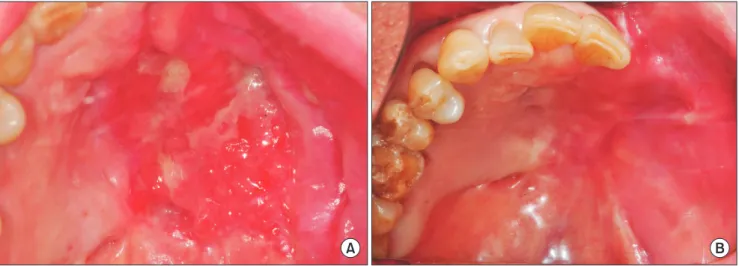
Fig. 5. Re-epithelialization of the grafted buccal fat pad 20 days (A) and 8 weeks (B) after surgery.
Hyun Seok et al: Reconstruction of partial maxillectomy defect with a buccal fat pad flap and application of 4-hexylresorcinol: a case report. J Korean Assoc Oral Maxillofac Surg 2016
to these areas6. The BFP provides a 7×4×3 cm pedicled tissue when proper dissection and advancement are performed11. Many authors recommend reconstructive surgery using the BFP with defects smaller than 4×5-cm size18. Our patient’s MEC specimen measured approximately 6×4×3 cm.(Fig. 3) The involved defect area was the left maxillary canine to the tuberosity, the hard palate, and the sinus mucosa floor. We carefully harvested and advanced the BFP flap and success- fully covered the defect without tension.(Fig. 4)
Epithelization of the grafted BFP begins 1 week after sur- gery, and complete healing is noted within 4 to 6 weeks6. This is accomplished by migration of the epithelial cells from the surrounding tissue in the flap margin19. At first, the su- perficial layer of the BFP is replaced with granulation tissue;
however, this eventually turns into stratified squamous epi- thelium18. Histologically, parakeratotic squamous epithelium covers the healed site, and dense fibrous connective tissue is present in the subcutaneous stroma18. To promote re-epithe- lialization of the large graft area, we applied 4HR ointment on the BFP area due to its antiseptic and broad-spectrum an- tibacterial properties10. 4HR inhibits foreign body giant cell formation via the diacylglycerol kinase (DAGK)-mediated pathway20 and suppresses the expression of TNF-α12. In a re- cent study, 4HR ointment was applied to a burn wound, and rapid epithelialization and collagen regeneration occurred12. In the present study, we applied 4HR ointment on the graft area for rapid re-epithelialization. The large graft area was re-epithelialized 20 days after surgery.(Fig. 5. A) During the healing process, minimal complications such as infection, hematoma, or partial necrosis were noted11. However, these complications have rarely been reported, and as in our previ- ous study, most cases of BFP flap successfully covered the saucerization defect without any complications14. In the pres- cysts1. High-grade MEC has local infiltration and grows ag-
gressively, similar to squamous cell carcinoma4. Radiologi- cally, high-grade MEC has irregular, poorly-defined margins and infiltrative adjacent tissue, while low-grade MEC dem- onstrates a well-delineated mass similar to a benign tumor3.
The MEC treatment varies based on the tumor location, clinical stage, and histological grade3. If MEC occurs in the parotid gland, a parotidectomy is performed, and the facial nerve will be excised4,13. MEC of the minor salivary gland in the hard palate is treated with wide local resection3. Low- grade MEC is surgically excised in a single piece along with the involved underlying mucosa and bone for an adequate tumor-free margin5. High-grade MEC has local infiltration and regional lymph node metastasis14,15, requiring wider surgical resection with postoperative radiotherapy or chemo- therapy1,16. Lymph node metastasis can occur in low-grade MEC1.
The biopsy result of the present case was low-grade MEC, and a reactive lymph node was suspected around the left submandibular gland based on contrast CT. Therefore, elec- tive neck dissection was performed along with tumor resec- tion. Low-grade MEC has a favorable prognosis; the 5-year survival rate is 76% to 95%, and recurrence has rarely been reported17.
The BFP has been widely used as a pedicled flap in the oral cavity for closure of an oroantral fistula, coverage of a sequestrectomy defect in osteomyelitis, regenerative treat- ment of peri-implantitis, and treatment of oral submucous fibrosis6,8. Reconstruction using the BFP is considered a reli- able technique due to an excellent blood supply, minimal morbidity, easy harvest, and simplicity18. The posterior max- illa, hard palate, and retromolar trigone are the ideal defects for reconstruction with the BFP due to anatomical proximity
A B
Fig. 6. Axial (A) and coronal (B) views showing the grafted buccal fat pad located in the left maxillary alveolus and hard palate defect (arrows) at the 8-week follow-up.
Hyun Seok et al: Reconstruction of partial maxillectomy defect with a buccal fat pad flap and application of 4-hexylresorcinol: a case report. J Korean Assoc Oral Maxillofac Surg 2016
ent case, the grafted BFP was completely epithelialized and covered the defect.(Fig. 5. B)
The hard palate is a frequently involved site of low-grade MEC14. We surgically resected low-grade MEC on the left maxillary alveolus and hard palate and successfully covered the large defect with a BFP flap. 4HR ointment was applied to the fat graft area for rapid re-epithelialization and wound healing. The grafted BFP was completely re-epithelialized without complication. Based on the results from this case, we confirmed that BFP grafting is a simple, reliable, and mini- mally complicated technique for reconstruction of posterior palatal defects, and 4HR is a clinically useful ingredient to promote re-epithelialization of fat.
Conflict of Interest
No potential conflict of interest relevant to this article was reported.
ORCID
Hyun Seok, http://orcid.org/0000-0002-5278-3807 Min-Keun Kim, http://orcid.org/0000-0002-5481-841X Seong-Gon Kim, http://orcid.org/0000-0001-5088-2732
References
1. Brandwein MS, Ivanov K, Wallace DI, Hille JJ, Wang B, Fahmy A, et al. Mucoepidermoid carcinoma: a clinicopathologic study of 80 patients with special reference to histological grading. Am J Surg Pathol 2001;25:835-45.
2. Caccamese JF Jr, Ord RA. Paediatric mucoepidermoid carcinoma of the palate. Int J Oral Maxillofac Surg 2002;31:136-9.
3. Baumgardt C, Günther L, Sari-Rieger A, Rustemeyer J. Mucoepi- dermoid carcinoma of the palate in a 5-year-old girl: case report and literature review. Oral Maxillofac Surg 2014;18:465-9.
4. Kokemueller H, Brueggemann N, Swennen G, Eckardt A. Muco- epidermoid carcinoma of the salivary glands--clinical review of 42 cases. Oral Oncol 2005;41:3-10.
5. Nabil S, Lo RC, Choi WS. Simultaneous radicular cyst and muco-
epidermoid carcinoma in the maxilla: a diagnostic nightmare. BMJ Case Rep 2013. doi: 10.1136/bcr-2013-010290.
6. Youn T, Lee CS, Kim HS, Lim K, Lee SJ, Kim BC, et al. Use of the pedicled buccal fat pad in the reconstruction of intraoral de- fects: a report of five cases. J Korean Assoc Oral Maxillofac Surg 2012;38:116-20.
7. Egyedi P. Utilization of the buccal fat pad for closure of oro-antral and/or oro-nasal communications. J Maxillofac Surg 1977;5:241-4.
8. Yeh CJ. Application of the buccal fat pad to the surgical treat- ment of oral submucous fibrosis. Int J Oral Maxillofac Surg 1996;
25:130-3.
9. Lee SW, Um IC, Kim SG, Cha MS. Evaluation of bone formation and membrane degradation in guided bone regeneration using a 4-hexylresorcinol-incorporated silk fabric membrane. Maxillofac Plast Reconstr Surg 2015;37:32.
10. Kim SG, Kim AS, Jeong JH, Choi JY, Kweon H. 4-hexylresorcinol stimulates the differentiation of SCC-9 cells through the suppres- sion of E2F2, E2F3 and Sp3 expression and the promotion of Sp1 expression. Oncol Rep 2012;28:677-81.
11. Rapidis AD, Alexandridis CA, Eleftheriadis E, Angelopoulos AP.
The use of the buccal fat pad for reconstruction of oral defects: re- view of the literature and report of 15 cases. J Oral Maxillofac Surg 2000;58:158-63.
12. Ahn J, Kim SG, Kim MK, Kim DW, Lee JH, Seok H, et al. Topical delivery of 4-hexylresorcinol promotes wound healing via tumor necrosis factor-α suppression. Burns 2016;42:1534-41.
13. Kim IK, Cho HW, Cho HY, Seo JH, Lee DH, Park SH. Facelift incision and superficial musculoaponeurotic system advancement in parotidectomy: case reports. Maxillofac Plast Reconstr Surg 2015;37:40.
14. Rapidis AD, Givalos N, Gakiopoulou H, Stavrianos SD, Faratzis G, Lagogiannis GA, et al. Mucoepidermoid carcinoma of the salivary glands. Review of the literature and clinicopathological analysis of 18 patients. Oral Oncol 2007;43:130-6.
15. Kim BG, Kim JH, Kim MI, Han JJ, Jung S, Kook MS, et al. Ret- rospective study on factors affecting the prognosis in oral cancer patients who underwent surgical treatment only. Maxillofac Plast Reconstr Surg 2016;38:3.
16. Kim CM, Park MH, Yun SW, Kim JW. Treatment of pathologic fracture following postoperative radiation therapy: clinical study.
Maxillofac Plast Reconstr Surg. 2015;37:31.
17. Mesolella M, Iengo M, Testa D, DI Lullo AM, Salzano G, Salzano FA. Mucoepidermoid carcinoma of the base of tongue. Acta Oto- rhinolaryngol Ital 2015;35:58-61.
18. Samman N, Cheung LK, Tideman H. The buccal fat pad in oral reconstruction. Int J Oral Maxillofac Surg 1993;22:2-6.
19. Hanazawa Y, Itoh K, Mabashi T, Sato K. Closure of oroantral com- munications using a pedicled buccal fat pad graft. J Oral Maxillo- fac Surg 1995;53:771-5; discussion 775-6.
20. Kweon H, Kim SG, Choi JY. Inhibition of foreign body giant cell formation by 4-hexylresorcinol through suppression of diacylglyc- erol kinase delta gene expression. Biomaterials 2014;35:8576-84.