J Korean Soc Surg Hand 2015;20(4):153-160.
http://dx.doi.org/10.12790/jkssh.2015.20.4.153
THE HAND
INTRODUCTION
Traffic accidents or industrial accidents may be the most frequent cause of large finger defect injury1, which can
hardly be treated by replantation since it can cause strip- ping of skin and soft tissues and involve vascular injuries due to several injury mechanisms. Regarding the flap in reconstruction of large finger defects, it is necessary to con-
The Usability of Medial Sural Artery Perforator Flap for Reconstruction of the Finger Defects
Min-Kyu Hwang, Sung-Chul Chu, So-Min Hwang, Hyung-Do Kim, Min-Wook Kim, Jong-Seo Lee
Aesthetic, Plastic and Reconstructive Surgery Center, Good Moonhwa Hospital, Busan, Korea
Received:June 26, 2015 Revised:[1] August 19, 2015
[2] September 25, 2015 Accepted:October 5, 2015
Correspondence to:Sung-Chul Chu Aesthetic, Plastic and Reconstructive Surgery Center, Good Moonhwa Hospital,
119 Beomil-ro, Dong-gu, Busan 48735, Korea TEL:+82-51-630-0199
FAX:+82-51-630-01459 E-mail:[email protected]
Purpose: Groin or abdominal flap, anterolateral thigh free flap, and radial fore- arm flap can typically be performed in large defects, however satisfactory results in functional recovery and aesthetic aspect have not been achieved using these methods. Medial sural artery perforator free flap is recommended as a comple- ment to these disadvantages, therefore we report the functional and aesthetic results of this flap for reconstruction of large finger defects.
Methods:From January 2008 to December 2013, 10 patients with large soft tis- sue defect of the fingers were treated with medial sural artery perforator free flap. Six months after the final surgery, metacarpophalangeal joint and proxi- mal interphalangeal joint range of motion was measured, and the circumfer- ence of the reconstructed finger was compared with that of the contralateral side. In addition, for assessment of the aesthetic satisfaction, the patients and three physicians compared the color of the reconstructed finger with that of adjacent skin on a five-point scale.
Results:The flaps survived without complications in all ten cases. Average flex- ion was 77 degrees in the metacarpophalangeal joint and 84 degrees in the proximal interphalangeal joints. The average circumference of the reconstruct- ed finger was measured as 12 percent larger than contralateral. The patient’s subjective satisfaction (4.1) and physicians’ objective satisfaction (4.2) regard- ing aesthetic aspect were very good.
Conclusion:Medial sural artery perforator free flap is a very thin, stable, fascio- cutaneous flap which has a tendon gliding effect and produces aesthetically good results. Therefore we consider medial sural artery perforator free flap as the flap which can solve the drawbacks of other techniques associated with large finger defect reconstruction.
Keywords:Finger defect, Medial sural artery perforator flap
This is an Open Access article distributed under the terms of the Creative Commons Attribution Non-Commercial License (http://creativecommons.org/licenses/by- nc/3.0/) which permits unrestricted noncommercial use, distribution, and reproduction in any medium, provided the original work is properly cited.
sider both the fact that a hand has a potential function, which involves tendon gliding, and the morphological aspect, which requires a thin flap.
Several types of flaps, which involve microsurgery, are used in reconstruction of large finger defects accompanied by functional limitations due to soft-tissue or bone defects.
In general, it is possible to use groin or abdominal flap, anterolateral thigh free flap, and radial artery forearm flap, any of which fails to produce satisfactory results in terms of functional recovery and aesthetics.
Since introduction of medial sural artery perforator free flap by Cavadas et al.2in 2001, many researchers have rec- ognized its clinical usefulness. The medial sural artery per- forator flap can be detailed by a perforator from the axis of the medial sural artery or from the medial sural artery itself.
First and second perforators can be harvested at approxi- mately 10 cm and 15 cm, respectively, along the axis from the mid-popliteal crease. A sizable perforator with accom- panying venae committant is precisely located with an average pedicle length exceeding 9.5 cm. It is a thin and pli- able flap compared to other perforator flap donor sites, and allows wide usage in hand soft tissue resurfacing.
Fasciocutaneous flap characteristic preserves the tendon gliding effect, which carries significant importance in func- tional aspect in hand reconstruction. In this study, we ana- lyze some cases of employing a medial sural artery perfora- tor free flap in reconstruction of large finger defects to determine its usability in terms of functional recovery and aesthetics.
MATERIALS AND METHODS
1. Subjects
This study was conducted in ten patients who had under- gone medial sural artery perforator free flap for large finger defects, which involved no injury in flexed or annular liga- ments of any finger, at this hospital between January 2008 and December 2013. They ranged in age from 21 to 51 (37 on average) and the male-to-female ratio was 8 to 2. The hand injuries were caused by a machinery accident in four cases and by a contact burn in one case. Each patient required coating due to skin and soft tissue defects in fingers.
2. Methods
1) Operative technique
History taking and basic examinations were performed to determine whether the patients could bear prolonged surgery and angiography was used to identify vascular injuries or variation in recipient and donor sites. Doppler and ultrasonography were used to mark a perforator of proper size in the calf, a donor site.
The patients were asked to lie in a supine position with hip abduction and knee joint flexion to elevate a flap. We designed the flap, giving consideration to the defect range at the recipient site, and had the perforator placed in the lat- eral area of the flap to obtain sufficient length of a pedicle.
The anterior border of the flap was incised and dissected and full dissection was performed on a perforator and accompanying veins.
The perforator made a musculocutaneous type of travel- ing in every case and the flap was shifted to the recipient site and vascular anastomosis was performed for free flap.
Primary suture could be performed at the donor site when the flap was 7 cm or smaller in diameter. Debulking was performed at least three months postoperatively as needed.
3. Postoperative assessment
The following items were measured for objective assess- ment of the level of functional improvement six months after the final operation. First, the range of motion of the metacarpal phalgeal and proximal interphalangeal joints of the operated finger were measured and compared with the unaffected side. The wrist joint was fixed and the fixed part of the protractor was placed parallel to the metacarpal bone, the metacarpophalangeal joint was flexed to the max- imum, the moving part of the protractor was placed parallel to the proximal bone, and the flexion angle of the metacar- pophalangeal joint was measured. Second, we measured the circumference of the proximal phalanx of the recon- structed fingers and compared the measurements with those of normal ones to determine the difference. Third, to assess the aesthetic satisfaction, the patients and three physicians compared the color of the reconstructed finger with adjacent skin on a five-point scale, which was modified with a visual analogue scale. Very well matching was 5
points, well matching was 4 points, moderate matching was 3 points, poor matching was 2 points, and very poor match- ing was 1 point.
RESULTS
All patients received free medial sural artery perforator free flap and the flaps survived without complication. All donor sites could be closed primarily. Debulking was performed postoperatively in two cases. Average flexion was 77 degrees in the metacarpophalangeal joint and 84 degrees in the proximal interphalangeal joints. The average circumference of the reconstructed finger was measured as 12 percent larger than contralateral (Table 1).
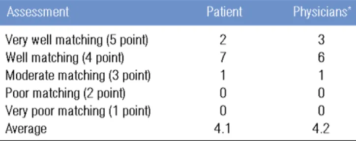
In results of the patient’s aesthetic satisfaction survey, 2 patients answered very well matching (5 points), 7 patients answered well matching (4 points), and 1 patient answered moderate matching (3 points), so that the aver- age patient’s subjective satisfaction was 4.1. In the aesthet- ic satisfaction survey of 3 physicians, 3 were very well matching (5 points), 6 were well matching (4 points), and 1 was moderate matching (3 points), so that the average physicians’ objective satisfaction was 4.2 (Table 2). Both patients’ and physicians’ satisfaction regarding aesthetic aspect was very good.
1. Case report 1) Case 1
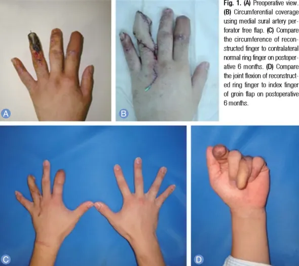
A 21-year-old male had a large defect on the index and ring fingers of his left hand, which was caught in a machine while he was working one month before he visited the hos- pital. He underwent groin flap in the first finger and com- posite graft in the third finger at the other hospital. At the time of his visit to this hospital, necrosis of the composite graft was in progress and skin and soft tissue defects with exposure of bone and tendon were observed after debride- ment of necrotic tissue. We planned to use a medial sural artery perforator free flap to coat the finger. Thus, a thin medial sural artery perforator free flap, 5×4 cm in size, was taken from his left medial calf to cover the exposed bones and tendons of his ring finger. The pedicle measured 6cm in length and was anastomosed end-to-end to the common digital artery. Six months postoperatively, the reconstructed finger was 7% greater circumference than the normal one
Table 1.Patient summary and results
1 Male/21 Lt. ring finger 5×4 cm, circumferential - 70 60 7 7.5
2 Male/24 Rt. ring finger 10×7 cm, radial side O 80 100 6 7
3 Female/41 Rt. index finger 5×4 cm, dorsum - 75 80 6.5 7.5
4 Male/49 Rt. ring finger 6×5 cm, circumferential - 90 100 6.5 7
5 Male/51 Lt. middle finger 8×6 cm, circumferential O 70 80 7 8
6 Female/43 Rt. long finger 6×ø6 cm, circumferential - 75 85 6.5 7.5
7 Male/31 Rt. index finger 6×5 cm, dorsum - 85 90 7 7.5
8 Male/37 Rt. index finger 5×5 cm, radial side - 85 90 6.5 7.5
9 Male/35 Lt. middle finger 7×6 cm, circumferential - 70 80 6 6.5
10 Male/39 Lt. index finger 6×6 cm, dorsum - 70 75 7 8
Average 77 84 6.6 7.4
MP, metacarpophalangeal; PIP, proximal interphalangeal; RF, reconstructed finger; Lt., left; Rt., right.
Sex/age Flexion angle (°) Finger PIP joint
Case (yr) Injured finger Debulking circumference (cm)
MP joint PIP joint Normal RF
Table 2.Aesthetic satisfaction
Very well matching (5 point) 2 3
Well matching (4 point) 7 6
Moderate matching (3 point) 1 1
Poor matching (2 point) 0 0
Very poor matching (1 point) 0 0
Average 4.1 4.2
*The results were assessed by three clinical physicians.
Assessment Patient Physicians*
and had better function with 70° flexion of the metacar- pophalangeal joint and 60° flexion of the proximal interpha- langeal joint than the index finger undergoing groin flap (Fig. 1).
2) Case 2
A 24-year-old male had a large defect in the region below the metacarpophalangeal joint of the right ring finger, which was caught in a sand mill before he visited the hospi- tal. Replantation was performed using the stump, however stump necrosis occurred 15 days postoperatively. We per- formed marginal tissue removal and planned to use a medi- al sural artery perforator free flap to coat the finger. Thus, a thin medial sural artery perforator free flap, 10×7 cm in size, was taken from his left medial calf to cover the exposed bones and tendons of his ring finger. The pedicle measured 8 cm in length and was anastomosed end-to-end to the
common digital artery. After debulking was performed five months postoperatively, the reconstructed finger had 16%
greater circumference than the normal one 12 months post- operatively and the joint range of motion improved: 80°
flexion for the metacarpophalangeal joint and 100° flexion for the proximal interphalangeal joint (Fig. 2).
DISCUSSION
Complete circumferential large defect of the digits usually results in a large cutaneous defect with tendinous struc- tures, bone and joint exposure. Traffic accidents or industri- al accidents may be the most frequent cause of large finger defects, and both can hardly be treated because it can involve a wide range of vascular injuries and because it is difficult to predict the range of viable tissue3. In general, it can be treated by making a sufficient excision to severely
Fig. 1. (A)Preoperative view.
(B) Circumferential coverage using medial sural artery per- forator free flap.(C)Compare the circumference of recon- structed finger to contralateral normal ring finger on postoper- ative 6 months.(D)Compare the joint flexion of reconstruct- ed ring finger to index finger of groin flap on postoperative 6 months.
injured or necrosed tissues and, then by performing skin grafting or flap according to the severity of the defect.
However circumferentially stripped fingers with circulatory disturbance constitute an unfavorable bed for skin grafts. In addition, skin grafting is not usually considered because of problems associated with wound contraction, non-gliding of tendons directly under skin grafts, and avascular necrosis of distal bones. Although local flaps offer the main advan- tage of similar texture, their use is limited by the size and location of the defect. The traditional type of treatment involves primary suture and coating with a local or distal pedicled flap and step-by-step reconstruction of injured tis- sues of ligaments, bones, and nerves. However, this has failed to produce satisfactory results because of delayed functional recovery in the hand. Development of micro- scopic surgery has enabled primary reconstruction of injured tissues, which involves free flap for transplanting composite tissues of bones, muscles, and nerves all at once.
This method can be most useful in reconstructing the hand in that it may shorten the duration of treatment and reduce
complications4. Many authors have reported that free flap is an excellent reconstruction method which can not only restore the joint motions by providing potential space for tendon and ligament gliding essential for functional recov- ery in the hand but also require a significantly shorter dura- tion of treatment than the existing ones4,5. For selection of an appropriate flap for a large finger defect, consider the type of defect tissue, condition of the recipient site, defect size, and shape. In general, it is possible to use groin or abdominal flap, anterolateral thigh free flap, and radial artery forearm flap for a digital large defect injury.
Groin or abdominal flap can cover the defect easily within a short period of time, and provide sufficient size for cover- age, with low donor morbidity and good aesthetic out- comes. However, there are many anatomical variations, and the length of the pedicle is short. The other disadvantage of this technique is that the hand must be attached to the dis- tant body part for at least 2 weeks, which usually causes joint stiffness resulting from prolonged immobilization.
Anterolateral thigh free flap, introduced by Song et al.6in Fig. 2. (A)Preoperative view.(B)Intrao-
perative finding of medial sural artery perforator free flap harvesting. (C) Circumferential coverage using medial sural artery perforator free flap. (D) Postoperative 12 months view.
1984, is widely used both because it can provide a combina- tion of diverse tissues and because a perforator of sufficient size can elevate a large skin flap7. However, as the vessels in the adipose layer run obliquely into the adipose tissue after penetrating the deep fascia, it is difficult to estimate the cor- rect point of the vessel in a very thin layer of adipose tissue.
If a wide thin flap is required, defatting after elevating the full thickness flap can be performed readily, but is an inac- curate procedure for elevation of a small, thin flap, which sometimes leaves vessels behind. In addition, it can cause anatomical vascular variation and require significant time for pedicle dissection. In addition, it may not only provide a lower level of aesthetic satisfaction because it is too thick to be used in reconstruction of a finger but also requires debulking. Such thickness of the flap can place restrictions on the motion of the finger joint8.
A radial artery forearm flap can be used as a sensory flap both because radial artery and cutaneous nerves make rela- tively constant gliding and because the flap can include nerves. However, it may have high donor morbidity and require sacrifice of principal vessels9.
Since introduction of the medial sural artery perforator free flap by Cavadas et al.2in 2001, many researchers have recognized its clinical usefulness. In this report, our experi- ence showed that it may not only produce aesthetically good results by elevating a thinner flap (average 5±2 mm) than any other type of flap since the skin in the calf is very thin but also obtain a long pedicle 5-12 cm in length (aver- age, 9.5 cm), and flap dimension was 90-120 cm2because vessels are constantly distributed. In addition, the major perforator of the medial sural artery is easier to identify than the perforators of other flaps, thus enabling a rather safe and rapid dissection between the deep fascia and the medi- al gastrocnemius muscle. The flap can be elevated only from the subcutaneous fat layer, consequently preserving the muscular layer with low donor morbidity. In addition, it can be elevated in the supine position without postural change during surgery and make the surgical procedure easy due to the use of a tourniquet. Elevation of a ≤7 cm flap may enable a primary suture at the donor site10,11.
This flap can be used for resurfacing large defects involv- ing one whole finger or almost half of a finger caused by
degloving injury or crushing injury. However the defect is small enough to cover with a local flap or regional flap or the dimension is larger than 90-120 cm2, then other meth- ods should be considered. As a fasciocutaneous flap effec- tive in tendon gliding, it is expected to solve problems asso- ciated with other operation techniques in reconstruction of digital circumferential large defect injuries. However, metic- ulous pedicle dissection is required in order to avoid pedicle injury, and caution is required for kinking of the pedicle during wrapping the circumferential large defect site with medial sural artery perforator free flap.
CONCLUSION
In this study, we used a medial sural artery perforator free flap for reconstruction of a digital large defect injury and measured the range of motion of the reconstructed finger:
the metacarpophalangeal and proximal interphalangeal joints had 77° and 84° flexion, respectively, on average. The results indicated a notable postoperative improvement in the range of motion since the range of motion of normal joints is 90° for the metacarpophalangeal joints and 90° to 100° for the proximal interphalangeal joints. It enabled reconstruction of fingers thinner than any other type of operation since the reconstructed fingers were 12% thicker than the normal ones. We used a medial sural artery perfo- rator free flap, which complements the weaknesses of the existing surgical procedures, and achieved functional and aesthetically satisfactory results in terms of reconstruction for digital large defect injuries.
REFERENCES
1. de Korte N, Dwars BJ, van der Werff JF. Degloving injury of an extremity. Is primary closure obsolete? J Trauma. 2009;67:E60-1.
2. Cavadas PC, Sanz-Gimenez-Rico JR, Gutierrez-de la Camara A, Navarro-Monzonis A, Soler-Nomdedeu S, Martinez-Soriano F. The medial sural artery perforator free flap. Plast Reconstr Surg. 2001;108:1609-15.
3. Slattery P, Leung M, Slattery D. Microsurgical arterial- ization of degloving injuries of the upper limb. J Hand Surg Am. 2012;37:825-31.
4. Hahn SB, Kim SH, Jung JH, Kang HJ. Free flap transfer for the reconstruction of injured hands. J Korean Soc Surg Hand. 2005;10:234-41.
5. Daniel RK, May JW Jr. Free flaps: an overview. Clin Orthop Relat Res. 1978;(133):122-31.
6. Song YG, Chen GZ, Song YL. The free thigh flap: a new free flap concept based on the septocutaneous artery.
Br J Plast Surg. 1984;37:149-59.
7. Ali RS, Bluebond-Langner R, Rodriguez ED, Cheng MH.
The versatility of the anterolateral thigh flap. Plast Reconstr Surg. 2009;124(6 Suppl):e395-407.
8. Koshima I, Nanba Y, Tsutsui T, Takahashi Y. New
anterolateral thigh perforator flap with a short pedicle for reconstruction of defects in the upper extremities.
Ann Plast Surg. 2003;51:30-6.
9. Ahn HC, Choi MS, Hwang WJ, Sung KY. The transverse radial artery forearm flap. Plast Reconstr Surg. 2007;
119:2153-60.
10. Xie RG, Gu JH, Gong YP, Tang JB. Medial sural artery perforator flap for repair of the hand. J Hand Surg Eur Vol. 2007;32:512-7.
11. Xie XT, Chai YM. Medial sural artery perforator flap.
Ann Plast Surg. 2012;68:105-10.
수지 연부조직 결손의 피복에서 내측 비복동맥 천공지 피판술의 유용성
황민규∙추성철∙황소민∙김형도∙김민욱∙이종서
좋은문화병원 미용성형재건센터
목적:수지의 넓은 연부조직 결손 시 일반적으로 서혜부 또는 복부 피판술, 대퇴유리피판술, 외측상완피판술 등을 시행할
수 있는데, 기능적 회복과 미용적 측면에서 만족스러운 결과를 얻지 못하였다. 내측 비복 천공지 유리피판술은 이러한 단 점들을 보완해주기에 만족스러운 기능적, 미용적 결과를 보고하는 바이다.
방법:2008년 1월부터 2013년 12월까지 본원에서 손가락의 넓은 면적의 연부조직 결손 환자 10명을 대상으로 내측 비
복 천공지 유리피판술을 시행하여 손가락을 재건하였다. 수술 후 6개월에 중수지 관절과 근위지 관절의 운동범위를 측 정하였고 재건수지의 근위지 둘레를 정상측과 비교하였고 재건수지와 주변 피부의 조화를 본인과 의사 3명이 5점 만점 으로 하여 만족도를 조사하였다.
결과:평균 중수지 관절 굴곡 77�, 평균 근위지 관절 굴곡 84�로 측정되었으며, 재건수지의 둘레는 정상측 수지보다 평
균 12% 더 두꺼운 것으로 측정되었다. 재건 수지에 대한 환자 개인의 주관적 만족도는 평균 4.1점, 의사 3명의 평가에 의한 객관적 만족도는 평균 4.2점으로 결과가 좋았다.
결론:내측 비복 천공지 피판술은 매우 얇고 안정적인 피판이라 수지의 넓은 연부조직 결손에 유용하며 기능적, 미용적
으로 우수한 피판술이라고 여겨진다.
색인단어:수지 결손, 내측 비복 천공지 피판술
접수일2015년 6월 26일수정일1차: 2015년 8월 19일, 2차: 2015년 9월 25일 게재확정일2015년 10월 5일
교신저자추성철 부산시 동구 범일로 119 좋은문화병원 미용성형재건센터
TEL051-630-0199 FAX051-630-0145 [email protected]