INTRODUCTION
Lumbosacral soft tissue defects are commonly encountered in the field of reconstructive surgery. Soft tissue defects in the lumbosacral area can be caused by pressure sores in hemi- or paraplegia patients, postoperative dehiscence, tumor removal, radiation ulcers, trauma, and burns, etc. Many surgical methods have been developed to correct lumbosacral defects, including primary closure, skin grafting, local random flaps, and muscle flaps. Muscle and myocutaneous flaps, which provide excellent padding, have been traditionally used as a coverage method
for skin and soft tissue defects in the sacral area.1 The gluteus maximus musculocutaneous flap is most commonly used because of its location relative to the defect and excellent blood supply and durability; however, the use of this flap might leave functional deficits in ambulatory patients and eliminate other reconstructive options in relapse cases. Moreover, the recurrence rate after surgical treatment for pressure sores has been reported to be 13% to 61%.2,3
Recently, with the advancement of microsurgical techniques, perforator flaps have been used in various cases. Perforator flaps based on the gluteal artery were first introduced by Koshima
Clinical Efficacy of Gluteal Artery Perforator Flaps for Various Lumbosacral Defects
Hyun June Park, Kyung Min Son*, Woo Young Choi, Ji Seon Cheon
Department of Plastic and Reconstructive Surgery, Chosun University College of Medicine, Gwangju, Korea
CC This is an open-access article distributed under the terms of the Creative Commons Attribution Non-Commercial License (http://creativecommons.org/licenses/by-nc/4.0) which permits unrestricted noncommercial use, distribution, and reproduction in any medium, provided the original work is properly cited.
Copyright © 2016 by the Korean Society for Microsurgery. All Rights Reserved.
Received October 24, 2016 Revised November 2, 2016 Accepted November 7, 2016
*Correspondence to: Kyung Min Son Department of Plastic and Reconstructive Surgery, Chosun University College of Medicine, 365 Pilmun-daero, Dong-gu, Gwangju 61452, Korea
Tel: +82-62-220-3978 Fax: +82-62-225-0996 E-mail: [email protected]
ORCID: http://orcid.org/0000-0001-5825-0270
Financial support: This work was supported by Chosun University Hospital.
Conflict of interest: None.
Purpose: Soft tissue defects in the lumbosacral area can be challenging to treat, and various methods to accomplish this have been proposed, including the use of perforator flaps. Herein, we present our experience with superior gluteal artery perforator (SGAP) and inferior gluteal artery perforator (IGAP) flaps for the reconstruction of lumbosacral defects.
Materials and Methods: From March 2013 to July 2016, 28 cases (27 patients) of lumbosacral defects were treated by reconstruction with SGAP or IGAP flaps. The defects were caused by pressure sores (21 cases), burns (3 cases), tumor resection (2 cases), scars (1 case), or foreign body infection (1 case). Reliable perforators around the defect were found using Doppler ultrasound. The perforator flaps were elevated with a pulsatile perforator and rotated to cover the defects.
Results: Twenty-three SGAP and 5 IGAP flap reconstructions were performed. The mean flap size was 9.2×6.1 cm2 (range, 5×3 cm2 to 16×10 cm2). Donor sites were closed by primary closure. Partial flap necrosis occurred in two cases, and minor complications of wound dehiscence occurred in 3 cases, which were healed by primary closure. The mean follow-up period was 4.4 months (range, 1~24 months).
Conclusion: Gluteal-based perforator flaps can be safely harvested due to pliability and reliable vascularity in the gluteal area, reducing donor site morbidity without sacrificing the underlying muscles. Thus, these flaps are useful options for the reconstruction of lumbosacral defects.
Key Words: Soft tissue injuries, Lumbosacral, Perforator flap
ARMS
Archieves of Reconstructive Microsurgery https://doi.org/10.15596/ARMS.2016.25.2.49et al.4 in 1993. Because gluteal artery perforator flaps provide a considerable amount of tissue with good vascularity, minimize donor-site morbidity, and preserve underlying muscles, superior gluteal artery perforator (SGAP) flaps may be excellent for the coverage of lumbosacral wounds.5-8 While SGAP flaps have been widely used, only a few reports with objective outcome data regarding the inferior gluteal artery perforator (IGAP) flap have been presented in Korea. Therefore, the aim of the current study was to present our experience with SGAP and IGAP flaps for the reconstruction of lumbosacral defects and discuss the usefulness of these flaps for lumbosacral defects.
MATERIALS AND METHODS
Patients
A retrospective review of 27 patients (18 men and 9 women;
mean age, 54.5 years; age range, 22~74 years) who underwent 28 cases of reconstruction with SGAP- or IGAP-based flaps at the Department of Plastic and Reconstructive Surgery of Chosun University Hospital from March 2013 to July 2016 was performed (Table 1). The mean defect size was 6.6×4.9 cm2 (ranging from 1×1 cm2 to 15×10 cm2). The defect etiologies included pressure sores (21 cases with one patient undergoing IGAP flap coverage for pressure sores on both ischial areas), postburn defect (3 cases), tumor resection (2 cases), scar (1
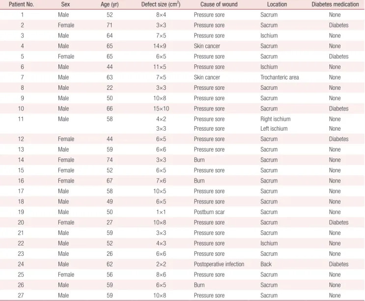
Table 1. Patients reconstructed with gluteal artery perforator flaps
Patient No. Sex Age (yr) Defect size (cm2) Cause of wound Location Diabetes medication
1 Male 52 8×4 Pressure sore Sacrum None
2 Female 71 3×3 Pressure sore Sacrum Diabetes
3 Male 64 7×5 Pressure sore Ischium None
4 Male 65 14×9 Skin cancer Sacrum None
5 Female 65 6×5 Pressure sore Sacrum Diabetes
6 Male 44 11×5 Pressure sore Ischium None
7 Male 63 7×5 Skin cancer Trochanteric area None
8 Male 22 3×3 Pressure sore Sacrum None
9 Male 50 10×8 Pressure sore Sacrum None
10 Male 66 15×10 Pressure sore Sacrum Diabetes
11 Male 58 4×2
3×3
Pressure sore Pressure sore
Right ischium Left ischium
None None
12 Female 44 6×5 Pressure sore Sacrum Diabetes
13 Male 59 6×6 Pressure sore Sacrum None
14 Female 74 3×3 Burn Sacrum None
15 Female 52 6×5 Pressure sore Sacrum None
16 Female 67 7×6 Burn Sacrum None
17 Male 58 10×5 Pressure sore Sacrum None
18 Male 49 6×5 Pressure sore Sacrum None
19 Male 50 1×1 Postburn scar Sacrum None
20 Female 27 10×8 Pressure sore Sacrum Diabetes
21 Male 59 3×3 Pressure sore Sacrum None
22 Male 52 4×3 Pressure sore Ischium None
23 Male 26 6×6 Pressure sore Sacrum None
24 Male 62 2×2 Postoperative infection Back Diabetes
25 Female 56 8×6 Pressure sore Sacrum None
26 Male 59 6×5 Burn Sacrum None
27 Male 59 10×8 Pressure sore Sacrum None
case), and foreign body infection (1 case).
Surgical technique
Surgery occurred once a negative bacterial culture of the wound was obtained. Patients were placed in the prone position.
During reconstruction of the sacral defect, potadine-soaked gauze was packed on anus to prevent infection associated with contamination. Preoperatively, adequate debridement of the bone and adjacent soft tissue was performed. If the bone was exposed, necrotic or nonviable bone was excised using a bone rongeur until bleeding from the bone occurred. Then, a hand- held Doppler assessment, guided by anatomical landmarks, was performed to mark the location of the gluteal perforators and the planned rotation flap was marked. SGAP perforators were situated mainly around the junction of the middle and medial third of the line drawn between the posterior superior iliac spine and the greater trochanter. IGAP perforators were located on a marked area around the horizontal middle third of the gluteal region parallel to the gluteal crease.9 Most of the flaps were elevated with one reliable perforator, and the width of the flap was designed to be 10% larger than the defect size. The incision was made down to the fascial layer of the gluteal muscle. Using electrocautery, subfascial dissection was performed working from lateral to medial. Once the selected perforator was identified, radical skeletonization dividing all the fascial strands around the perforator was performed to prevent kinking.
Circulation was verified with capillary reaction after rotation and drains were placed beneath the flap, which were removed after 48 to 72 hours. The flap was inset into the defect area and the donor site was covered layer by layer. Color, temperature, bleeding, and venous refill of the flap were carefully evaluated at the end of the suture and in the first 72 hours.
RESULTS
Twenty-three cases involved SGAP flaps (one was combined with a split thickness skin graft) and 5 cases involved IGAP flaps (Table 2). The average flap size was 9.2×6.1 cm2 (range, from 5×3 cm2 to 16×10 cm2). Perforator flap survival was complete, with the exception of two cases of partial flap necrosis in patients with diabetes mellitus; the flap loss sites were subsequently covered with a contralateral V-Y advancement
flap. Three patients developed minor complications of partial dehiscence in the wound edge, which was closed by delayed primary closure. All donor sites were closed by primary intent, and there were no complications on the suture margins.
Case 1 (Case 25)
A 56-year-old female with paraplegia due to a traumatic fracture on the thoracic spine in 2008 visited to our department for pressure sores in the sacral area. She had a 8×6 cm2-sized pressure sore, and the muscles were exposed with a significant amount of discharge from a suspected infection. She was treated
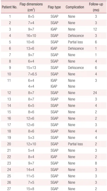
Table 2. Summary of results Patient No. Flap dimensions
(cm2) Flap type Complication Follow-up (mo)
1 8×5 SGAP None 3
2 7×4 SGAP None 3
3 9×7 IGAP None 12
4 16×10 SGAP Dehiscence 3
5 10×6 SGAP Partial loss 6
6 13×6 IGAP Dehiscence 1
7 9×7 SGAP None 1
8 6×4 SGAP None 4
9 15×13 SGAP Dehiscence 6
10 7×6.5 SGAP None 4
11 6×4
4×4
IGAP IGAP
None None
3
12 8×7 SGAP None 24
13 8×7 SGAP None 3
14 6×5 SGAP None 4
15 8×6 SGAP None 2
16 12×6 SGAP None 2
17 12×6 SGAP None 3
18 8×6 SGAP None 4
19 5×3 SGAP None 4
20 12×10 SGAP Partial loss 2
21 5×4 SGAP None 3
22 8×4 IGAP None 2
23 9×7 SGAP None 8
24 14×4 SGAP None 3
25 11×5 SGAP None 3
26 7×5 SGAP None 3
27 15×8 SGAP None 2
SGAP: superior gluteal artery perforator, IGAP: inferior gluteal artery perforator.
via IV antibiotics and debridement of the unhealthy tissue.
After the infection on the wound site was controlled, a one- stage reconstruction using a SGAP flap was performed. The skin island, which measured 11×5 cm2, was rotated to cover the defect. The donor site was closed primarily. No complications occurred at the donor and recipient sites. At the 1-month follow-up, no sign of infection was seen at the operation site (Fig.
1).
Case 2 (Case 24)
A 62-year-old male with a history of surgical intervertebral fusion due to a herniated lumbar disc visited our department for persistent discharge on the operation site. Once a negative bacterial culture of the wound was obtained, primary closure was performed. However, persistent discharge and delayed wound healing due to a suspected foreign body infection occurred. Therefore, a combined operation with
a neurosurgeon was conducted for the removal of fixation devices. The defect size was 2×2 cm2 after removal of the device;
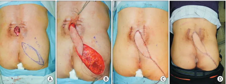
however, a large undermining space was found. Therefore, a 14×4 cm2 de-epithelialized SGAP flap was designed to cover the dead space caused by the foreign body infection. During flap elevation, a reliable perforator was found in the medial margin of the flap. Thus, only perforator skeletonization was performed without intramuscular dissection. After flap elevation, the flap was rotated 180o, and the distal part was de-epithelialized and inset into the defect site to provide adequate coverage. The donor site was closed primarily. The flap survived without any complications. During 3 months of follow-up, his postoperative course was uneventful (Fig. 2).
Case 3 (Case 22)
A 52-year-old male patient with paraplegia due to a traumatic fracture in the thoracic spine in 1995 was referred to us with
A B C D
Fig. 2. (A) The preoperative image, about 14×4 cm2 sized flap was designed in elliptical shape. (B) The intraoperative image, de-epithelialized flap was rotated 180o and inset on the defect. (C) The immediate postoperative image. (D) Follow-up image after 3 months, it didn’t showed infection sign.
A B C
Fig. 1. (A) The preoperative image, superior gluteal artery perforator marking near the defect is shown. Flap size was 11×5 cm2. (B) The intraoperative image, flap was inset on the defect and donor site was primarily closed. (C) The postoperative image after 2 weeks, there are no complications on flap and donor site.
a grade 3 left ischium pressure sore (size, 4×3 cm2). He had undergone previous advancement flap surgery twice because of an ischium sore at same site. Debridement, antibiotic therapy, and wound care were initiated. After achieving control of the infection, we performed reconstruction using an IGAP flap rather than an advancement flap. A hand-held Doppler was used to trace the IGAP around the wound site and an elliptical flap (8×4 cm2), was designed to cover the defect. The flap was rotated 90o and the donor site was closed primarily. The patient showed no recurrence at the 8-month follow-up (Fig. 3).
DISCUSSION
The common causes of lumbosacral defects include pressure sores in paraplegic patients and postoperative dehiscence following spinal surgery. Delayed wound coverage of defects can trigger progressive infections and wound pain. Thus, surgical debridement and subsequent wound reconstructions are the best treatment options for most patients with lumbosacral defects. Pressure sores in paraplegic patients present a particularly difficult challenge because of high rates of wound complication and recurrence.2,3 Therefore, the gluteus muscle or musculocutaneous advancement flaps have been considered to be the standard first-line treatment for lumbosacral reconstruction as they are reliable and involve a short learning curve for surgeons.10-12 However, a major weakness of this method is that it causes a disturbance in gait motion in ambulatory patients due to the removal of gluteus muscle from its original body insertion. Other disadvantages include a bulky appearance, limited flap transposition, and unnecessary blood loss when splitting the muscle.
In 1993, Koshima et al.4 described 20 to 25 perforators supplying the entire gluteal region and used gluteal perforator
flaps to cover sacral pressure sores. Gluteal perforator flaps are large and safe and, moreover, can be raised unilaterally with minimal bleeding, leaving the muscle intact with little donor-site morbidity. With the development of this technique, ambulatory patients can be spared from any difficulty in walking since the gluteus muscle is not sacrificed. In paraplegic patients, preserving the gluteus maximus muscle provides an opportunity to repair recurrent pressure sores. Additional reports of sacral-coccyx reconstruction using a gluteus maximus muscle perforator pedicled flap exist.6,13,14 Based on cadaver dissection studies, of the 7 to 19 perforators dispersed in the gluteal region,9 the length and diameter of the pedicle is reported to be 3.0 to 9.1 cm and 0.6 to 1.6 mm, respectively.4,9 In studies based on Korean populations, the average number of perforators from the gluteus maximus muscle has been reported to be 12.2, with 37% of the perforators originating from the superior gluteal artery, while others originate from the inferior gluteal artery. In our series, the mean size of the gluteal artery flaps was 60.5 cm2 (ranging from 15 to 195 cm2) and the maximum flap size supplied by one gluteal artery perforator could reach 15×13 cm2.
The conventional rotation flap (i.e., a fully undermined rotation fasciocutaneous flap) has been used to preserve the gluteus maximus muscle; however, this design pattern does not have a sufficient blood supply due to a random pattern of pedicles.15 We performed an island-type flap design, with a designed elliptical-shaped skin paddle, and the donor site underwent primary repair. Because a perforator-based flap has a better blood supply compared to that based on a random pedicle pattern, there is an advantage in flap survival rate.
Depending on the location of defect, we used SGAP flaps for the reconstruction of lumbosacral defects, and IGAP flaps for coverage of ischial area defects. SGAP flaps, which are used for
A B C
Fig. 3. (A, B) Intraoperative images, inferior gluteal artery perforator flap was designed and inset on the defect with 90o rotational type. (C) The immediate postoperative image, donor site underwent primary repair.
the sacral area, are inadequate for coverage of the ischial area because of an insufficient pedicle length. Traditionally, many experts have used latissimus dorsi and paraspinous muscle flaps to reconstruct defects of the lumbar area induced by postoperative infections or mass removal.16,17 Recently, experts have started using lumbar artery or posterior intercostal artery perforator flaps due to the development of the microsurgical field.17 Moreover, SGAP has also been shown to be an effective treatment option for lumbar area defects; Moon et al.8 covered the undermining space of the lumbar area (e.g., pseudomeningocele repair, etc.) using de-epithelialized SGAP flaps. In the present study, we provided our experience with a similar case (Case 2).
The inferior gluteal artery is the other dominant blood supply to the gluteal region. Le-Quang16 first reported the use of an inferior gluteal musculocutaneous free flap in 1979. IGAP flaps have similar features to SGAP flaps; however, IGAP flaps have a larger cutaneous territory.9 This has clinical significance for the elevation of bulky flaps on gluteal regions in cases of breast reconstruction. Since pedicled IGAP flaps were used for ischial area pressure sores in 2002 by Higgins et al.,17 several similar studies have been reported.18,19 Particular attention must be paid to wound dehiscence in the treatment of bedsores using IGAP flaps, as the ischial area is very mobile and vulnerable to pressure in a sitting position. The present study included 5 cases using IGAP flaps for the reconstruction of defect and postoperative dehiscence occurred in one case. However, the dehiscence was completely recovered via subsequent primary closure.
The present study has several limitations. First, the follow-up period was relatively short. As most patients were paraplegic, frequent hospital visits were difficult unless the pressure sore recurred. Second, 2 cases of flap necrosis occurred in patients with chronic diabetic mellitus. Because patients with diabetic mellitus generally have many vascular complications, such as atherosclerosis and vasoconstriction, we suggest that surgeons should be aware of the patency of the perforator via preoperative computed tomographyangiography. Third, because IGAP cases were fewer in number compared to SGAP cases, the objective comparison between SGAP and IGAP flaps is difficult. However, with accumulated experience, the survival rate and complications of the two flaps may be evaluated.
CONCLUSION
The SGAP and IGAP flaps provide valuable options for challenging defects on the lumbosacral area. Harvesting these flaps without sacrifice of the underlying muscle means not only reduced donor site morbidity, but also more freedom in composing and tailoring the flap. Because of the pliability and reliable vascularity in the gluteal area, wide and long perforator propeller flaps can be safely harvested and the redundant portion of the flap can be useful in clinical situations such as pressure sores. Therefore, we suggest that these flaps should be considered as useful treatment options for various lumbosacral defects.
REFERENCES
1. Ger R, Levine SA. The management of decubitus ulcers by muscle transposition. An 8-year review. Plast Reconstr Surg 1976;58:419-28.
2. Disa JJ, Carlton JM, Goldberg NH. Efficacy of operative cure in pressure sore patients. Plast Reconstr Surg 1992;89:272-8.
3. Tavakoli K, Rutkowski S, Cope C, Hassall M, Barnett R, Richards M, et al. Recurrence rates of ischial sores in para- and tetraplegics treated with hamstring flaps: an 8-year study. Br J Plast Surg 1999;52:476-9.
4. Koshima I, Moriguchi T, Soeda S, Kawata S, Ohta S, Ikeda A.
The gluteal perforator-based flap for repair of sacral pressure sores. Plast Reconstr Surg 1993;91:678-83.
5. Verpaele AM, Blondeel PN, Van Landuyt K, Tonnard PL, Decordier B, Monstrey SJ, et al. The superior gluteal artery perforator flap: an additional tool in the treatment of sacral pressure sores. Br J Plast Surg 1999;52:385-91.
6. Coşkunfirat OK, Ozgentaş HE. Gluteal perforator flaps for coverage of pressure sores at various locations. Plast Reconstr Surg 2004;113:2012-7.
7. Zeng A, Jia Y, Wang X, Liu Z. The superior gluteal artery perforator flap for lumbosacral defect repair: a unified approach.
J Plast Reconstr Aesthet Surg 2013;66:e56-7.
8. Moon SH, Choi JY, Lee JH, Oh DY, Rhie JW, Ahn ST. Feasibility of a deepithelialized superior gluteal artery perforator propeller flap for various lumbosacral defects. Ann Plast Surg 2015;74:
589-93.
9. Ahmadzadeh R, Bergeron L, Tang M, Morris SF. The superior and inferior gluteal artery perforator flaps. Plast Reconstr Surg 2007;120:1551-6.
10. Fisher J, Arnold PG, Waldorf J, Woods JE. The gluteus maximus musculocutaneous V-Y advancement flap for large sacral defects. Ann Plast Surg 1983;11:517-22.
11. Ramirez OM, Orlando JC, Hurwitz DJ. The sliding gluteus maximus myocutaneous flap: its relevance in ambulatory patients. Plast Reconstr Surg 1984;74:68-75.
12. Gould WL, Montero N, Cukic J, Hagerty RC, Hester TR. The
"split" gluteus maximus musculocutaneous flap. Plast Reconstr Surg 1994;93:330-6.
13. Ao M, Mae O, Namba Y, Asagoe K. Perforator-based flap for coverage of lumbosacral defects. Plast Reconstr Surg 1998;101:
987-91.
14. Roche NA, Van Landuyt K, Blondeel PN, Matton G, Monstrey SJ. The use of pedicled perforator flaps for reconstruction of lumbosacral defects. Ann Plast Surg 2000;45:7-14.
15. Wong CH, Tan BK, Song C. The perforator-sparing buttock rotation flap for coverage of pressure sores. Plast Reconstr Surg
2007;119:1259-66.
16. Le-Quang C. Two new free flaps proceeding from aesthetic surgery: the lateral mammary flap and the inferior gluteal flap.
Paper presented at: Transactions of the Seventh International Congress of Plastic and Reconstructive Surgery; 1979 May 20- 25; Rio de Janeiro, Brazil.
17. Higgins JP, Orlando GS, Blondeel PN. Ischial pressure sore reconstruction using an inferior gluteal artery perforator (IGAP) flap. Br J Plast Surg 2002;55:83-5.
18. Meltem C, Esra C, Hasan F, Ali D. The gluteal perforator-based flap in repair of pressure sores. Br J Plast Surg 2004;57:342-7.
19. Kim YS, Lew DH, Roh TS, Yoo WM, Lee WJ, Tark KC. Inferior gluteal artery perforator flap: a viable alternative for ischial pressure sores. J Plast Reconstr Aesthet Surg 2009;62:1347-54.