C A S E R E P O R T Open Access
Reconstruction of cheek mucosal defect with a buccal fat pad flap in a squamous cell carcinoma patient: a case report and literature review
Dae-Seok Hwang 1,2,3 , Jinyoung Park 1,3* , Uk-Kyu Kim 1,3 , Hae-Ryoun Park 2 , Gyoo-Cheon Kim 2 and Mi-Heon Ryu 2
Abstract
Background: Squamous cell carcinoma (SCC) is the most commonly occurring malignant tumor in the oral cavity.
In South Korea, it occurs most frequently in the mandible, tongue, maxilla, buccal mucosa, other areas of the oral cavity, and lips. Radial forearm free flap (RFFF) is the most widely used reconstruction method for the buccal mucosal defect. The scar of the forearm donor, however, is highly visible and unsightly, and a secondary surgical site is needed when such technique is applied. For these reasons, buccal fat pad (BFP) flap has been commonly used for closing post-surgical excision sites since the recent decades because of its reliability, ease of harvest, and low complication rate.
Case presentation: In the case reported herein, BFP flap was used to reconstruct a cheek mucosal defect after excision. The defect was completely covered by the BFP flap, without any complications.
Conclusion: Discussed herein is the usefulness of BFP flap for the repair of the cheek mucosal defect. Also, further studies are needed to determine the possibility of using BFP flap when the defect is deep, and the maximum volume that can be harvested considering the changes in volume with age.
Keywords: Buccal fat pad flap, Buccal mucosal defect, Buccal fat pad, Oral cavity reconstruction, Pedicled buccal fat pad flap
Background
Squamous cell carcinoma (SCC) accounts for about 90%
of the malignant tumors occurring in the oral cavity [1, 2].
SCC can occur in any part of the mouth, but in South Korea, it occurs most frequently in the mandible, tongue, maxilla, buccal mucosa, other areas of the oral cavity, and lips. The incidence of oral cancer in the mandible and buccal mucosa has been increasing since 2001 [3].
The treatment of SCC in the buccal mucosa consists of a wide surgical excision, and it is essential to achieve negative margins [4]. After a wide surgical excision, the
buccal mucosal defect is most commonly reconstructed through radial forearm free flap (RFFF). This technique, however, leaves a scar on the forearm donor that is highly visible and unsightly. Moreover, it requires a sec- ondary surgical site [5–9]. For these reasons, buccal fat pad (BFP) flap has been commonly used for closing sur- gical excision sites since the recent decades because of its reliability, ease of harvest, and low complication rate [10–17]. A buccal mucosa defect can be successfully covered with a pedicled BFP flap, which was first de- scribed by Egyedi in 1977 for the closure of oroantral and oronasal communications secondary to oncologic resections [10].
Since then, many studies have reported the anatomy of BFP with its blood supply and the large number of cases with a small number of complications [11–22]. Presented herein is a patient diagnosed with SCC on the left-cheek
* Correspondence: [email protected]
1
Department of Oral and Maxillofacial Surgery, School of Dentistry, Pusan National University Dental Hospital, 20, Geumo-ro, Mulgeum-eup, Yangsan, Gyeongsangnam-do, South Korea
3
Dental Research Institute, Pusan National University Dental Hospital, Yangsan, South Korea
Full list of author information is available at the end of the article
© The Author(s). 2018 Open Access This article is distributed under the terms of the Creative Commons Attribution 4.0
International License (http://creativecommons.org/licenses/by/4.0/), which permits unrestricted use, distribution, and
reproduction in any medium, provided you give appropriate credit to the original author(s) and the source, provide a link to
the Creative Commons license, and indicate if changes were made.
mucosa. The malignant tumor was excised, and the defect was covered with a simple pedicled BFP flap.
Case presentation
In October 2015, a 55-year-old female patient was referred to the author’s hospital because of the recurrence of a le- sion in the left buccal mucosa. The patient was diagnosed with squamous epithelial hyperplasia after excisional bi- opsy and histological examination in February 2015, and recurrence was confirmed during the follow-up in Sep- tember 2015. Upon the initial examination at Pusan Na- tional University Dental Hospital, a 2-cm exophytic lesion was observed in the left buccal mucosa (Fig. 1a), and the patient did not have any symptom. Also, no invasion of the left mandible was observed on the panoramic view.
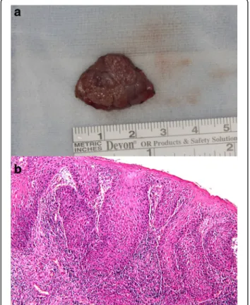
Excision and reconstruction by BFP flap was planned under general anesthesia considering the clinical and radiological findings obtained on November 11, 2015. The size of the removed lesion with a safety margin was 2.1 × 2.0 × 0.9 cm (Fig. 2a), and the buccal mucosa defect was reconstructed as a pedicled BFP flap (Fig. 1b–d). The buc- cal mucosal defect was successfully covered without any tension. The specimen was examined via H&E staining and immunostaining for Ki-67 and p53. The histological examination showed that the atrophic epithelium was
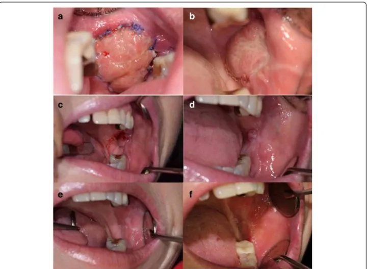
Fig. 1 a Clinical photograph on first visit. b Preoperative lesion. c Lesion was excised. d The immediate post-operative result
Fig. 2 a Gross anatomy. Macroscopically, the excised specimen had
dimensions of 2.1 × 2.0 × 0.3 cm. b Microscopic examination (H&E
stain) confirmed well-differentiated SCC
composed of dysplastic squamous cells with no matur- ation pattern. Moreover, the epithelial cells displayed ac- tive invasion of the underlying stromal tissue (Fig. 2b).
Upon immunohistochemical examination, both Ki-67 and p53 were found to be positive. Therefore, the lesion was diagnosed as a well-differentiated SCC.
After the diagnosis of malignancy, the patient was sub- jected to magnetic resonance imaging (MRI), computed tomography (CT), and positron emission tomography with 2-deoxy-2-fluorine-18-fluoro-D-glucose integrated with computed tomography (18-FDG PET/CT) to deter- mine the treatment direction and cancer stage (Fig. 3). It
Fig. 3 Re-epithelialization of the grafted buccal fat pad after operation. a. The ninth day. b After 3 weeks. c After 1 month. d After 2 months. e After 3 months. f After 1 year
Fig. 4 Radiological findings showing post-op state change on left buccal area. a 18-FDG PET/CT coronal view showing hot FDG spot on left
buccal area because of post-operative change. b T2-weighted magnetic resonance image, coronal view
was found from the 18-FDG PET/CT, MRI, and CT that there was no remnant tumor or no significant cervical lymphadenopathy.
Epithelialization of the grafted BFP was observed on 9 days, 3 weeks, 1 month, 2 months, 3 months, and 1 year after surgery (Fig. 4). The defect was completely covered, without any complications. There was no evidence of re- currence or distant metastasis at every follow-up visit until 2 years after the surgery.
Discussion
In the case reported herein, a buccal mucosal defect was reconstructed with BFP flap. The oral defect was covered by the BFP in the posterior maxilla, hard and soft palate, and retromolar region after teeth extraction, sauceriza- tion, and tumor excision [14, 15, 17].
BFP is an encapsulated mass of adipose tissue in the oromaxillofacial region located in the buccal space be- tween the buccinator muscle and the mandibular ramus and masseter muscle. The BFP has four extensions of the central body: the buccal, pterygoid, pterygopalatine, and temporal extensions [11, 14, 17]. The central body and buccal extension account for approximately 50% of the BFP and are the most clinically significant parts.
These are most commonly transposed to cover oral de- fects [13].
BFP has three sources of blood supply: the maxillary artery (buccal and deep temporal branches), superficial temporal artery (transverse facial branch), and facial ar- tery (small branches). Due to its rich blood supply, the use of BFP flap has a high success rate [12, 15].
Many authors have introduced BFP flap as the safest reconstructive method for small to medium-sized intraoral defects. Martin-Granizo et al. [17] reported that compared to RFFF, the most notable advantages of BFP flap are that it requires a simple and rapid surgical tech- nique, has a low complication rate, and has predictable results without any esthetic sequela. Furthermore, the risk of infection is reduced because BFP is located in the same surgical area as the defect to be covered. Tideman et al. [11] reported the ability to close defects up to 60 × 50 × 30 mm in size using BFP. Moreover, it covers the areas from the premolar area to the posterior tuberosity in the maxilla, retromolar trigone, buccal mucosa, and anterior tonsillar pillar. It must be sutured to the mar- gins of the defect without tension, however, to prevent necrosis of the flap [15].
Pedicled BFP flap, however, has several complications.
Past studies described cheek depression after transfer- ring a large volume of buccal fat [13, 14]. Also, the un- expected complication of mouth opening limitation reported by the previous literatures may result from the dense fibrous connective tissue in the subepithelial stroma lacking lamina propria and submucosa [23].
To minimize the incidence of postoperative complica- tions, it is suggested that the patient receive a liquid or soft, non-chewy diet until BPF epithelialization [15]. Epi- thelialization takes place within 4–6 weeks. Loukas et al.
[24] found that the mean volume of BPF is 10.2 ml in males and 8.9 ml in females, with a 6 mm thickness and a 9.7 g mean weight. Based on a review of the literature, the use of BFP has increased due to its advantages. Few studies, however, have investigated the volumetric varia- tions in BPF among age and gender groups.
Before the use of pedicled BFP flap for the reconstruc- tion of defects, the individual volume of BFP needs to be calculated from radiographic images such as CT or MRI images to assess if coverage is possible. Further studies are needed to determine the possibility of using BFP flap when the defect is deep, and the maximum volume that can be harvested considering the changes in volume with age.
Conclusion
When reconstructing a buccal defect in the oral cavity, pedicled buccal fat pad (BFP) flap is useful. If the defect is small, as in the patients described herein, reconstruc- tion with BFP flap may produce good results. More studies are needed, however, to determine the maximum volume that can be harvested and the size that can be covered by BFP flap considering gender, age, and indi- vidual variations.
Abbreviations
18-FDG PET/CT: Positron emission tomography with 2-deoxy-2-fluorine-18- fluoro- D-glucose integrated with computed tomography; BFP: Buccal fat pad; CT: Computed tomography; MRI: Magnetic resonance imaging;
RFFF: Radial forearm free flap; SCC: Squamous cell carcinoma
Acknowledgements
I would like to thank MB Lee for his proofreading and advice.
Funding
This research was conducted with the support of Clinical Research Grant, Pusan National University Dental Hospital, Republic of Korea.
Authors ’ contributions
JYP obtained data and wrote the manuscript. UKK and GCK carefully helped in drafting the manuscript. HRP and MHR interpreted a slide of resected tissue and took a picture of the slide. DSH participated in its design and coordination and carefully reviewed and revised the manuscript. All authors read and approved the final manuscript.
Ethics approval and consent to participate
This case report was reviewed by Institutional Review Board (IRB) of Pusan National University Dental Hospital and was approved from deliberation.
(PNUDH-2018-001).
Consent for publication
Written informed consent was obtained from the patient for publication of this case report and accompanying images.
Competing interests
The authors declare that they have no competing interests.
Publisher ’s Note
Springer Nature remains neutral with regard to jurisdictional claims in published maps and institutional affiliations.
Author details
1