ISSN 0378-6471 (Print)⋅ISSN 2092-9374 (Online)
https://doi.org/10.3341/jkos.2018.59.7.657
Original Article
인공수정체 낭내 탈구에서 시행된 유리체절제술 및 새로운 인공수정체 공막내고정술의 임상 결과
Clinical Outcomes of Combined Vitrectomy and Intrascleral Fixation of New Intraocular Lenses in In-the-bag Dislocations
이민수⋅채수혜⋅방찬우⋅전혜민⋅윤희성
Min Soo Lee, MD, Sue Hey Chae, MD, Chan Woo Bang, MD, Hye Min Jeon, MD, Hee Seong Yoon, MD, PhD
성모안과병원
Sungmo Eye Hospital, Busan, Korea
Purpose: To investigate the clinical outcomes of combined vitrectomy and intrascleral fixation of a new posterior chamber intra- ocular lens (PC IOL) as a treatment for IOL dislocation.
Methods: We conducted a retrospective interventional study at our medical facility from January 2015 to January 2017.
Posteriorly dislocated IOLs were removed with pars plana vitrectomy. Two intrascleral tunnels, 2.0 mm in length, were created 1.5 mm to the limbus at 6 and 12 o’clock positions. Both haptics of new foldable acrylic 3-piece IOLs were inserted into the tunnel until the IOL was secured in a central position. We analyzed the preexisting ocular condition, visual acuity (VA), and refractive er- ror preoperatively and postoperatively, and recorded postoperative complications.
Results: Forty-nine patients (50 eyes) were enrolled in the study. The mean follow-up period was 12.8 ± 6.6 months. A best-cor- rected VA of 6/12 or better was achieved in 43 eyes (86%). The mean VA significantly improved from 0.32 logarithm of the mini- mum angle of resolution (logMAR) at baseline to 0.18 logMAR at last follow-up (p = 0.03). The refractive status after intrascleral fixation of the PC IOL revealed a mean hyperopic shift of +1.09 ± 1.28 diopters from the predicted spherical equivalent.
Postoperative vitreous hemorrhages occurred in six cases and were cleared without visual compromise. Cystoid macular edema was well-controlled by topical nonsteroidal anti-inflammatory drugs (NSAID) medications in two cases. In two cases, IOL dis- location recurred and required re-operation. There were no serious adverse events of suture-related complications, retinal de- tachment, corneal compromise, or endophthalmitis in any of the patients.
Conclusions: Our data revealed that use of combined vitrectomy and intrascleral fixation of PC IOLs is a safe and efficient tech- nique to correct IOL dislocation. We observed good visual outcomes with only minor complications.
J Korean Ophthalmol Soc 2018;59(7):657-664
Keywords: Intraocular lens dislocation, Intrascleral fixation, Pars plana vitrectomy, Posterior chamber intraocular lens
■Received: 2018. 2. 14. ■ Revised: 2018. 5. 9.
■Accepted: 2018. 6. 22.
■Address reprint requests to Hee Seong Yoon, MD, PhD Sungmo Eye Hospital, #409 Haeun-daero, Haeundae-gu, Busan 48064, Korea
Tel: 82-51-743-0775, Fax: 82-51-743-0776 E-mail: [email protected]
* Conflicts of Interest: The authors have no conflicts to disclose.
ⓒ2018 The Korean Ophthalmological Society
This is an Open Access article distributed under the terms of the Creative Commons Attribution Non-Commercial License (http://creativecommons.org/licenses/by-nc/3.0/) which permits unrestricted non-commercial use, distribution, and reproduction in any medium, provided the original work is properly cited.
인공수정체 탈구는 백내장수술 후 드물지 않은 합병증으 로 전체 백내장수술의 0.2-2.8% 정도의 빈도로 발생한다.1 특히 섬모체소대의 약화와 연관이 깊은 인공수정체 낭내 (in-the-bag) 탈구는 외상, 거짓비늘 증후군, 망막수술의 과 거력 혹은 결체조직질환 등이 위험인자로 알려져 있고, 특 별한 기저 원인 없이 발생하는 경우도 많다.2-4
인공수정체 낭내 탈구를 치료하기 위한 이차 인공수정체
삽입수술에는 다양한 방법이 있으며, 전방 인공수정체, 홍 채 고정 인공수정체, 인공수정체 공막봉합고정술이 그 대표 적인 예이다.5-16 이 중 인공수정체 공막봉합고정술은 다른 방법들에 비해 각막내피세포 손실이 적고, 녹내장의 위험이 적으며, 홍채앞유착의 위험이 적다는 장점이 있다.17-20 하지 만 고정실과 관련된 부작용이 단점으로 남아있는데, 흔한 부작용으로는 고정실에 의한 자극, 감염, 고정실 풀림 등이 있다. 특히 고정실 풀림은 재수술의 적응증이 되기도 하며, 수술 후 6년 내 27.9%의 비교적 높은 빈도로 발생이 보고된 바 있다.21
고정실을 사용하지 않고 이차 인공수정체를 공막내에 고 정하는 인공수정체 공막내고정술은 여러 변형된 방법이 소 개되었다.22-27 섬모체고랑 위치에 공막터널을 만들어 고정 하기도 하고, 피브린글루를 이용하여 고정하기도 하며, 다 양한 모양의 공막절개를 만들어 지지부를 고정하기도 한다.
모든 공막내고정술은 인공수정체의 고정에 고정실을 사용 하지 않기 때문에 고정실과 관련된 부작용이 없다는 장점 이 있다. 하지만 기존 소개된 공막내고정술은 공막터널의 크기가 충분히 크지 않고, 봉합실을 전혀 쓰지 않는 무봉합 고정(sutureless fixation)을 시도하였기에 인공수정체를 지지 하는 고정력이 충분한가에 대한 의문이 든다. 더욱이 기존 의 보고들은 새로운 수술 방법의 소개 및 단기간의 수술 결 과를 보고하는데 그쳐, 비교적 장기간의 안정과 임상 결과 에 대한 고찰은 부족하다. 이에 저자들은 인공수정체 낭내 탈구에서 유리체절제술을 함께 시행하고 공막터널에 새로 운 인공수정체 공막내고정술을 시행하여 6개월 이상의 안 정과 임상 결과에 대해 보고하고자 한다.
대상과 방법
본원에서 2015년 1월부터 2017년 1월까지 인공수정체 낭 내 탈구로 유리체절제술 및 인공수정체 공막내고정술을 시 행받고 최소 6개월 이상 추적 관찰이 가능하였던 환자 49명 (50안)을 대상으로 후향적으로 연구를 진행하였다. 모든 수 술은 한 명의 술자에 의해 시행되었고, 본 연구는 본원 연구 윤리심의위원회의 승인을 받았다(승인번호 2018-BM-02-1).
모든 환자는 수술 전과 후에 세극등현미경검사, 안압측정, 자동굴절검사, 최대교정시력을 포함한 일반적 안과 검사를 받았다. 수술 후 경과관찰은 술 후 1일, 1주, 1, 3, 6개월째 하였으며, 이후 경과관찰은 필요에 따라 최대 6개월 이내로 경과관찰하였다. 수술 후 초기 부작용은 1개월 이내에 발생 한 경우로 정의하였고, 후기 부작용은 1개월 이후에 발생한 경우로 정의하였다. 수술 후 저안압과 고안압은 각각 6 mmHg 이하, 25 mmHg 이상으로 정의하였다. 각막내피세포
밀도는 비접촉 경면 현미경(SP-8000®, Konan Medical, Inc., Hyogo, Japan)을 이용하여 수술 전 및 수술 후 6개월에 측정 하였다. 그리고 부분결합간섭 원리의 OA2000® (Tomey, Nagoya, Japan)을 이용하여 각막 곡률, 안축장 길이 등 생체 계측을 무수정체안 설정(aphakia mode)하에 시행하고 인공 수정체의 도수를 수술 전에 계산하였다.
수술 방법
모든 대상자들은 구후마취하에 23게이지 유리체절제술 을 시행하였다. 탈구된 인공수정체가 연성 재질일 경우 3.0 mm 각공막 절개창을 통해 제거하였고, 탈구된 인공수정체 의 재질이 Polymethyl methacrylate (PMMA)일 경우 6.0 mm 각공막 절개창을 통해 제거하고 10-0 nlyon으로 절개창을 봉합하였다.
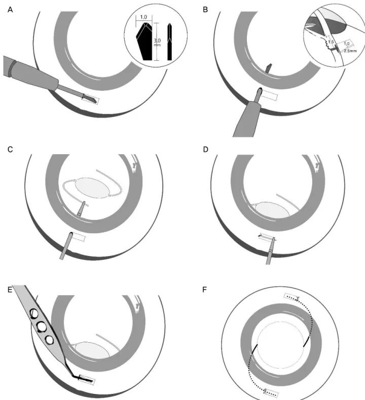
새로 고정할 인공수정체는 모든 예에서 삼체형 인공수정 체인 MN60MA® (Alcon, Fort Worth, TX, USA)를 이용하였 고, 공막내고정은 다음과 같이 시행되었다. 6시와 12시 방 향의 각막윤부 주변에 결막편을 만들어 공막을 노출시키고 전방천자용 절개칼(trifacet paracentesis incision diamond knife 4.1305®, Huco®, Hauterive, Switzerland)을 이용하여 각막윤 부에서 1.5 mm 떨어진 위치에서 층판절개로 두 개의 공막 터널을 각막윤부와 평행하게 만든다(Fig. 1A). 이후 같은 위 치에서 공막터널에 직각을 이루면서 공막을 관통하는 공막 통과절개를 만든다(Fig. 1B). 고정할 인공수정체를 각공막 절 개창을 통해 유리체강 내로 떨어뜨리고, 공막통과절개를 통 해 눈속집게(serrated jaw microforceps 123008®, Optikon®, Rome, Italy)를 넣어 인공수정체 지지부를 잡아 안구 밖으로 빼내고(Fig. 1C, D), 반대편 인공수정체 지지부도 마찬가지 의 방법으로 빼낸다. 맥퍼슨 집게를 이용하여 인공수정체 지지부를 두 군데 공막터널 내로 광학부 중심이 잘 유지됨 을 확인하며 충분히 밀어 넣어 위치시킨다(Fig. 1E). 이때 공막터널 내의 인공수정체 지지부가 비춰보이므로 정확한 위치 확인이 가능하다. 공막터널은 10-0 nylon을 이용하여 단속봉합을 시행하고 그 위로 결막편을 봉합하여 수술을 마무리한다(Fig. 1F).
통계 분석
모든 환자들은 수술 후 1일, 1주, 1개월, 3개월, 6개월 후 에 각각 시력, 안압 검사, 자동굴절검사기와 구면렌즈대응 치를 측정하였으며, 이를 바탕으로 최대교정시력을 측정하 였고, 수술 후 합병증을 관찰하였다. 시력은 logarithm of the minimal angle resolution (logMAR)으로 분석하였고, 수술 후 굴절률과 수술 전 목표 굴절률의 차이를 굴절 오차(spherical refractive error)로 정의하고 분석하였다. 통계학적 분석은
A B
C D
E F
Figure 1. Technique for the intrascleral fixation of intraocular lens (IOL). (A) Scleral tunnel created 1.5 mm from the limbus using
a paracentesis incision diamond knife. Detailed view (inset) of the diamond knife. (B) Transscleral incision created perpendicular to the scleral tunnel. 3-dimensional view (inset) of the scleral tunnel and transscleral incision. (C) The leading haptic of the secondary IOL is grasped with 23-gauge intraocular forceps. (D) The leading haptic is pulled through the transscleral incision, and left externalized. (E) The McPherson forceps grasps the externalized tip, inserts the IOL haptic into the tunnel. (F) The tailing haptic is then inserted into the second scleral tunnel, and the two tunnels were closed in a conventional manner.SPSS 21.0 (IBM Corp., Armonk, NY, USA)을 사용하였으며 수술 전과 후 결과값의 비교는 paired t-test로 분석하였다.
모든 경우 p값이 0.05 이하일 때 유의하다고 하였다.
결 과
49명의 환자(50안)의 나이는 평균 62.1세였으며 남성이
Baseline Final p-value* BCVA (logMAR) 0.34 ± 0.47 0.18 ± 0.32 0.03
IOP (mmHg) 16.0 ± 5.1 14.4 ± 3.3 0.01
ECD (cells/mm2) 2,316.1 ± 541.5 2,206.5 ± 536.0 0.00 Refraction
(SE, diopter)
+8.3 ± 4.4 -0.2 ± 1.8 0.00
Values are presented as mean ± SD unless otherwise indicated.
BCVA = best corrected visual acuity; IOP = intraocular pressure;
ECD = endothelial cell density; SE = spherical equivalent.
*Paired t-test.
Table 2. Changes of BCVA, IOP, ECD and refraction after surgery
Complications No. Eyes (%) Early complicationsVitreous hemorrhage Hypotony
IOP elevation IOL dislocation
Late complications (>1 month) CME
6 (12) 2 (4) 2 (4) 2 (4)
2 (4) Values are presented as n (%).
IOP = intraocular pressure; IOL = intraocular lens; CME = cys- toid macular edema.
Table 3. Postoperative complications
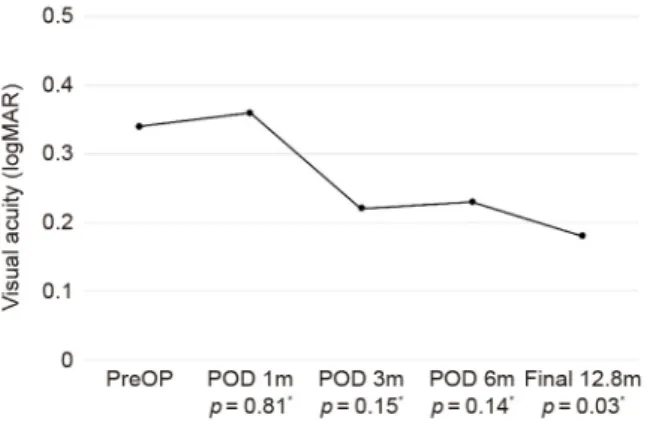
Figure 2. Mean best corrected visual acuity (BCVA) changes
after surgery. The mean BCVA before surgery was 0.34 ± 0.47 (logMAR) and the BCVA improved to 0.18 ± 0.32 at final fol- low-up. Note the temporary deterioration of BCVA at the first month after surgery. PreOP = preoperative; POD = post- operative day; m = month(s). *Paired t-test.Characteristic Data
No. of eyes (patients) 50 (49)
Age (years) (range) 62.1 ± 9.7 (44-80)
Sex (male/female) 42/7
Time to IOL dislocation (years) (range) 11.3 ± 7.3 (0.1-28.0) Follow-up (months) (range) 12.8 ± 6.6 (6-27) Axial length (mm) (range) 24.3 ± 1.8 (21.54-30.30)
BCVA (logMAR) 0.34 ± 0.47
IOP (mmHg) 16.0 ± 5.1
ECD (cells/mm2) 2,316.1 ± 541.5
Refraction (SE, diopter) +8.3 ± 4.4
Values are presented as mean ± SD (range) unless otherwise indicated.
IOL = intraocular lens; BCVA = best corrected visual acuity; IOP
= intraocular pressure; ECD = endothelial cell density; SE = spherical equivalent.
Table 1. Preoperative clinical characteristics
42명, 여성이 7명이었다. 첫 백내장수술 후 인공수정체 낭 내 탈구가 발생하기까지 평균 기간은 11.3 ± 7.3년이었다.
인공수정체 공막내고정술 후 평균 추적관찰 기간은 12.8개 월로 최소 6개월부터 최대 27개월까지 추적검사를 시행하 였다(Table 1).
최대교정시력(logMAR)은 수술 전 0.34 ± 0.47이었으며 수술 후 1개월, 3개월, 6개월 그리고 최종 0.36 ± 0.47, 0.22
± 0.33, 0.23 ± 0.38 그리고 0.18 ± 0.32로 변화하였고 수술 전과 최종 시력의 차이만 통계학적으로 유의하였다(p=0.03) (Table 2, Fig 2).
구면렌즈대응치는 수술 전 +8.3 ± 4.4디옵터였으며 수술 후 1개월, 3개월, 6개월 그리고 최종 -0.3 ± 2.0, -0.3 ± 1.9, -0.3 ± 1.9 그리고 -0.2 ± 1.8디옵터로 변화하였고 수술 전과 수술 후의 차이는 통계학적으로 모두 유의하였다(p<0.05) (Table 2). 굴절 오차는 수술 후 최종 +1.1 ± 1.3디옵터 더 원시로 편위되어 있었다. 각막내피세포 밀도(cells/mm2)는 수술 전 2,316.1 ± 541.5에서 수술 후 6개월째 2206.5 ± 536.0, 평균 -7.0 ± 20.7%로 통계학적으로 유의하게 감소하 였다(p<0.05) (Table 2).
수술 후 초기 합병증으로 유리체 출혈이 6안(12%)으로 제일 흔하였으며, 일시적 안압 하강 2안(4%), 일시적 안압
상승 2안(4%), 인공수정체 재탈구 2안(4%)이 발생하였다.
수술 후 후기 합병증으로 낭포황반부종이 2안(4%)에서 발 생하였다. 수술 후 경과관찰 기간 동안 각막 부전, 망막 박 리, 인공수정체 광학부의 동공 포획, 안내염 등의 합병증이 발생한 경우는 없었다(Table 3).
고 찰
인공수정체 낭내 탈구를 치료하는 방법에는 크게 낭내 탈 구된 인공수정체를 그대로 이용하여 공막에 봉합하여 재위 치(IOL reposition)시키거나 탈구된 인공수정체를 제거하고 새로운 인공수정체를 고정(IOL exchange)하는 방법으로 나 뉜다. 두 방법에 따른 임상 결과의 차이는 유의하지 않은 것 으로 알려져 있고, 이에 술자의 선호에 따라 수술 방법을 선 택하게 된다.28 탈구된 인공수정체를 제거하고 새로운 인공 수정체를 고정하는 방법에는 전방인공수정체를 이용하거나 홍채 또는 공막에 후방인공수정체를 고정하는 것이 있다.
이 중 후방인공수정체를 공막에 고정하는 것은 수정체 고유
의 위치에 인공수정체가 위치하기 때문에 이론적으로 각막 내피세포의 손상과 녹내장이 생길 위험이 적어 다른 이차 인공수정체 삽입술보다 선호되는 방법이다.29 이차 인공수 정체를 공막에 고정하는 방법은 크게 고정실을 이용하여 공 막에 봉합하여 고정하는 공막봉합고정술과 고정실을 사용 하지 않고 공막내에 인공수정체 지지부를 고정하는 공막내 고정술로 나뉜다. 기존 공막봉합고정술의 가장 흔한 단점은 고정실과 관련된 합병증인데, 고정실에 의한 염증 및 공막 위축, 고정실의 노출, 고정실의 분해 및 끊어짐에 따른 인공 수정체 위치 이동 등이다. 이 중 인공수정체 공막고정실 매 듭의 노출은 비교적 흔히 발생하는 합병증으로, 안구 내 세 균 침입의 통로로 작용하여 안내염까지 합병될 수 있다.30 인공수정체 공막내고정술은 기존의 공막봉합고정술의 단점을 극복할 수 있어 최근 다양한 수술 방법이 소개되었
다.24,31-34 공막내고정술은 다양한 위치에, 다양한 술기를 이
용하여 공막터널을 만들고 여기에 인공수정체 지지부를 고 정하기 때문에 고정실을 사용하지 않는다. 공막터널을 만드 는 방법에는 24게이지 캐뉼라를 이용하는 방법, 부분층 공 막 덮개를 이용하는 방법, 27게이지 주사침을 이용하는 방 법 등이 있다.22-24 하지만 이와 같은 술기를 실제로 적용하 기에는 수술기구의 제약이 있고, 술기가 직관적이지 않아 습득하기에도 많은 어려움이 예상된다. 더욱이 이 술기들에 관한 장기간의 안전이나 임상적 결과에 대한 보고도 아직 없다. 본 저자들이 소개한 공막내고정술은 공막터널을 만드 는 방법이 평소 흔히 사용하는 수술 기구로만 시행할 수 있 고 직관적이어서 술기 습득이 비교적 용이할 것으로 생각 된다. 또한 장기간 추적 관찰 결과 큰 부작용 없이 안전하고 좋은 임상 결과를 보였다.
인공수정체 낭내 탈구는 치료하지 않을 경우 낭포황반부 종, 만성 전방 염증, 인공수정체 움직임에 의한 망막 열공, 망막 박리 등을 일으킬 수 있다.35-38 현대의 유리체망막수술 기술의 발전에 힘입어 유리체강 내로 탈구된 인공수정체를 어렵지 않게 제거할 수 있게 되었다. 본 저자들처럼 탈구된 인공수정체를 눈속집게를 이용하여 잡아 전방으로 끌어올 린 후 각공막절개창을 통해 제거하는 방법이 가장 흔하게 사용되는 방법이며, 경우에 따라서 과불화탄소를 주입하여 탈구된 인공수정체를 망막으로부터 띄운 뒤 집게로 잡아 제거하는 것이 더 안전한 방법이 될 수도 있다.
인공수정체 탈구를 치료함에 있어서 유리체절제술을 함 께 시행하는 것에는 여러 장점이 있다. 인공수정체 탈구와 이에 합병된 유리체망막 질환을 동시에 해결할 수 있어 시 력의 회복이라는 수술의 목적을 빠르게 달성할 수 있는 것 이 가장 큰 장점이다. 수술 중 주입액을 통해 안압을 안정적 으로 유지할 수 있으며, 전방을 유지, 안정시켜주어 수술 시
야가 좋아져 공막고정술을 보다 정확하고 안전하게 시행할 수 있다. Althaus and Sundmacher39는 인공수정체 공막봉합 고정술 시 수술 중 안압이 정상적으로 유지될 때 보다 더 정확한 봉합고정이 가능하다 하였는데, 이는 공막터널을 만 들고 공막내고정을 함에 있어서도 마찬가지일 것으로 생각 된다. 더욱이 공막고정술 전에 유리체절제술을 시행하게 되 면 홍채 후면에 유리체가 남아있지 않아, 이후 인공수정체 고정을 위한 조작 과정에서 유리체 견인에 의한 망막 손상 의 위험을 줄여 줄 수 있다. 본 연구에서는 모든 환자에 대 해 인공수정체 공막내고정술과 유리체절제술을 함께 시행 하였는데 수술 후 부작용으로 낭포황반부종의 발생 빈도가 낮았던 점(4%), 망막박리가 없었던 점도 이와 관련이 있다 고 생각된다. Bading et al40은 인공수정체 공막봉합고정술 과 함께 유리체절제술을 시행한 결과 큰 부작용이 없이 안 전한 결과를 보였다 하고, 젊은 환자에 있어서도 무수정체 안을 치료함에 있어 이차 인공수정체 삽입술과 유리체절제 술을 동시에 시행하는 것이 더 적절하다 하였다. 하지만 유 리체절제술을 동시에 시행하는 것은 수술 자체가 복잡하고, 많은 경험과 능숙한 기술이 필요하기에 주의가 필요하다.
본 연구에서 인공수정체 공막내고정술 후 구면렌즈대응 치 굴절 오차는 예측 값에 비해 1.1 ± 1.3디옵터 원시로 편 위된 결과를 보였다. 다양한 방법의 인공수정체 공막내고정 술에 관한 과거 연구들에서 굴절 오차에 관한 언급은 없었
다.24,31-34 공막봉합고정술 후 굴절 오차에 관한 연구 결과는 있
었는데, Yang and Chao41은 -1.18디옵터의 근시화, Donaldson et al42은 -1.32디옵터의 근시화, Suto et al43은 -0.78디옵터의 근시화로 다수의 연구에서 근시화되는 경향을 보고하였다.
본 연구의 결과와 뚜렷한 차이가 존재하는데, 그 이유에 대 해 두 가지 정도로 생각해 보았다. 첫째로 지지부의 고정 위 치에 따른 차이가 존재한다. 앞서 언급한 연구 모두 각막 윤 부에서 1.5 mm 떨어진 위치에 봉합고정술을 시행했다는 점 에서 본 연구와 차이가 없으나, 본 연구의 경우 공막 터널의 위치를 윤부의 후방경계를 기준으로 삼았다는 점이다. 각막 윤부의 두께는 일반적으로 2 mm로 알려져 있는데, 각막 윤 부의 기준점을 어떻게 하느냐에 따라 유효렌즈위치(effective lens position, ELP)가 변하고 본 연구의 경우 유효렌즈위치 가 타 연구에 비해 후방에 존재할 것으로 생각된다. 두 번째 이유로 지지부의 고정 방법에 따른 차이가 존재한다. 본 연 구의 경우 폭 1.0 mm, 길이 2.5 mm의 공막터널에 인공수정 체 지지부를 삽입하였다. 고정실로 지지부의 중간 부위를 묶어 공막 안쪽 지점에 고정하는 봉합고정술의 경우보다 지지부의 움직임에 공간적 여유가 있다. 이와 같은 차이는 인공수정체에 과도한 장력이 작용하는 것을 줄여줄 수 있 는데, Ma et al44은 공막봉합고정술 시 인공수정체에 과도한
장력이 작용할 경우 인공수정체의 전방 이동을 유발한다 하였다. 이와 같은 이유로 본 연구의 경우, 인공수정체의 고 정 위치가 예상보다 후방에 있다고 해석할 수 있고, 타 연구 에서 5.4-13.2%의 확률로 보고된 바 있는 수술 후 광학부 동 공포획이 본 연구에서 발생하지 않았던 것도 같은 이유라 생각된다.41-43 이에 저자들은 본 연구의 수술 방법으로 수술 을 계획할 때에는 수술 후 -1.0~-1.5디옵터로 목표 굴절력을 설정하는 것을 추천한다.
저자들은 본 수술법의 장기 임상관찰에서 몇 가지 수술 후 합병증을 경험하였다. 초기 합병증으로 유리체 출혈이 6 안(12%)에서 발생하였는데, 최근의 타 연구에서 공막내고 정술 후 유리체 출혈의 발생률 0%, 3.2%, 5.0%, 7.0%보다 현저히 높은 발생률이었다.31,45-47 이는 본 수술 시 시행한 공 막통과절개와 연관이 높다고 생각된다. 저자들은 이차 인공 수정체의 지지부를 안구 밖으로 빼낼 때 공막통과절개를 통 해 눈속집게를 이용하였다. 또한 이 과정에서 눈속집게 조 작에 의한 절개창 주변의 앞섬모체동맥과 같은 혈관의 손상 가능성이 있다. 타 연구에서는 본 과정에서 공막통과절개가 아닌 주사침을 이용하였기 때문에 본 연구보다 낮은 유리체 출혈 빈도를 보였다 생각된다.31,45,46 하지만 본 연구에서 발 생한 수술 후 유리체 출혈은 모두 치료 없이 자연적으로 호 전되었고, 수술 후 일시적 시력 악화 후에 호전을 보인 것과 연관이 있다(Fig 2). 유리체 출혈이 별다른 치료 없이 호전 된 것은 모든 예에서 유리체절제술을 함께 시행했다는 점과 공막통과절개에 단순 봉합을 시행했다는 점에 기인한다고 생각된다. 인공수정체 재탈구는 2안(4%)에서 발생하였는데, 모두 한쪽의 지지부만 이탈되었고, 2안 모두 안축장 길이가 각각 29.31 mm, 30.30 mm로 고도 근시안에서 발생하였으 며, 이 중 1안에서는 수술 후 일시적 저안압이 동반되었다.
고도 근시안의 경우 안구 직경이 크고, 공막이 얇아 공막터 널에 의한 인공수정체 지지부의 고정력이 충분하지 않았으 리라 생각되며, 더욱이 저안압이 동반될 경우 고정력은 더 약화되었을 것이라 생각된다. 재탈구된 예를 치료함에 있어 서도 본 수술법은 이점이 있다. 기존의 공막터널과 공막통 과절개 부위를 박리하고 눈속집게를 이용하여 재탈구된 인 공수정체 지지부를 끄집어내어 다시 고정함으로써 쉽게 재 탈구를 해결할 수 있었기 때문이다. 수술 후 각막내피세포 수는 수술 전에 비해 유의하게 감소하였다. 각막내피세포 수의 감소는 안구 내 수술 자체만으로 유발될 수 있고, 또 탈구된 인공수정체를 제거하는 과정에서 각막내피세포의 손상이 가중되어 발생하였을 것으로 생각된다. 본 수술 후, 수술 전에 비해 평균 -7.0 ± 20.7%의 폭으로 각막내피세포 수가 감소하였는데, 이는 유리체절제술 후(-9.0 ± 14.6%), 유 리체절제술 및 백내장수술 후(-13.9 ± 15.5%) 관찰된 각막내
피세포 수의 감소폭보다 적은 양이었고, 기존의 전방인공수 정체 고정술에 비해서도 적은 양이었다.48,49
본 연구는 의무기록을 후향적으로 분석한 연구로서 정확 한 연구 디자인이 되지 못한 한계점이 있다. 특히 장기간의 임상 결과를 알아보기 위해 6개월 이상 경과관찰이 가능했 던 경우만 분석에 포함하였기에 선택 편향(selection bias)의 가능성이 존재한다. 수술 후 결과에 영향을 미치는 요인으로 인공수정체의 기울어짐이나 중심이탈 등을 분석에 포함하 지 못한 것도 한계점이다. 전안부빛간섭단층촬영(CASIA SS-1000, Tomey Corporation, Nagoya, Japan)을 거의 모든 예 에서 시행은 하였으나, 수술 후 일정한 시기에 시행하지 않 았기에 수술 후 결과에 포함시켜 분석하기에는 한계가 있었 다.
저자들은 모든 예에서 한 종류의 삼체형 인공수정체 (MN60MA®, Alcon)로 수술을 진행하였기에 다양한 인공수 정체에 따른 결과를 비교하지 못하였다는 점이 아쉬움으로 남는다. 다만 현재 국내에서 통용되는 대부분의 삼체형 인 공수정체는 MN60MA® (Alcon)와 마찬가지로 광학부 직경 6.0 mm, 전체 직경 13.0 mm의 형태여서 인공수정체 종류에 따른 수술 후 결과에 큰 차이가 존재할 가능성은 낮다고 생 각된다. 추가적으로 Matsuki et al50의 실험 연구에 따르면, 인공수정체 공막내고정술 시에 인공수정체의 전체 직경은 13.9-14.9 mm의 길이로 잡아당겨지게 되고 고위수차와 난 시가 다소 발생하지만 수술 후 시력에는 큰 영향이 없다고 하였다. 고도근시안에서 14.0 mm 크기의 인공수정체를 사 용하면 고정력의 안정 측면에서는 도움이 되리라 생각되며, 이에 추후 추가적인 연구가 필요하다.
결론적으로 인공수정체 낭내 탈구를 치료함에 있어 유리 체절제술 및 인공수정체 공막내고정술은 장기간의 임상관 찰 결과에서 안전하고 효과적인 방법이라 하겠다. 본 수술 방법은 특별한 수술 기구나 어려운 술기 없이 공막터널을 만들어 인공수정체를 고정하고, 그와 함께 유리체절제술을 시행함으로써 장기간 좋은 시력 결과를 보였고, 심각한 합 병증도 발생하지 않았다. 이에, 전방인공수정체 고정술 및 공막봉합고정술의 단점을 보완할 수 있는 좋은 대안이라 생각된다. 본 연구진의 결과로는 수술 후 -1.0~-1.5디옵터로 목표 굴절력을 설정하는 것이 도움이 될 것이라 생각된다.
추후 본 수술 방법에 따른 기울어짐과 중심이탈에 대한 평 가와 보다 더 정교하고 안정적인 방법에 대해 지속적인 연 구가 필요할 것으로 생각된다.
REFERENCES
1) Gross JG, Kokame GT, Weinberg DV; Dislocated In-The-Bag
Intraocular Lens Study Group. In-the-bag intraocular lens dislocation. Am J Ophthalmol 2004;137:630-5.
2) Krėpštė L, Kuzmienė L, Miliauskas A, Janulevičienė I. Possible predisposing factors for late intraocular lens dislocation after rou- tine cataract surgery. Medicina (Kaunas) 2013;49:229-34.
3) Fernández-Buenaga R, Alio JL, Pérez-Ardoy AL, et al. Late in-the-bag intraocular lens dislocation requiring explantation: risk factors and outcomes. Eye (Lond) 2013;27:795-801; quiz 802.
4) Davis D, Brubaker J, Espandar L, et al. Late in-the-bag sponta- neous intraocular lens dislocation: evaluation of 86 consecutive cases. Ophthalmology 2009;116:664-70.
5) Zeh WG, Price FW Jr. Iris fixation of posterior chamber intraocular lenses. J Cataract Refract Surg 2000;26:1028-34.
6) Por YM, Lavin MJ. Techniques of intraocular lens suspension in the absence of capsular/zonular support. Surv Ophthalmol 2005;
50:429-62.
7) Azar DT, Wiley WF. Double-knot transscleral suture fixation tech- nique for displaced intraocular lenses. Am J Ophthalmol 1999;128:
644-6.
8) Bloom SM, Wyszynski RE, Brucker AJ. Scleral fixation suture for dislocated posterior chamber intraocular lens. Ophthalmic Surg 1990;21:851-4.
9) Chan CK. An improved technique for management of dislocated posterior chamber implants. Ophthalmology 1992;99:51-7.
10) Chang S, Coll GE. Surgical techniques for repositioning a dis- located intraocular lens, repair of iridodialysis, and secondary in- traocular lens implantation using innovative 25-gauge forceps. Am J Ophthalmol 1995;119:165-74.
11) Friedberg MA, Pilkerton AR. A new technique for repositioning and fixating a dislocated intraocular lens. Arch Ophthalmol 1992;110:413-5.
12) Kokame GT, Yamamoto I, Mandel H. Scleral fixation of dislocated posterior chamber intraocular lenses: temporary haptic external- ization through a clear corneal incision. J Cataract Refract Surg 2004;30:1049-56.
13) Maguire AM, Blumenkranz MS, Ward TG, Winkelman JZ. Scleral loop fixation for posteriorly dislocated intraocular lenses.
Operative technique and long-term results. Arch Ophthalmol 1991;109:1754-8.
14) Schneiderman TE, Johnson MW, Smiddy WE, et al. Surgical man- agement of posteriorly dislocated silicone plate haptic intraocular lenses. Am J Ophthalmol 1997;123:629-35.
15) Koh HJ, Kim CY, Lim SJ, Kwon OW. Scleral fixation technique using 2 corneal tunnels for a dislocated intraocular lens. J Cataract Refract Surg 2000;26:1439-41.
16) Lewis JS. Ab externo sulcus fixation. Ophthalmic Surg 1991;22:
692-5.
17) Drolsum L. Long-term follow-up of secondary flexible, open loop, anterior chamber intraocular lenses. J Cataract Refract Surg 2003;29:498-503.
18) Biro Z. Results and complications of secondary intraocular lens implantation. J Cataract Refract Surg 1993;19:64-7.
19) Downing JE. Ten-year follow up comparing anterior and posterior chamber intraocular lens implants. Ophthalmic Surg 1992;23:308-15.
20) Evereklioglu C, Er H, Bekir NA, et al. Comparison of secondary implantation of flexible open-loop anterior chamber and scler- al-fixated posterior chamber intraocular lenses. J Cataract Refract Surg 2003;29:301-8.
21) Vote BJ, Tranos P, Bunce C, et al. Long-term outcome of combined pars plana vitrectomy and scleral fixated sutured posterior chamber intraocular lens implantation. Am J Ophthalmol 2006;141:308-12.
22) Gabor SG, Pavlidis MM. Sutureless intrascleral posterior chamber intraocular lens fixation. J Cataract Refract Surg 2007;33:1851-4.
23) Agarwal A, Kumar DA, Jacob S, et al. Fibrin glue-assisted suture- less posterior chamber intraocular lens implantation in eyes with de- ficient posterior capsules. J Cataract Refract Surg 2008;34:1433-8.
24) Yamane S, Inoue M, Arakawa A, Kadonosono K. Sutureless 27-gauge needle-guided intrascleral intraocular lens implantation with lamellar scleral dissection. Ophthalmology 2014;121:61-6.
25) Totan Y, Karadag R. Trocar-assisted sutureless intrascleral posteri- or chamber foldable intra-ocular lens fixation. Eye (Lond) 2012;26:788-91.
26) Ohta T, Toshida H, Murakami A. Simplified and safe method of su- tureless intrascleral posterior chamber intraocular lens fixation:
Y-fixation technique. J Cataract Refract Surg 2014;40:2-7.
27) Takayama K, Akimoto M, Taguchi H, et al. Transconjunctival su- tureless intrascleral intraocular lens fixation using intrascleral tun- nels guided with catheter and 30-gauge needles. Br J Ophthalmol 2015;99:1457-9.
28) Kristianslund O, Råen M, Østern A, Drolsum L. Late in-the-bag in- traocular lens dislocation: a randomized clinical trial comparing lens repositioning and lens exchange. Ophthalmology 2017;124:151-9.
29) Güell JL, Barrera A, Manero F. A review of suturing techniques for posterior chamber lenses. Curr Opin Ophthalmol 2004;15:44-50.
30) Heilskov T, Joondeph BC, Olsen KR, Blankenship GW. Late en- dophthalmitis after transscleral fixation of a posterior chamber in- traocular lens. Arch Ophthalmol 1989;107:1427.
31) Scharioth GB, Prasad S, Georgalas I, et al. Intermediate results of sutureless intrascleral posterior chamber intraocular lens fixation. J Cataract Refract Surg 2010;36:254-9.
32) Hayashi K, Hayashi H, Nakao F, Hayashi F. Intraocular lens tilt and decentration, anterior chamber depth, and refractive error after trans- scleral suture fixation surgery. Ophthalmology 1999;106:878-82.
33) Durak A, Oner HF, Koçak N, Kaynak S. Tilt and decentration after primary and secondary transsclerally sutured posterior chamber in- traocular lens implantation. J Cataract Refract Surg 2001;27:227-32.
34) Sinha R, Bansal M, Sharma N, et al. Transscleral suture-fixated versus intrascleral haptic-fixated intraocular lens: a comparative study. Eye Contact Lens 2017;43:389-93.
35) Flynn HW Jr. Pars plana vitrectomy in the management of sub- luxed and posteriorly dislocated intraocular lenses. Graefes Arch Clin Exp Ophthalmol 1987;225:169-72.
36) Smiddy WE. Dislocated posterior chamber intraocular lens. A new technique of management. Arch Ophthalmol 1989;107:1678-80.
37) Campo RV, Chung KD, Oyakawa RT. Pars plana vitrectomy in the management of dislocated posterior chamber lenses. Am J Ophthalmol 1989;108:529-34.
38) Mittra RA, Connor TB, Han DP, et al. Removal of dislocated intra- ocular lenses using pars plana vitrectomy with placement of an open-loop, flexible anterior chamber lens. Ophthalmology 1998;105:1011-4.
39) Althaus C, Sundmacher R. Endoscopically controlled optimization of trans-scleral suture fixation of posterior chamber lenses in the ciliary sulcus. Ophthalmology 1993;90:317-24.
40) Bading G, Hillenkamp J, Sachs HG, et al. Long-term safety and functional outcome of combined pars plana vitrectomy and scler-
= 국문초록 =
인공수정체 낭내 탈구에서 시행된 유리체절제술 및 새로운 인공수정체 공막내고정술의 임상 결과
목적: 인공수정체 낭내(in-the-bag) 탈구에서 시행된 유리체절제술 및 새로운 인공수정체 공막내고정술의 임상 결과를 알아보고자 한다.
대상과 방법: 2015년 1월부터 2017년 1월까지 인공수정체 낭내 탈구로 본원에서 유리체절제술 및 인공수정체 공막내고정술을 시행받 고 6개월 이상 경과관찰이 가능했던 증례를 후향적으로 연구하였다. 수술 방법은 유리체절제술을 먼저 시행하고 탈구된 인공수정체를 제거하였다. 공막내고정술은 각막 윤부로부터 1.5 mm 떨어진 6, 12시 부위에 공막터널을 만들고, 새로운 인공수정체 지지부(haptic)를 공막터널에 삽입하여 고정하였다. 수술 전 상태, 수술 전후 시력 및 굴절력, 수술 후 합병증을 관찰하였다.
결과: 49명 50안이 연구 대상에 포함되었으며, 평균 경과관찰 기간은 12.8 ± 6.6개월이었다. 수술 후 결과에서 최종 관찰의 최대교정 시력이 6/12 또는 그 이상을 보인 환자가 43안(86%)이었으며, 최대교정시력(logMAR)은 수술 전 0.32에서 수술 후 최종 0.18로 유의한 시력 호전을 보였다(p=0.03). 수술 후 굴절 오차는 +1.09 ± 1.28의 원시화를 보였다. 수술 후 부작용으로 유리체 출혈이 6안, 낭포황 반부종이 2안에서 발생하였고 보존적 치료로 모두 회복되었다. 2안에서 탈구가 재발하여 재수술하였고, 고정실 합병증, 망막 박리, 각막 부전, 안내염 등과 같은 심각한 부작용은 없었다.
결론: 인공수정체 낭내 탈구를 치료함에 있어서 유리체절제술 및 인공수정체 공막내고정술은 안전하고 효과적인 방법이며, 고정실과 관련된 부작용이 없는 좋은 수술법이라 하겠다.
<대한안과학회지 2018;59(7):657-664>
al-fixated sutured posterior chamber lens implantation. Am J Ophthalmol 2007;144:371-7.
41) Yang CS, Chao YJ. Long-term outcome of combined vitrectomy and transscleral suture fixation of posterior chamber intraocular lenses in the management of posteriorly dislocated lenses. J Chin Med Assoc 2016;79:450-5
42) Donaldson KE, Gorscak JJ, Budenz DL, et al. Anterior chamber and sutured posterior chamber intraocular lenses in eyes with poor capsular support. J Cataract Refract Surg 2005;31:903-9.
43) Suto C, Hori S, Fukuyama E, Akura J. Adjusting intraocular lens power for sulcus fixation. J Cataract Refract Surg 2003;29:1913-7.
44) Ma DJ, Choi HJ, Kim MK, Wee WR. Clinical comparison of cil- iary sulcus and pars plana locations for posterior chamber intra- ocular lens transscleral fixation. J Cataract Refract Surg 2011;37:1439-46.
45) Kumar DA, Agarwal A, Packiyalakshmi S, et al. Complications and visual outcomes after glued foldable intraocular lens im-
plantation in eyes with inadequate capsules. J Cataract Refract Surg 2013;39:1211-8.
46) Yamane S, Sato S, Maruyama-Inoue M, Kadonosono K. Flanged intrascleral intraocular lens fixation with double-needle technique.
Ophthalmology 2017;124:1136-42.
47) Khan MA, Gupta OP, Smith RG, et al. Scleral fixation of intra- ocular lenses using Gore-Tex suture: clinical outcomes and safety profile. Br J Ophthalmol 2016;100:638-43.
48) Koushan K, Mikhail M, Beattie A, et al. Corneal endothelial cell loss after pars plana vitrectomy and combined phacoemulsification –vitrectomy surgeries. Can J Ophthalmol 2017;52:4-8.
49) Numa A, Nakamura J, Takashima M, Kani K. Long-term corneal endothelial changes after intraocular lens implantation. Anterior vs posterior chamber lenses. Jpn J Ophthalmol 1993;37:78-87.
50) Matsuki N, Inoue M, Itoh Y, et al. Changes in higher-order aberra- tions of intraocular lenses with intrascleral fixation. Br J Ophthalmol 2015;99:1732-8.