Detection of Helicobacter pylori in Saliva of Patient with Oral Lichen Planus
Ji-Won Ryu, D.D.S.,M.S.D., Seung-Woo Kang, D.D.S.,M.S.D., Chang-Lyuk Yoon, D.D.S.,M.S.D.,Ph.D., Jong-Mo Ahn, D.D.S.,M.S.D.,Ph.D.
Department of Oral Medicine, College of Dentistry, Chosun University.
Lichen planus is a common, chronic inflammatory disease of the skin and mucous membrane for which no precise causes have been confirmed. But it is often connected with infections.
Helicobacter pylori (H. pylori) among various bacteria has been associated with the cause of gastritis, peptic ulcer and gastric cancer. Considering the similarities of histological features between gastric ulcer and oral ulcers, it is resonable to assume that H. pylori might also be involved in the development oral mucosal ulceration. So we employed this study to investigate the possible involvement of H. pylori in the aetiology of erosive oral lichen planus.
We analyzed detection rate of H. pylori in saliva of patients with erosive oral lichen planus by nested PCR. As a result, it revealed a significant difference statistically by showing positivity in 16 to 21 (76.2%) saliva samples of patients group and in 11 of 44 (25%) saliva samples of control group (P>0.001). We were able to suppose that H. pylori in saliva can be related to cause of erosive oral lichen planus.
Key wards: Erosive oral lichen planus, Helicobacter pylori, saliva
1)Ⅰ. INTRODUCTION
Helicobacter pylori (H. pylori) has been associated with development of chronic gastritis, peptic ulcer and gastric cancer.1,2) H. pylori, microaerophilous and gram-negative bacteria, exist mainly stomach in human and act important role of
Corresponding author: Jong-Mo Ahn
Department of Oral Medicine, College of Dentistry, Chosun University 421, Seosuk-Dong, Dong-Gu, Gwang-Ju, 501-825, Korea
Tel : 82-62-220-3896 Fax : 82-62-234-2119 E-mail : [email protected] Received: 2008-05-10 Accepted: 2008-06-25
* This study was supported by research funds from Chosun University, 2008.
development of gastrointestinal disease. But finding of H. pylori in saliva, plaque and periodontal pockets mean that oral cavity can be another reservoir of H. pylori.3)
The potential ability of H. pylori to colonize in oral cavity is related with selective and specific adhesion between H. pylori, Fusobacterium nucleatum and Fusobacterium periodontium in human dental plaque. Such selective coaggregations show that dental plaque is may be provided as a reservoir for pathogens outside of human stomach.4) Also, additional characteristic of H. pylori is that it combine with salivary mucins which cover the oral epithelium. This is because sulfated oligosaccharide on salivary mucins provides the receptor structure for adhesion of H. pylori on oral surface.5) Li et al6) reported that they had found a specific DNA of H. pylori by PCR in 30 of 40 (75%) saliva samples of adults infected H. pylori, and
Czésnikiewicz-Guzik et al7) reported that as results of research about existence of H. pylori, they had found H. pylori in 51 (51%) gastric mucosa sample and in 54 (54%) saliva sample.
Because H. pylori exist in stomach and oral cavity like this, a causative role for H. pyloriin the pathogenesis of oral mucosal ulcers has been suggest. Birek et al8) proved that it is relation of oral aphthous ulcer and H. pylori, Sanli et al9) reported results of endoscopy and histopathological research in upper gastrointestinal tract of patients with oral lichen planus.
But, Riggio et al10) reported that results of research do not support a definitive aetiological role for H. pylori in recurrent aphthous ulcer, Shimoyama et al11) asserted that H. pylori might not have a direct association with oral ulcerations.
The purpose of our study was to investigate detection rate of H. pylori in saliva of patient with oral lichen planus, oral ulcerative disease, because of the similarities of histological feature between gastric ulcer and oral ulcers.
Ⅱ MATERIALS AND METHODS 1. MATERIALS
21 Patients with oral lichen planus of erosive type diagnosed in Department of Oral Medicine, Dental Hospital, Chosun University were selected as the patients group, and 44 individuals whose oral condition was good clinically were selected as control group. The age of patients group was from 42 to 81 years ( 8 males and 13 females). The age of control group was 40 and over years ( 19 males and 25 females).
For the study, we collected saliva from them into 1.5ml microcentrifuge tube and the secreted saliva was stored frozen -20℃ prior to experiments.
2. METHODS 1) DNA extraction
using AccuPrepⓇ Genomic DNA Extraction Kit(Bioneer, Daejeon, Korea). First, we mixed 200㎕
of phosphate buffered saline(PBS), 20㎕ of Proteinase K(20mg/ml), and 200㎕ of Binding Buffer(GC) into 1.5ml microcentrifuge which contains saliva, then executed vortexing. After heating it for 10 minutes in 60℃, we added 100㎕
of isopropanol into a tube then vortexed it for 5 seconds. Such mixed solution was poured into prepared binding column tube to conduct centrifuge in 8,000rpm for one minute, then threw away the filtered solution.
We had repeated this process until the mixed solution had finally disappeared. 500㎕ of W1 Buffer was added, then conducted centrifuge in 8,000rpm for one minute, then we poured out the filtered solution, then 500㎕ of W2 Buffer was added, then conducted centrifuge in 8,000rpm for one minute, then poured out the filtered solution, then finally conducted centrifuge in 12,000rpm for one minutes.
Binding column tube was moved to new sterilized 1.5ml tube, and 50㎕ EL Buffer was added into Binding column tube. After making it reaction in room temperature for one minute, centrifuge was conducted in 8,000rpm for one minutes. Then, filtered solution was stored in -20℃ before being used in polymerase chain reaction(PCR).
2) The amplification by PCR
For the first PCR of H. pylori genomic DNA, 10
㎕ template DNA, and 1㎕ of each primer (20pmole/
㎕) in HEPY Primer set (Bioneer, Daejeon, Korea) were added into AccuPower PCR Premix (Bioneer, Daejeon, Korea) which contains 10mM of Tris-HCl(pH9.0), 40mM of KCl, 1.5mM of MgCl2, 250㎛ of dNTP, and 1U of Tag DNA Polymerase, and to make its total volume of 20㎕, we added 8-mop in HEPY Primer set (Bioneer, Daejeon, Korea).
The PCR was performed under conditions of 94℃
5minutes, 1cycle: 94℃ for 30 seconds, 62℃ for 30 seconds, 72℃ for 60 seconds, 33cycles: 72℃ for 5 minutes, 1cycle in PCR thermocycler(MinicyclerTM,
The mixture for second PCR of H. pylori genomic DNA, contained AccuPower PCR Premix (Bioneer, Daejeon, Korea) containing 10mM of Tris-HCl(pH9.0), 40mM of KCl, 1.5mM of MgCl2, 250㎛ of dNTP, and 1U of Tag DNA Polymerase, with 2㎕ of first PCR product, and 1㎕ of each Primer(20pmole/㎕) in HEPY Primer set (Bioneer, Daejeon, Korea), and to make its total volume of 20
㎕, we added 8-mop in HEPY Primer set (Bioneer, Daejeon, Korea). The PCR was performed under conditions of 94℃ 5minutes, 1cycle: 94℃ for 30seconds, 58℃ for 30 seconds, 72℃ for 60 seconds,
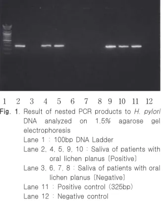
1 2 3 4 5 6 7 8 9 10 11 12 Fig. 1. Result of nested PCR products to H. pylori
DNA analyzed on 1.5% agarose gel electrophoresis
Lane 1 : 100bp DNA Ladder
Lane 2, 4, 5, 9, 10 : Saliva of patients with oral lichen planus [Positive]
Lane 3, 6, 7, 8 : Saliva of patients with oral lichen planus [Negative]
Lane 11 : Positive control (325bp) Lane 12 : Negative control
Group Frequency % P-value
Patients (N=21) 16 76.2
< 0.001
Control (N=44) 11 25
N : Number of individuals
Table 1. The detection of H. pylori in patients with oral lichen planus and control group
33cycles: 72℃ for 5 minutes, 1cycle in PCR thermocycler(MinicyclerTM, MJ Research, U.S.A).
Besides, for every PCR amplification, negative control and positive control in HEPY Primer set (Bioneer, Daejeon, Korea) had also amplified simultaneously. Therefore we excluded the possibility of contamination and false positive.
3) Electrophoresis
We applied on 3㎕ of PCR product on 1.5%
agarose gel which includes ethidium bromide(0.5㎍/
㎖) and after electrophoresis, we analyzed the size of PCR product using UV Transilluminator(Fig. 1).
For Marker, 100 base pairs(bp) Ladder (Bioneer, Daejeon, Korea) was used.
4) Statistical analysis
To compare the detection rate of patients with oral lichen planus and control group, statistical analysis was executed χ2 test with SPSS Windows 12.0 program.
Ⅲ. RESULTS
Nested PCR amplification was executed with template DNA, which was extracted from saliva of 21 patients with oral lichen planus and 44 adults as control group.
From 1.5% agarose gel electrophoresis, we were able to observe amplified PCR product of 325base pair(bp) (Fig. 1). 16 patients (76.2%) with oral lichen planus and 11 adults from control group show positivity, showing statistical significance (Table 1).
Ⅳ. DISCUSSION
Lichen planus is a common, chronic inflammatory disease of the skin and mucous membranes.
The disease has been classified into five groups, namely reticular, erosive, atrophic, hypertrophic and bullae form. The reticular form is the most common variant and is characterized by small white papule (Wickham's striae). The erosive form is the second most frequent variant and is characterized by small or extensive painful erosions with isolated papules or lines at the periphery.12)
The aetiology of lichen planus is unknown, but it is often connected with infections.13) H. pylori among various bacteria lives beneath the gastric mucous layer, on the surface of epithelial cells and produce various enzymes in abundance-urease is one of the most common enzymes associated with H. pylori that has an essential role in the colonization. Urease creates a non-acidic microambiance that allows the bacteria to survive the sterilizing effect of chlorhydric acid. The bacteria travel through the viscous mucus film that protects the gastric epithelium, adhere to the epithelial cells, and begin to replicate. An immune response is subsequently initiated by neutrophils, which results in gastric tissue alteration.14) Considering the similarities of histological features between gastric ulcer and oral ulcers, it is reasonable to assume that H. pylori might also be involved in the development of oral mucosal ulcers.10,15) So a causative role for H. pylori in the pathogenesis of oral mucosal ulcers has been suggested previously.8) Leimola-Virtanen et al15) reported H. pylori DNA can be found in separate oral mucosal ulcers in apparently immunocompetent adults, Long et al16) reported that H. pylori might in some way associated with recurrent aphthous ulcer, which in turn is associated with an increased incidence of digestive disease, and Sanli et al9) asserted that patients with lichen planus should be evaluated for possible gastrointestinal involvement with endoscope. But Vainio et al13) reported that the
lichen planus was not significantly different from that in patients with other skin disease, Daudén et al17) reported that the more virulent cytotoxin-associated gene A(Cag A) positive strains of H. pylori are not strongly associated with lichen planus.
Our study aimed to investigate the possible involvement of H. pylori in the aetiology of erosive oral lichen planus. In our study, H. pylori was detected in 16 of 21 (76.2%) saliva samples of patients with oral lichen planus and in 11 of 44 (25%) saliva samples of control group.
Therefore, we were able to assume that H. pylori in saliva can be relate to cause of erosive lichen planus.
Saliva is a relatively simple biological sample for diagnosis of H. pylori infection. Recently, many analysis methods using PCR have been developed to detect H. pylori in oral cavity. The PCR offers the advantage of detecting H. pylori in area outside the stomach where cultures usually fail, or when biopsy material is not available. PCR performed on DNA extracted from bile juice, stools and oral secretions can detect as few as two, or even a single, H. pylori organism.8,18)
Most of PCR assays are based on arrangement of urease gene and 16S ribosomal RNA gene.19) Lamaroon et al20) studied the association H.
pylori and recurrent aphthous ulcer by a highly sensitive technique, nested PCR and Song et al21) reported than nested PCR showed superior detection ability to H. pylori than [13C] urea breath test after research in saliva and dental plaque. We also were able to get satisfying result by nested PCR using manufactured HEPY Primer set with control DNA (Bioneer, Daejeon, Korea).
Ⅴ. CONCLUSION
We analyzed detection rate of H. pylori in saliva of patients with oral lichen planus by nested PCR to investigate the possible involvement of H. pylori in the aetiology of erosive oral lichen planus. As a
statistically by showing positivity in 16 of 21 (76.2%) saliva samples of patients group and in 11 of 44 (25%) saliva samples of control group.
In conclusion, we were able to suppose that H.
pylori in saliva can be related to cause of erosive oral lichen planus.
REFERENCES
1. Tytgat GN, Noach LA, Rauwa EA. Helicobacter pylori infection and deuodenal ulcer disease.
Gastroenterol Clin North Am 1993;22:127-139.
2. Wotherspoon AC, Ortiz-Hidalgo C, Falzon MR, Lsaacson PG. Helicobacter pylori-associated gastritis and primary B-cell gastic lymphoma. Lancet 1991;338:1175-1176.
3. Dowsett SA, Kowolik MJ. Oral Helicobacter pylori : Can we stomach it ? Crit Rev Oral Biol Med 2003;14:226-233.
4. Andersen RN, Ganeshkumar N, Kolenbrander PE.
Helicobacter pylori adheres selectively to Fusobacterium spp. Oral Microbiol Immunol 1998;
13:51-54.
5. Veerman EC, Bank CN, Namavar F, Appelmelk BJ, Bolscher JG, Nieuw Amerongen AV. Sulfated glycans on oral mucin as receptors for Helicobacter pylori. Glycobiology 1997;7(6):737 -743.
6. Li C, Musich PR, Ha T et al. High prevalence of Helicobacter pylori in saliva demonstrated by a novel PCR assay. J Clin Pathol 1995;48(7):662-666.
7. Czésnikiewicz-Guzik M, Karczewska E, Bieloński W.
Association of the presence of Helicobacter pylori in the oral cavity and in the stomach. J Physiol Pharmacol 2004;15:187-194.
8. Birek C, Grandhi R, McNeill K, Singer D, Ficarra G, Bowden G. Detection of Helicobacter pylori in oral pahthous ulcers. J Oral Pathol Med 1999;28(5):
197-203.
9. Sanli H, Cetinkaya H, Türsen U, Kaya M, Kuzu I, Gürler A. Upper gastrointestinal findings in oral lichen planus. Turk J Gastroentrol 2002;13(1):31-34.
10. Riggio MP, Lennon A, Wray D. Detection of Helicobacter pylori DNA in recurrent aphthous stomatitis tissue by PCR. J Oral Pathol Med 2000;29(10):507-513.
11. Shimoyama T, Horie N, Kato T, Kaneko T, Komiyama K. Helicobacter pylori in oral ulceration.
J Oral Sci 2000;42(4):225-229.
12. Laskaris G. Color Atlas of Oral Disease. New York, 1988, Thieme Medical Publishers, Inc., pp.184-186.
13. Vainio E, Huovinen S, Liutu M, Uksila J, Leino R.
Peptic ulcer and Helicobacter pylori in patients with lichen planus. Acta Derm Venereol 2000;80(6):
427-429.
14. Bussac G. Helicobacter pylori and the oral environment. Practical periodontics and aesthetic dentistiry 1999;11(8):918-922.
15. Leimola-Virtanen R, Happonen RP, Syrjänen S.
Cytomegalovirus (CMV) and Helicobacter pylori (HP) found in oral mucosal ulcers. J Oral Pathol Med 1995;24(1):14-17.
16. Long BJ, Chen K, Wu BL, Duan JM. Detection of Helicobacter pylori in oral cavity of patients with recurrent aphthous ulcer. Nan Fang Yi Ke Da Xue Xue Bao 2007;27(4):477-478.
17. Daudén E, Cabrero MM, Oᾕate MJ, Pajares JM, Garcia-Dĺez A. Helicobacter pylori Cag A seropositivity is not strongly associated with lichen planus. J Am Acad Dermatol 2003;49(6):1199.
18. Westblom TU. Molecular diagnosis of Helicobacter pylori. Immunol Invest 1997;26:163-174.
19. Kang SW, Yoon CL, Ahn JM, Lee HJ, Hur W. Effect of vomiting in the intraoral incidence of Helicobacter pylori. Korean J Oral Med 2003;28(4):447-453.
20. Lamaroon A, Chaimano Ṥ, Linpisarn S, Pongsiriwet S, Phornphutkul K. Detection of Helicobacter pylori in recurrent aphthous ulceration by nested PCR. J Oral Sci 2003;45(2):107 -110.
21. Song Q, Spahr A, Schmid RM, Adler G, Bode G.
Helicobacter pylori in the oral cavity : high prevalence and great DNA diversity. Dig Dis Sci 2000;45(11):2162-2167.
국문요약
구강 편평태선 환자의 타액에서 Helicobacter pylori의 검출
조선대학교 치과대학 구강내과학교실
유지원․강승우․윤창륙․안종모
편평태선은 피부와 점막에 발생하는 흔한 만성 염증성 질환으로 정확한 원인은 잘 알려져 있지 않으나 종종 감염과 관련 되어있다. 다양한 박테리아 중 Helicobacter pylori (H. pylori)는 위염, 위․십이지장 궤양 그리고 위암과 관련되어 있다.
위궤양과 구강 궤양들의 조직학적 특징의 유사성을 고려할 때 H. pylori는 구강 점막궤양의 발생과 관련 있음을 추론할 수 있다.
따라서 미란성 구강편평태선의 발생에 H. pylori가 관련 있는지를 조사하기위해 이 연구를 수행하였다.
미란성 구강편평태선을 가진 환자의 타액을 중합효소연쇄반응에 의해 분석한 결과 21명의 환자 중 16명 (76.2%)에서 H.
pylori가 검출되었고, 대조군은 44명 중 11명 (25%)에서 H. pylori가 검출되어 통계적 유의성을 나타내었다(P>0.001). 이상 의 결과를 종합해 볼 때 타액내 H. pylori는 미란성 구강편평태선의 발생에 원인이 될 수 있음을 추론할 수 있었다.
주제어: 미란성 구강편평태선, Helicobacter pylori, 타액