Journal of Bacteriology and Virology 2008. Vol. 38, No. 4 p.227 – 234
Expression of CCL18 (Dendritic Cell-Derived Chemokine) mRNA in Gastric Mucosa Infected with Helicobacter pylori
Hoon-Young Park, Eun-Kyeong Jo, Hwa-Jung Kim and Jeong-Kyu Park
*Department of Microbiology, College of Medicine, Cancer Research Institute, Chungnam National University, Daejeon, Korea
Received : October 16, 2008 Revised : December 15, 2008 Accepted : December 18, 2008
Helicobacter pylori (H. pylori) is a major etiologic agent causing chronic gastritis, along with other features, including
lymphoid follicles, surface epithelial degeneration with mucous depletion and intestinal metaplasia. To clarify the mechanism by which H. pylori induces gastric mucosal injury and development of lymphoid follicles, in this study we examined the expression of the iNOS, cagA, CCL3 and CCL18 mRNA in biopsy materials obtained from severe and mild chronic gastritis. Reverse transcription-PCR analysis showed that the rate of iNOS expression in mucosa of severe or mild chronic gastritis was 95.7% or 92.9, respectively. The expression of cagA mRNA in mucosa of severe chronic gastritis was 63.8%, but cagA mRNA was not found in mucosa of mild chronic gastritis. The expression of CCL3 mRNA in mucosa of severe chronic gastritis was 95.7%, but CCL3 mRNA was not found in mucosa of mild chronic gastritis. The expression of CCL18 mRNA was 53.2% in lymphoid follicle of gastric mucosa, but CCL18 mRNA was found in gastric mucosa without lymphoid follicle (46.8%). The prevalence of expression of both cagA and CCL18 mRNA was 59.6% and cagA mRNA expression without CCL18 was 16.7% in lymphoid follicle of gastric mucosa.
These results suggest that the expression of iNOS mRNA in both mild and severe chronic gastritis is very high, therefore, the NO pathway is suspected of being an important factor in the etiology of chronic gastritis. The expression of cagA mRNA in gastric mucosa is associated with severity of chronic gastritis. Almost all of the CCL18 mRNA positive gastric mucosa were also expressing cagA, therefore CCL18 mRNA expression is closely related to the cagA mRNA expression (p<0.0001).
Key Words: Helicobacter pylori, Lymphoid follicle, cagA, CCL18
서 론
위염은 위점막에 침윤하는 염증세포가 주로 호중구로 이루어지는 급성위염과 림프구 및 형질세포로 이루어지 는 만성위염으로 구별된다. 만성위염은 위점막의 만성적
인 염증변화로서 종국에는 점막의 위축과 상피세포의 장 형화생 (intestinal metaplasia)에 이르는 병변으로 정의될 수 있으며 일반적으로 미란 (erosion)이 없는 것이 상례이 다. 상피세포의 변화는 형성이상 (dysplasia)으로 발전하 여 위암 발생의 소지가 될 수 있다. 만성위염의 주된 원 인은 Helicobacter pylori (H. pylori)의 만성감염이며 전정 부 만성위염 (antral chronic gastritis) 환자의 90%에서 H.
pylori가 발견된다 (7,20).
H. pylori는 위 점액층, 상피세포의 내강 쪽 표면과 측 면의 상피세포간 접합부에 존재하며 점막조직 내로 침습 하는 경우는 드물다. H. pylori 감염에 의한 위점막 손상
227
*Corresponding author: Jeong-Kyu Park, M.D., PhD. Department of Microbiology, College of Medicine, Chungnam National University, 6 Munhwa-Dong, Jung-Gu, Daejeon 301-747, Korea.
Phone: +82-42-580-8244, Fax: +82-42-585-3686 e-mail: [email protected]
**This study was financially supported by research fund of Chungnam National University in 2006.
은 균이 생산하는 urease, phospholipase, 독소관련 단백질 (cytotoxin associated protein; cagA) 및 공포형성 세포독소 (vacuolating cytotoxin; vacA)에 의한 직접적인 손상과 계 속적인 염증반응에 의한 간접적인 손상이 있다 (5).
H. pylori 감염에 의한 숙주의 반응으로 위점막에 림프 구, 대식세포 및 호중구의 침윤이 특징적이다 (9). cagA 는 위점막에 국소염증반응을 더 심하게 일으켜 cagA가 양성인 H. pylori에 의한 위염의 경우 cagA가 음성인 H.
pylori에 의한 위염에 비하여 병리조직학적으로 염증세포 의 침윤이 더 심하고 interleukin-8 (IL-8)과 같은 시토카인 분비가 더 많이 일어난다고 한다 (6,13,27,28). 그러므로 위점막 염증반응의 유도와 조절에 시토카인이 중요한 역 할을 한다.
IL-8을 포함한 백혈구 화학주성 활성을 보이는 시토 카인을 케모카인이라 하며, 4개의 보존된 시스테인 (cysteine) 잔기의 처음 2개의 배열을 기준으로 2개의 주 요 집단으로 분류된다. C-X-C 케모카인은 처음 2개의 시스테인이 하나의 다른 아미노산으로 나뉘어져 있으며 C-C 케모카인은 처음 2개의 시스테인이 서로 연결되어 있다. C-X-C 케모카인에는 IL-8, CXCL1 (growth related oncogene α, GRO-α) 등이 있으며 주로 호중구에 대한 화 학주성 활성이 있다. C-C 케모카인은 단핵구와 림프구에 대한 화학주성 활성이 있으며 CCL2 (monocyte chemo- attractant protein-1; MCP-1), CCL3 (monocyte inflammatory protein-1α; MIP-1α), CCL5 (regulated on activation, normal T cell expressed and secreted; RANTES) 등이 있다 (32). 위점 막으로의 염증세포의 침윤과 활성은 여러 종류의 시토 카인과 케모카인의 발현과 관련되어 있다. H. pylori에 감염된 위유문부 점막에서 IL-8, IL-1 α/β, tumor necrosis factor-α (TNF-α), CXCL1, CCL2, CCL3, CCL5 등의 mRNA 발현이 증가된다고 한다 (2,11,14,22,24,25). 케모카인은 염 증세포의 침윤을 유도할 뿐만 아니라 iNOS 활성을 유도 할 수 있으며 iNOS 활성에 의하여 생산된 NO는 조직 손상을 일으킨다. 이러한 만성염증은 계속적으로 상피세 포에 손상을 주어 위축성 위염으로 진행되고 장상피화생 에 이른다 (9,10,19). 그러므로 H. pylori에 의한 만성염증 반응에서 C-C 케모카인이 관여할 것으로 추정되어 왔다.
정상 위점막에는 생리학적으로 점막연관 림프조직이 없다. 그러나 H. pylori의 항원 자극으로 발생될 수 있다.
위생검 조직에서 림프여포 (lymphoid follicles)는 H. pylori 에 의한 위염에서 흔히 볼 수 있는 아주 중요한 소견이
다. 또한 이는 위점막연관 림프조직형 림프종의 발생에 중요한 역할을 하는 것으로 알려져 있다. 정상 위점막의 면역감시계는 상피세포층내에 CD8+ T 림프구와 고유층 (lamina propria)에 적은 수의 CD4+ T 림프구로 이루어져 있다. 이들 T 림프구와 더불어 항원제시세포인 대식세포 와 매우 적은 수의 CD1+ Langerhans' cells이 동반된다. H.
pylori 항원이 면역계를 자극하면 케모카인과 시토카인에 의하여 호중구 침윤과 상피세포의 변성을 유도한다. 이 어서 국소 면역의 초기단계를 의미하는 점막목 (mucous neck) 부위의 CD1+ 항원제시세포 주위에 T 림프구의 집단이 이어지고 B 림프구와 여포수지상세포가 침윤되어 점막 림프여포가 형성된다 (8,26,31). 만성 C형 간염에서 가장 특징적인 것은 간문맥 림프여포와 간문맥 주위의 염증이며 간문맥 림프여포에서 C-C 케모카인인 CCL18 mRNA 발현이 관찰된다고 한다 (18). 이 CCL18은 배중 심 (germinal center)에 있는 수지상세포에서 생산되며 미 감작 T 림프구에 대한 화학주성 활성이 있다고 한다 (1).
H. pylori는 만성위염의 가장 중요한 원인이며, 림프여 포, 점막의 손실을 동반한 상피세포의 변성과 장상피화 생의 원인이 된다 (4). 그러나 H. pylori에 의한 만성위염 의 병인에서 균의 인자와 숙주반응에 대하여는 잘 알려 져 있지 못하다 (10). 이에 본 연구에서는 H. pylori의 감 염이 있으며 염증세포의 침윤, 림프여포의 형성 및 장상 피화생이 있는 중증의 만성위염환자군과 H. pylori의 감 염이 없으며 림프여포, 장상피화생이 없는 경증의 만성 위염환자군을 대상으로 중증 만성위염의 병인을 추구하 고자 하였다. 즉 H. pylori의 병인에 관련이 있는 cagA mRNA 발현과 염증세포의 침윤과 관련이 되는 C-C 케 모카인 mRNA와 iNOS mRNA의 발현으로 NO에 의한 상피세포의 손상에 대한 원인을 규명하려 하였다. 또한 만성위염의 특징적인 소견이 되는 림프여포의 발생에 관 련된 CCL18의 역할을 규명하고자 하였다.
재료 및 방법
1. 대상
상부위장관 내시경검사로 만성위염으로 진단된 환자 75명을 대상으로 하였다. 5개의 생검조직을 채취하여 한 개는 Campylobacter-like organism (CLO) test, 2개는 역전사 효소 중합연쇄반응 (reverse transcriptase-polymerase chain reaction, RT-PCR), 다른 2개는 hematoxylin-eosin staing
(H&E 염색)을 위하여 사용되었다. 이 중에서 CLO 검사 양성이며 림프여포가 있는 47명과 CLO 검사 음성이며 림프여포가 없는 만성위염환자 28명을 대조군으로 하였 다. 대상 환자에게 실험의 목적, 방법 및 결과에 대한 통 보 등에 대하여 설명하고 동의서 (informed consent)를 받 았다.
2. 방법
1) CLO 검사
생검겸자로 채취한 한 개의 조직을 CLO 젤에 담아 24시간 동안 관찰하여 색깔이 노란색에서 분홍색으로 변 하면 양성으로, 변하지 않으면 음성으로 판정하였다.
2) 병리조직검사
통상적인 H&E 염색으로 만성염증의 정도를 판정하였 다. 염증은 위점막 고유층에 림프구와 형질세포가 침윤된 경우로 단핵구의 침윤 정도에 따라 세 등급으로 구분하 였고, 중성구가 침윤된 정도를 기준으로 중성구의 침윤 이 위소와 (foveolar)의 1/3에 국한된 경우를 경증, 2/3까 지를 중등증, 그 이상의 경우를 중증으로 정하였다.
3) Total RNA의 분리
위에서 분리된 생검조직으로부터 total RNA를 RNAgent kit (Promega, Madison, WI, USA)로 분리하였다. 방법을 간 단히 기술하면, 먼저 생검조직 단편에 0.5 ml의 변성용 액을 가하고 무균적인 18 G 주사용 바늘을 이용하여 단 편을 완전히 용해하였다. 이에 1/10 용량의 2 M sodium acetate와 동량의 phenol/chlorform/isoamylalcohol 용액을 가하고 10초간 혼합하였다. 얼음위에서 15분간 기다린 후 4℃, 10,000 g에서 20분간 원침하였다. 상층액을 모아 새 시험관에 넣고 동량의 isopropanol을 가하고 -80℃에서 30
분 이상 세워두었다. 이를 다시 4℃, 10,000 g에서 20분간 원침하여 RNA 침사를 얻은 후 변성용액 0.5 ml에 재부 유하고 동량의 isopropanol을 가한 후 -80℃에서 30분 이 상 세워두었다. 4℃, 10,000 g에서 20분간 재원침한 후 얻 은 RNA 침사를 차가운 75% ethanol 1 ml를 가하여 세척 한 후 상청액을 버리고 4℃, 10,000 g에서 15분간 원침하 였다. RNA 침사를 15~20분간 공기 중에 건조한 다음 30 μl의 RNAse free water에 부유하여 완전히 용해시켰다.
이러한 과정으로 추출한 RNA는 RT-PCR에 이용하기까 지 -70℃에 보관하였고, UV spectrophotometer로 260 nm에 서 그 흡광도를 측정하여 정량하였다.
4) RT-PCR
Total RNA를 역전사반응에 의하여 cDNA로 만들고 이 어서 PCR에 의하여 DNA를 증폭하였다. RT-PCR은 각 검사물의 10 μg의 total RNA를 이용하였다. 먼저 RNA를 65℃에서 5분간 가열하여 변성시키고 얼음에 잠겨서 급 냉시킨 후 4 μl의 10 X RT Buffer (0.5 M Tris-Cl, pH 8.0, 0.5 M KCl, 0.1 M DTT, 0.05 M MgCl2), 4 μl의 10 X dNTP (2.5 mM dATP, dTTP, dCTP, dGTP; Boehringer Mannheim, Indianapolis, IN, USA)을 가하고 20 μM의 oligo-dT16와 5 U의 AMV (avian myeloblastosis virus) reverse transcriptase (Poscochem, Ulsan, Korea)를 가하고 총 반응액이 40 μl가 되도록 증류수를 첨가하여 42℃에서 1시간 반응하여 cDNA를 합성하였다.
cDNA 산물은 99℃에서 5분간 가열하여 역전사효소를 불활화시킨 다음 얼음에 잠겨서 급냉하였다. 그 후 cDNA 산물에 5 μl의 10 X PCR buffer (0.1 M Tris-Cl, pH 8.0, 0.5 M KCl, 0.015 M MgCl2), 5 μl의 10 X dNTP, 20 μM의 5'-primer 및 3'-primer, 1 U Taq DNA polymerase를 각 tube에 첨가하
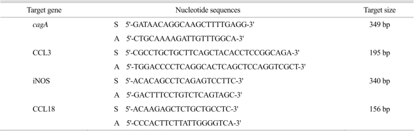
Table 1. Primers used for analysis of gene expression
Target gene Nucleotide sequences Target size
cagA S 5'-GATAACAGGCAAGCTTTTGAGG-3' 349 bp
A 5'-CTGCAAAAGATTGTTTGGCA-3'
CCL3 S 5'-CGCCTGCTGCTTCAGCTACACCTCCGGCAGA-3' 195 bp
A 5'-TGGACCCCTCAGGCACTCAGCTCCAGGTCGCT-3'
iNOS S 5'-ACACAGCCTCAGAGTCCTTC-3' 340 bp
A 5'-GACTTTCCTGTCTCAGTAGC-3'
CCL18 S 5'-ACAAGAGCTCTGCTGCCTC-3' 156 bp
A 5'-CCCACTTCTTATTGGGGTCA-3'
여 총반응액이 50 μl가 되도록 증류수를 가하였고, DNA thermal cycler (Biometra, Gōttingen, Germany)를 사용하여 PCR을 수행하였다.
5) cagA, iNOS, CCL3 및 CCL18 mRNA의 발현 분석 시토카인 및 케모카인 유전자 발현 정도를 알아보기 위해 실험에 이용된 primer는 제노텍 (Daejeon, Korea)에 주문 합성하여 사용하였으며, 각 primer의 sequence 및 primer가 표적으로 하는 DNA의 크기는 Table 1과 같다.
cDNA 증폭조건을 간단히 기술하면, 94 ℃에서 1분간 변 성하고, CCL3는 57℃에서, cagA는 55℃, iNOS는 54℃에 서 각각 1분간 annealing한 다음 72℃에서 1분간 확장하 였다. CCL18은 94℃에서 30초간 변성, 60℃에서 30초간 annealing, 72℃에서 30초간 확장하였으며 이들 조건에서 모두 30주기 반복하였다.
6) 전기영동에 의한 PCR 산물의 검색
PCR이 끝난 후, 18 μl의 증폭된 PCR 산물을 취하여 10 X gel loading buffer (0.25% bromophenol blue/0.25% xylene cyanol FF/50% glycerol)와 잘 섞은 다음 1 μg/ml의 ethidium bromide가 포함된 2% agarose gel 상에 Tris-Acetate-EDTA (TAE) buffer 하에서 전기영동하였으며 이동한 DNA band 의 위치를 자외선 투사기 (ultraviolet transilluminator) 상에 서 관찰하였다.
7) 통계학적 분석
집단 간의 상관관계 분석은 Fisher's exact test로 하였으 며 p<0.05를 의미가 있는 것으로 하였다.
결 과
1. 만성위염환자의 위점막 생검조직에서 iNOS mRNA의 발현
CLO 검사 양성이며 림프여포가 있는 47명 중에서 iNOS mRNA가 발현된 경우는 45명 (95.7%)으로 매우 높
았으며, 발현되지 않는 경우는 4.3%로 매우 낮았다 (Table 2). 대조군인 CLO test 음성이며 림프여포가 없는 만성위 염환자 28명 중 26명 (92.9%)에서 발현되어 만성위염환 자는 H. pylori 감염 여부와 림프여포의 존재 여부에 관 계없이 iNOS mRNA가 대부분 발현되었다.
2. 만성위염환자의 위점막 생검조직에서 cagA mRNA의 발현
CLO 검사 양성이며 림프여포가 있는 47명 중에서 cagA mRNA가 발현된 경우는 30명으로 63.8%이었으며 발현되지 않는 경우는 36.2%이었다. CLO 검사 음성이며 림프여포가 없는 만성위염환자 28명은 모두 cagA mRNA 를 발현하지 않았다 (Table 3). 그러므로 H. pylori 감염과 cagA 발현과는 높은 상관관계가 있었다 (p<0.0001).
3. 만성위염환자의 위점막 생검조직에서 CCL3 mRNA의 발현
CLO 검사 양성이며 림프여포가 있는 47명 중에서 CCL3 mRNA가 발현된 경우는 45명으로 95.7%로 매우 높았으며 발현되지 않는 경우는 4.3%로 매우 낮았다. 대 조군인 CLO 검사 음성이며 림프여포가 없는 만성위염 환자 28명은 모두 CCL3 mRNA를 발현하지 않았다 (Ta- Table 2. The expression of iNOS mRNA in lymphoid follicle of
gastric mucosa with CLO test positive iNOS mRNA
aL.F and bCLO Yes (%) NO (%) Total (%) Present and positive 45 (95.7) 2 (4.3) 47 (100) Absent and negative 26 (92.9) 2 (7.1) 28 (100)
a LF: lymphoid follicle, b CLO: CLO test
The two sided p value is 0.6265, considered not significant by Fisher's exact test.
Table 4. The expression of CCL3 mRNA in lymphoid follicle of gastric mucosa with CLO test positive
CCL3 mRNA
aL.F and bCLO Yes (%) NO (%) Total (%) Present and positive 45 (95.7) 2 (4.3) 47 (100) Absent and negative 0 ( 0) 28 (100) 28 (100)
a LF: lymphoid follicle, b CLO: CLO test
The two sided one-side p value is <0.0001, considered extremely significant by Fisher's exact test. There is a significant association between rows and columns.
Table 3. The expression of cagA mRNA in lymphoid follicle of gastric mucosa with CLO test positive
cagA mRNA
aL.F and bCLO Yes (%) NO (%) Total (%) Present and positive 30 (63.8) 17 (36.2) 47 (100) Absent and negative 0 ( 0) 28 (100) 28 (100)
a LF: lymphoid follicle, b CLO: CLO test
The two sided p value is <0.0001, considered extremely significant by Fisher's exact test. There is a significant association between rows and columns.
ble 4). 그러므로 H. pylori 감염과 CCL3 발현과는 높은 상관관계가 있었다 (p<0.0001).
4. H. pylori에 감염된 위점막 생검조직에서 CCL3와 cagA mRNA 발현과의 상관관계
CCL3와 cagA mRNA 발현과 림프여포 형성과의 관계 를 알아보기 위하여 CLO 검사 양성이며 림프여포가 있 는 47명을 대상으로 조사하였다 (Table 5). CCL3와 cagA mRNA가 동시에 발현된 경우는 28명 (59.6%)이었다. 그 러나 cagA mRNA는 발현되었으나 CCL3 mRNA가 발현 되지 않은 경우가 4.2%이고, 반대의 경우 즉, cagA mRNA 는 발현이 없고 CCL3 mRNA가 발현되는 경우가 36.2%
로 상관관계가 적었다.
5. 만성위염환자의 위점막 생검조직에서 CCL18 mRNA 의 발현
CLO 검사 양성이며 림프여포가 있는 47명 중에서 CCL18 mRNA가 발현된 경우는 25명으로 53.3%이었으 며 발현되지 않는 경우는 46.8%로 유의한 차이가 없었다 (Table 6). 대조군인 CLO 검사 음성이며 림프여포가 없는 만성위염환자 28명은 모두 CCL18 mRNA를 발현하지 않 았다. 그러므로 H. pylori 감염이 있으며 림프여포가 있는
만성위염환자의 위점막은 CCL18 유전자 발현이 매우 높 았다 (p<0.0001).
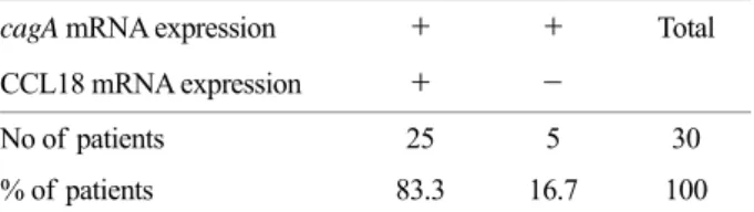
6. H. pylori에 감염된 위점막 생검조직에서 CCL18과 cagA mRNA 발현과의 상관관계
CCL18과 cagA mRNA 발현과 림프여포 형성과의 관계 를 알아보기 위하여 CLO 검사 양성이며 림프여포가 있 는 47명 중에서 cagA mRNA가 발현된 30명을 대상으로 조사하였다 (Table 7). CCL18과 cagA mRNA가 동시에 발 현된 경우는 25명 (83.3%) 이었다. 그러나 cagA mRNA는 발현되었으나 CCL18 mRNA가 발현되지 않은 경우도 16.7%이었다. 그러므로 H. pylori 감염이 있으며 림프여 포가 있는 만성위염환자 위점막의 CCL18 유전자 발현 과 cagA 유전자 발현과는 매우 높은 상관관계가 있었다 (p<0.0001).
고 찰
H. pylori는 만성위염의 가장 중요한 원인의 하나이며, 림프여포, 점막의 손실을 동반한 상피세포의 변성과 장 상피화생의 원인이 된다 (8). 그러나 H. pylori에 의한 만 성위염의 병인에서 균의 인자와 숙주반응에 대하여는 잘 알려져 있지 못하다 (10). 이에 본 연구에서는 H. pylori의 감염이 있으며 염증세포의 침윤, 림프여포의 형성 및 장 상피화생이 있는 중증의 만성위염환자군과 H. pylori의 감염이 없으며 림프여포, 장상피화생이 없는 경증의 만성 위염환자군을 대상으로 만성위염의 병인을 cagA mRNA 발현과 염증세포의 침윤과 관련이 되는 C-C 케모카인 mRNA와 iNOS mRNA의 발현으로 추구하고자 하였다.
H. pylori는 다량의 urease를 생산하며, 많은 균주는 vacA와 cagA 등과 같은 외독소를 생산한다. Urease는 위 Table 5. Interrelationships between cagA mRNA and CCL3 mRNA
expression in lymphoid follicle of gastric mucosa with CLO test positive
cagA mRNA expression + + - Total
CCL3 mRNA expression + - +
No of patients 28 2 17 47
% of patients 59.6 4.2 36.2 100 The two sided p value is 0.5282, considered not significant by Fisher's exact test.
Table 7. Interrelationships between cagA and CCL18 mRNA expression in lymphoid follicle of gastric mucosa with CLO test positive
cagA mRNA expression + + Total
CCL18 mRNA expression + -
No of patients 25 5 30
% of patients 83.3 16.7 100
The two sided p value is <0.0001, considered extremely significant by Fisher's exact test. There is a significant association between rows and columns.
Table 6. The expression of CCL18 mRNA in lymphoid follicle of gastric mucosa with CLO test positive
CCL18 mRNA
aL.F and bCLO Yes (%) NO (%) Total (%) Present and positive 25 (53.2) 22 (46.8) 47 (100) Absent and negative 0 ( 0) 28 (100) 28 (100)
a LF: lymphoid follicle, b CLO: CLO test
The two sided p value is <0.0001, considered extremely significant by Fisher's exact test. There is a significant association between rows and columns.
내강에 존재하는 요소를 암모니아와 이산화탄소로 분해 한다. 이렇게 하여 생산된 암모니아는 세균 주위의 위산 을 중화시킴으로서 강한 산성환경에서도 세균이 살아갈 수 있게 한다. 암모니아는 또한 직접적으로 위점막 상피 세포에 작용하여 세포손상을 일으킨다. vacA는 위점막 상피세포의 세포질에 공포가 생기게 하며, cagA는 위점 막 상피세포로 하여금 호중구에 대하여 강한 화학주성을 가진 IL-8을 분비하게 하는 것으로 알려져 있다. 침윤된 호중구 및 단핵구는 NO를 생산하여 위점막을 손상시킨 다 (11).
조직학적으로 H. pylori 감염에 의한 숙주의 반응으로 위점막에 림프구, 대식세포 및 호중구의 침윤이 특징적이 다 (22). H. pylori에 의한 만성위염에서 NO는 대식세포와 위 상피세포에서도 iNOS 활성에 의하여 생산되며 iNOS 활성의 증가는 점막손상과 관련이 있다고 한다 (25). CLO 검사 양성이며 림프여포가 있는 중증의 만성위염환자에 서는 iNOS mRNA의 발현이 95.7%로 매우 높았으며 대 조군인 CLO 검사 음성이며 림프여포가 없는 경증의 만 성위염환에서도 92.9%로 대부분 발현되었다. Li 등 (19) 은 iNOS mRNA의 발현은 정상 또는 소수의 단핵구가 침윤된 경우보다 중등도 또는 심한 단핵구 침윤이 있는 조직에서 매우 높았으며, H. pylori 감염조직에서 iNOS 발현비율이 56%이었다고 한다. 또한 iNOS mRNA 발현 양성 조직의 92%가 H. pylori 양성으로 in vivo에서 H.
pylori 감염은 iNOS 발현을 자극할 수 있다고 하였다.
Tatemichi 등 (25)은 23명의 만성위염환자 중에서 iNOS 발현비율이 43.5%이었다고 보고하였다. 본 연구의 결과 는 이들의 결과에 비하여 iNOS의 발현비율이 매우 높았 으며, 만성위염환자는 H. pylori 감염 여부와 조직학적인 염증의 경중에 관계없이 매우 높게 발현되어 이 원인에 대한 연구가 추후 필요하리라 생각된다.
cagA는 위점막에 국소염증반응을 더 심하게 일으켜 cagA가 양성인 H. pylori에 의한 위염의 경우 cagA가 음 성인 H. pylori에 의한 위염에 비하여 병리조직학적으로 염증세포의 침윤이 더 심하고 IL-8과 같은 케모카인 분 비가 더 많이 일어난다고 보고되어 H. pylori의 병원성인 자로서의 cagA의 역할이 제안되어 왔다 (9,13,28). CLO 검사 양성이며 림프여포가 있는 47명 중에서 cagA mRNA 가 발현된 경우는 30명으로 63.8%이었으며 발현되지 않 는 경우는 36.2%이었다. CLO 검사 음성이며 림프여포가 없는 만성위염환자 28명은 모두 cagA mRNA를 발현하
지 않았다. 상부위장관 내시경 생검조직을 이용한 CLO 검사의 예민도는 85~95%, 특이도는 약 92%로 예민도와 특이도가 모두 높아 우리나라에서는 내시경검사가 가능 한 의료기관에서 H. pylori 감염의 진단에 있어 1차 검사 로 추천된다. 배양검사는 H. pylori 감염의 진단목적으로 는 임상에서 이용되지 않는다 (17). 위장관 생검조직에서 추출한 total DNA로부터 PCR을 이용한 H. pylori 감염의 진단은 추천되지 않지만, 항생제 내성에 관련된 유전자의 변이, 유전형의 분류 및 cagA와 같은 병독력 인자의 발현 에 유용하게 이용되고 있다. 그러나 ureA와 cagA primer 를 이용한 RT-PCR법은 위점막 생검조직에서 H. pylori 유 전자를 검출하는 예민하고 특이성이 있는 유용한 검사법 으로 제안되었다 (21). 국내의 만성위염환자에서 PCR에 의한 경우 cagA 양성율이 89.5%이었으며 (15), 비궤양성 소화불량증환자의 64.3%에서 cagA가 양성이었다 (27). 중 국에서 cagA+ 균주 감염율이 십이지장궤양에서 79%이었 고 만성위염에서 62%이었다 (19). cagA와 vacA의 양성률 은 십이지장궤양 환자와 만성위염환자간에 유의한 차이 가 없어, H. pylori 감염시 서로 다른 점막변화를 나타내 는 데에는 관여하지 않는다고 하였다 (16). 그러나 외국 의 보고들에 의하면 cagA 유전자가 양성인 H. pylori에 의한 위염의 경우 cagA 유전자 음성 예에 비하여 병리조 직학적 위염의 정도가 심하였다고 보고하였다 (13,30). 본 연구의 결과에서도 CLO 검사 음성이며 림프여포가 없 는 만성위염환자의 위생검 조직에서 모두 cagA mRNA가 발현되지 않았으며 CLO 검사 양성이며 림프여포가 있는 경우에서 63.8%가 발현되었음으로 cagA는 만성위염의 중증도에 관여된다고 생각된다.
CCL3는 H. pylori가 감염된 위점막에 단핵, 대식세포 및 림프구의 침윤에 주도적인 역할을 한다고 한다 (12,23, 29). 그러나 최 등 (3)은 C-C 케모카인인 CCL3와 CCL4 mRNA 발현은 H. pylori 감염 여부, H. pylori density 및 호중구 침윤 정도에 따라 유의한 차이를 나타내지 않았 을 뿐만 아니라, 만성염증 정도와도 유의한 상관관계가 없었다고 상반된 보고를 하였다. 본 연구 결과에서는 대 조군인 CLO 검사 음성이며 림프여포가 없는 만성위염 환자의 위생검 조직에서 모두 CCL3 mRNA를 발현되지 않았던 것과는 대조적으로 중증의 만성위염환자의 경우 95.7%로 매우 높게 발현되어 만성위염의 정도와 관계가 있었다. 그러나 cagA가 CCL3 mRNA 발현에 미치는 영향 에 있어서는 CCL3와 cagA mRNA가 동시에 발현된 경
우가 59.6%이었으나 cagA mRNA는 발현되었으나 cagA mRNA는 발현이 없고 CCL3 mRNA가 발현되는 경우가 36.2%로 상관관계가 적었다. 그러므로 CCL3 mRNA의 발현에는 cagA 이외의 다른 인자가 관여되었을 것으로 생각되었다.
위점막연관 림프조직형 림프종의 발생에 중요한 역할 을 하는 것으로 알려진 림프여포는 H. pylori에 의한 위 염에서 흔히 볼 수 있는 아주 중요한 소견이다 (31). H.
pylori 항원이 면역계를 자극하면 케모카인과 시토카인에 의하여 항원제시세포 주위에 T 림프구의 집단이 이어지 고 B 림프구와 여포수지상세포가 침윤되어 점막 림프여 포가 형성된다고 한다 (26). 또한 만성 C형 간염에서 가 장 특징적으로 간문맥 림프여포가 형성되는데 CCL18 mRNA의 발현이 관찰된다고 한다 (18). 이 CCL18은 배 중심에 있는 수지상세포에서 생산되며 미감작 T 림프구 에 대한 화학주성 활성이 있다고 한다. 본 연구에서도 림프여포의 형성에 CCL18이 영향을 미치는지의 관계를 CCL18 mRNA 발현으로 조사하였다. 림프여포가 있는 환자 중에서 53.2%가 CCL18 mRNA를 발현하였다. 그러 나 림프여포가 있는 환자에서도 46.8%가 발현이 되지 않아 유의한 차이는 없었다. 그러나 림프여포가 없는 만 성위염환자 모두는 CCL18 mRNA가 발현되지 않아 특이 성은 매우 높았다. cagA가 CCL18 mRNA 발현에 미치는 영향에 있어서는 CCL18과 cagA mRNA가 동시에 발현 된 경우는 83.3%이었으며 cagA mRNA는 발현되었으나 CCL18 mRNA가 발현되지 않은 경우가 16.7%로 cagA 가 CCL18 mRNA 발현에 미치는 영향은 매우 유의하였 다 (p<0.0001). 그러나 H. pylori의 cagA 이외의 독력인자 들도 CCL18발현 유도에 관여하는지에 관한 연구가 더 요구된다.
이상의 결과로 만성위염환자에 있어서 H. pylori 감염 여부와 조직학적인 염증의 경중에 관계없이 NO가 매우 높게 발현되어 만성위염의 병인에 NO는 중요한 역할을 한다고 생각된다. cagA는 만성위염의 중증도에 관여된다 고 생각된다. C-C 케모카인인 CCL3는 만성위염의 정도 와 관계가 있었으나 CCL3 발현에는 cagA 이외의 다른 인자가 관여되었을 것으로 생각되었다. 림프여포의 형성 에 CCL18은 특이성은 매우 높았으나 림프여포가 있는 환자에서도 46.8%가 발현이 되지 않아 유의한 차이는 없었다. 그러나 cagA가 CCL18 mRNA 발현에 미치는 영 향은 매우 유의하였다 (p<0.0001).
참 고 문 헌
1) Adema GJ, Hartgers F, Verstraten R, de Vries E, Marland G, Menon S, Foster J, Xu Y, Nooyen P, McClanahan T, Bacon KB, Figdor CG: A dendritic-cell-derived C-C chemo- kine that preferentially attracts naive T cells. Nature 387: 713- 717, 1997.
2) Bayerdrffer E, Lehn N, Hatz R, Mannes GA, Oertel H, Sauerbruch T, Stolte M: Difference in expression of Helico- bacter pylori gastritis in antrum and body. Gastroenterology 102: 1575-1582, 1992.
3) Choi IJ, Kim JS, Jung HC, Kim JM, Lee KL, Son IS, Kim CY: Expression of CXC and CC chemokines in the gastric mucosa infected with Helicobacter pylori. Korean J Gastro- enterol 36: 163-174, 2000.
4) Correa P: Chronic gastritis: a clinico-pathological classification.
Am J Gastroenterol 83: 504-509, 1988.
5) Crabtree JE: Role of cytokines in pathogenesis of Heloco- bacter pylori-induced mucosal damage. Dig Dis Sci 43: 46S -55S, 1998.
6) Crabtree JE, Farmery SM, Lindley IJ, Figura N, Peichl P, Tompkins DS: CagA/cytotoxic strains of Helicobacter pylori and interleukin-8 in gastric epithelial cell lines. J Clin Pathol 47: 945-950, 1994.
7)Crawford JM: The gastrointestinal tract. pp 797-875. In Robbins and Cotran Pathologic Basis of Disease, 7th ed, Kumar V, Abbas AK and Fausto N (Ed), Elsevier Saunders, Philadelphia, 2005.
8) Dixon MF, Genta RM, Yardley JH, Correa P: Classification and grading of gastiritis: the update Sydney system. International Workshop of Gastritis, Houston, 1994. Am J Surg Pathol 20:
1161-1181, 1996.
9) Drake IM, Mapstone NP, Schorah CJ, White KL, Chalmers DM, Dixon MF, Axon AT: Reactive oxygen species activity and lipid peroxidation in Helicobacter pylori-associated gastritis: relation to gastric mucosal ascorbic acid concentrations and effect of H. pylori eradication. Gut 42: 768-771, 1998.
10) Hansen PS, Go MF, Varming K, Andersen LP, Genta RM, Graham DY, Nielsen H: Proinflammatory activation of neutrophils and monocytes by Helicobacter pylori in patients with different clinical presentations. Infect Immun 67: 3171 -3174, 1999.
11) Hatz RA, Rieder G, Stolte M, Bayerdrffer E, Meimarakis
G, Schildberg FW, Enders G: Pattern of adhesion molecule expression on vascular endothelium in Helicobacter pylori- associated antral gastritis. Gastroenterology 112: 1908-1919, 1997.
12) Hofman VJ, Moreilhon C, Brest PD, Lassalle S, Brigand KL, Sicard D, Raymond J, Lamarwue D, Hbuterne XA, Mari B, Barbry PJP, Hofman PM: Gene expression profiling in human gastric mucosa infected with Helicobacter pylori.
Modern Pathol 20: 974-989, 2007.
13) Husson MO, Gottrand F, Vachee A, Dhaenens L, de la Salle EM, Turck D, Houcke M, Leclerc H: Importance in diagnosis of gastritis of detection by PCR of the cagA gene in Helicobacter pylori strains isolated from children. J Clin Microbiol 33: 3300-3303, 1995.
14) Jung HC, Kim JM, Song IS, Kim CY: Helicobacter pylori induced an array of pro-inflammatory cytokines in human gastric epithelial cells: quantification of mRNA for interleukin- 8, -1 alpha/beta, granulocyte-macrophage colony-stimulting factor, monocyte chemoattractant protein-1 and tumor necrosis factor-alpha. J Gastroenterol Hepatol 12: 473-480, 1997.
15) Kim DY: The positive rates of Helicobacter pylori cagA gene in gastric biopsy specimens of the patients with gastritis, gastric ulcer, duodenal ulcer, and gastric cancer and comparison of the degree of the gastritis. Korean J Gastroenterol 32: 24-31, 1998.
16) Kim SW, Chung IS, Lee KM, Lee DS, Yoon JG, Kim SS, Yang YS, Choi MG, Han SW, Choi KY, Park DH:
Comparison of intestinal metaplasia and serum pepsinogen levels between Helicobacter pylori-infected duodenal ulcer and chronic gastritis. Korean J Gastroenterol 36: 155-162, 2000.
17) Korean Helicobacter pylori study group: Diagnosis and treatment of Helicobacter pylori infection in Korea. Korean J Gastroenterol 32: 275-289, 1998.
18) Kusano F, Tanaka Y, Marumo F, Sato C: Expression of C-C chemokines is associated with portal and periportal inflammation in the liver of patients with chronic hepatitis C.
Lab Invest 80: 415-422, 2000.
19) Li CQ, Pignatelli B, Ohshima H: Coexpression of interleukin- 8 and inducible nitric oxide synthase in gastric mucosa infected with cagA Helicobacter pylori. Dig Dis Sci 45: 55-62, 2000.
20) Marshall BJ, Warren JR: Unidentified curved bacilli in the stomach of patients with gastritis and peptic ulceration. Lancet 1: 1311-1315, 1984.
21) Peek RM Jr, Miller GG, Tham KT, Prez-Prez GI, Cover TL, Atherton JC, Dewey Dunn G, Blaser MJ: Detection of Helicobacter pylori gene expression in human gastric mucosa.
J Clin Microbiol 33: 28-32, 1995.
22) Price AB: The Sydney system: histological division. J Gastro- enterol Hepatol 6: 209-222, 1991.
23) Sato Y, Sugimura K, Mochizuki T, Honma T, Suriki H, Tashiro K, Ishizuka K, Narisawa R, Ichida T, Van Thiel DH, Asakura H: Regionl differences on production of chemokines in gastric mucosa between Helicobacter pylori- positive duodenal ulcer and gastric ulcer. Dig Dis Sci 44:
2390-2396, 1999.
24) Shimoyama T, Everett SM, Dixon MF, Axon AT, Crabtree JE: Chemokine mRNA expression in gastric mucosa is associated with Helicobacter pylori cagA positivity and severity of gastritis. J Clin Pathol 51: 765-770, 1998.
25) Tatemichi M, Ogura T, Nagata H, Esumi H: Enhanced expression of inducible nitric oxide synthase in chronic gastritis with intestinal metaplasia. J Clin Gastroenterol 27: 240-245, 1998.
26) Tursi A, Gasbarrini G: Acquired gastric mucosa-associated lymphoid tissue (MALT): a review with special emphasis on association with extragastric diseases and management pro- blems of gastric MALT. J Clin Gastroenterol 29: 133-137, 1999.
27) Yamaoka Y, Kita M, Kodama T, Sawai N, Imanishi J:
Helicobcter pylori cagA gene and expression of cytokine messenger RNA in gastric mucosa. Gastroenterology 110:
1744-1752, 1996.
28) Yamaoka Y, Kita M, Kodama T, Sawai N, Kashima K, Imanishi J: Induction of various cytokines and development of severe mucosal inflammation by cagA gene positive Helicobcter pylori strains. Gut 41: 442-451, 1997.
29) Yamaoka Y, Kita M, Kodama T, Sawai N, Tanahashi T, Kashima K, Imanishi J: Chemokines in the gastric mucosa in Helicobcter pylori infection. Gut 42: 609-617, 1998.
30) Yang US, Cho M, Kang DH, Song CS, Song GA, Park SK:
The prevalence of cagA+Helicobcter pylori in the peptic ulcer diseases. Korean J Gastroenterol 31: 184-191, 1998.
31) Zaitoun AM: The prevalence of lymphoid follicles in Helico- bacter pylori associated gastritis in patients with ulcers and non-ulcer dyspepsia. J Clin Pathol 48: 325-329, 1995.
32) Zlotnik A, Yoshie O: Chemokines: a new classification system and their role in immunity. Immunity 12: 121-127, 2000.