pISSN 1598-9992 eISSN 2233-6869
REVIEW ARTICLE
한국의 헬리코박터 유병률 개선에 따른 소화기질환의 변화
박수헌
가톨릭대학교 의과대학 내과학교실
Changes in Upper Gastrointestinal Diseases according to Improvement of Helicobacter pylori Prevalence Rate in Korea
Soo-Heon Park
Department of Internal Medicine, College of Medicine, The Catholic University of Korea, Seoul, Korea
Helicobacter pylori can cause variety of upper gastrointestinal disorders such as peptic ulcer, mucosa associated lymphoid tissue (MALT)-lymphoma, and gastric cancer. The prevalence of H. pylori infection has significantly decreased in Korea since 1998 owing to active eradication of H. pylori. Along with its decrease, the prevalence of peptic ulcer has also decreased.
However, the mean age of gastric ulcer increased and this is considered to be due to increase in NSAID prescription. Gastric cancer is one of the leading causes of cancer deaths in Korea and Japan, and IARC/WHO has classified H. pylori as class one carcinogen of gastric cancer. Despite the decreasing prevalence of H. pylori infection, the total number of gastric cancer in Korea has continuously increased from 2006 to 2011. Nevertheless, the 5 year survival rate of gastric cancer patients significantly increased from 42.8% in 1993 to 67% in 2010. This increase in survival rate seems to be mainly due to early detection of gastric cancer and endoscopic mucosal dissection treatment. Based on these findings, the prevalence of peptic ulcer is expected to decrease even more with H. pylori eradication therapy and NSAID will become the main cause of peptic ulcer. Although the prevalence of gastric cancer has not changed along with decreased the prevalence of H. pylori, gastric cancer is expected to decrease in the long run with the help of eradication therapy and endoscopic treatment of precancerous lesions. (Korean J Gastroenterol 2015;65:199-204)
Key Words: Helicobacter; Stomach neoplasms; Peptic ulcer; Eradication therapy
CC This is an open access article distributed under the terms of the Creative Commons Attribution Non-Commercial License (http://creativecommons.org/licenses/
by-nc/3.0) which permits unrestricted non-commercial use, distribution, and reproduction in any medium, provided the original work is properly cited.
Copyright © 2015. Korean Society of Gastroenterology.
교신저자: 박수헌, 150-713, 서울시 영등포구 63로 10, 가톨릭대학교 여의도성모병원 소화기내과
Correspondence to: Soo-Heon Park, Division of Gastroenterology, Department of Internal Medicine, The Catholic University of Korea, Yeouido St. Mary’s Hospital, 10 63-ro, Yeongdeungpo-gu, Seoul 150-713, Korea. Tel: +82-2-3779-2093, Fax: +82-2-786-0803, E-mail: [email protected]
Financial support: None. Conflict of interest: None.
서 론
헬리코박터(Helicobacter pylori) 감염은 다양한 위염, 위궤 양, 십이지장궤양, mucosa associated lymphoid tissue (MALT) 림프종 및 위암 등의 소화기질환을 일으킨다. 한국에 서 헬리코박터제균 치료는 거의 30년 동안 소화성궤양과 MALT 림프종 및 일부 위염(기능성소화불량)에 대한 치료로 이루어졌다. 헬리코박터의 유병률은 1998년 처음 보고된 이 후 지속적으로 감소되어 왔으며 이에 따른 소화기질환의 양상 도 변화되어 가고 있다. 헬리코박터제균 치료는 장기간 위산
분비 억제제를 투여하는 치료에 비해 의료비용이 저렴하고 효 과적이기 때문이다.1 기능성소화불량 환자의 경우도, 헬리코 박터제균 치료가 비용이 많이 들기는 하지만 위산분비 억제 치료보다는 더 효과적이고 증상 소실률대비 비용이 대부분의 나라에서 저렴하다. 헬리코박터를 치료하는 것은 다양한 소화 기질환에 대한 의료비용을 줄이는 좋은 모델이기도 한데, 과 연 모든 국민을 대상으로 헬리코박터를 치료하여 위암 발생을 줄이고 의료사회비용을 줄일 수 있는지에 대한 연구도 필요한 실정이다.
Fig. 1. Trends of seroprevalence of Helicobacter pylori infection in asymptomatic subjects without a history of H. pylori eradication in 1998, 2005, and 2011 (*p<0.05, 1998 vs. 2011). Cited from the article of Lim et al.4 (BMC Gastroenterol 2013;13:104).
본 론
1. 헬리코박터 유병률
헬리코박터 감염의 유병률은 1998년 66.9%에서 2005년 59.6%, 2011년에는 54.4%로 감소하였다(Fig. 1). 각각의 조 사기간별로 분석해도 2005년과 1998년 사이와 2011년에서 2005년 사이에 헬리코박터 유병률의 유의한 감소가 있었 다.2-4 지역적으로는 경상도와 강원도를 제외한 전국의 모든 지역에서 유의하게 감소되었다. 또한 모든 연령에서 헬리코박 터 감염이 감소되었으며, 특히 1998년에서 2005년 사이에는 40대에서 현저한 감소가 있었다. 이런 헬리코박터 유병률의 감소는 급속한 경제성장, 사회 환경개선 및 헬리코박터제균 치료 등에서 영향을 받은 것으로 생각된다.
2. 소화성궤양
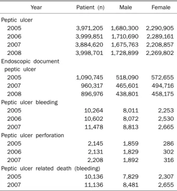
소화성궤양의 원인 중 가장 중요한 것이 헬리코박터 감염 으로, 헬리코박터제균 치료를 하면 궤양의 재발률이 현저히 줄어든다. 2010년 건강보험 심사평가원에서 발간한 소화성궤 양에 대한 연구보고서에 의하면 상병코드 분류에 의한 소화성 궤양 환자는 2005-2008년에 매년 397만 명, 399만 명, 388만 명, 399만 명으로 변화가 없었다. 그러나 이는 내시경 검사와 관계없이 소화성궤양 질병코드를 사용하였기 때문으로, 내시 경 검사를 한 후 진단된 소화성궤양 환자의 수는 2005-2007 년에매년 109만 명에서 96만 명, 89만 명으로 점차 감소하고 있다(Table 1).5
한국에서 소화성궤양 유병률은 2007년 보고에 의하면 2.4%로, 이중 위궤양은 61.2%, 십이지장궤양은 38.8%였다.6 국내에서 입원한 환자를 대상으로 소화성궤양이 발생하는 평
Table 1. Total Number of Patients with Peptic Ulcer and Peptic Ulcer Related Complications5
Year Patient (n) Male Female
Peptic ulcer
2005 3,971,205 1,680,300 2,290,905
2006 3,999,851 1,710,690 2,289,161
2007 3,884,620 1,675,763 2,208,857
2008 3,998,701 1,728,899 2,269,802
Endoscopic document peptic ulcer
2005 1,090,745 518,090 572,655
2007 960,317 465,601 494,716
2008 896,976 438,801 458,175
Peptic ulcer bleeding
2005 10,264 8,011 2,253
2006 10,602 8,072 2,530
2007 11,478 8,813 2,665
Peptic ulcer perforation
2005 2,145 1,859 286
2006 2,131 1,829 302
2007 2,208 1,892 316
Peptic ulcer related death (bleeding)
2005 10,136 7,829 2,307
2007 11,136 8,481 2,655
균연령은 1990년 47.8세, 1996년 50.8세, 2006년 58.1세로 증가되었다.7 또한 17개 병원의 연구에 의하면 위궤양의 유병 률은 각각 9.6%, 10.5%, 및 12.0%로 증가되고 있는 반면 십 이지장 궤양의 유병률은 8.4%, 8.7%, 8.2%로 변화가 없었다.8 다른 다기관 연구에서도 1994년과 2004년 10년 후의 위궤양 의 유병률은 73.1%에서 66.1%로 감소되었고 십이지장 궤양 의 유병률은 79.3%에서 68.1%로 감소되었다.9
2007년 상병코드 분류에 의한 비스테로이드성 소염제를 60 일 이전에 사용한 비스테로이드성 소염제로 인한 궤양은 전체 환자 1,165,444명(30.0%)이었고 위궤양 환자 704,078명, 십이 지장궤양 152,762명, 궤양출혈 환자 2,839명이었다(Table 1).
소화성궤양의 합병증으로 발생하는 궤양 출혈 환자는 2007년 11,478명으로 내시경으로 확인된 소화성궤양 환자 960,317 명의 1.2%에서 발생하였다. 다른 합병증인 궤양 천공은 2,208명에서 발생하여 전체 내시경으로 확인된 소화성궤양 환자의 0.2%였다(Table 1). 또한 출혈로 인한 소화성궤양 사 망률은 11,136명으로 내시경으로 확인된 소화성궤양 환자의 1.2%이다(Table 1).
요약하면 내시경으로 확인된 소화성궤양의 전체 수는 감소 되고 있으며 이는 헬리코박터 유병률의 감소 및 제균 치료로 인한 궤양 재발 감소가 한 원인으로 생각된다. 소화성궤양 중 위궤양은 점차 증가하고 있으며 십이지장궤양은 유병률에 변 화가 없다고 할 수 있다. 또한 궤양이 발생하는 연령이 점차 높아진다는 것은 비스테로이드성 소염제나 아스피린과 같은
약제 사용으로 인한 것으로 보이며, 헬리코박터 감염으로 인 한 궤양 발생이 줄어들고 있다는 근거가 될 수 있다.
3. 위염
헬리코박터 감염은 위염을 일으키고 소화불량 증상을 유발 한다. 이러한 위염환자에서 헬리코박터 감염의 유병률은 2012 년 48.9%로 알려져 있다.10 이중 로마기준에 따른 소화불량 분류에서 명치고통 증후군(epigastric pain syndrome) 환자 가 43.2%, 식후고통증후군(postprandial distress syndrome) 은 56.3%로 식후고통증후군 환자가 약간 더 많은 반면, 헬리 코박터 감염은 명치고통 증후군 환자군에서 더 높다. 그러나 헬리코박터 감염에 따른 위염의 내시경소견은 차이가 없었 다.10 2012년 소화불량을 호소하는 환자들의 내시경 소견 중 14.1%는 정상소견을 보였다. 내시경 검사에서 이상소견은 표재 성 위염이 31.3%로 가장 많았고, 위축성 위염 27.1%, 미란성 위염 23.7%, 장상피화생 7.1%의 순으로 많았다.11 반면 1998년 발표된 문헌에서도 소화불량 환자에서 정상소견이 8.4%인 것 을 감안하면 헬리코박터 유병률이 감소하면서 정상소견으로 보이는 내시경 환자수도 증가하는 것으로 생각된다.12 이는 서 구에서 발표된 수치인 평균 51%에 비해서는 아직 많이 낮은 데, 여기에는 높은 헬리코박터 유병률, 짜게 먹는 식습관, 음 주, 흡연 등과 같은 여러 요인이 원인이 될 수 있다.13
4. 헬리코박터제균 치료
헬리코박터제균 치료는 소화성궤양의 재발을 감소시키고 위암의 재발을 방지하며 MALT 림프종을 치료하므로 건강보 험의 급여로 인정되고 있다. 기능성 소화불량 환자의 경우에 도 미국이나 유럽에서 치료비용을 감소시키기 위하여 제균 치 료를 사용하고 있으며, 2013년 3월부터는 일본에서도 위암의 발생을 줄이기 위해 일본 후생성의 허가를 얻어 시행하고 있 다. 헬리코박터제균에 사용되는 약제는 1차 치료에는 양성자 펌프억제제, 아목시실린 및 클라리스로마이신 또는 메트로니 다졸의 3제 요법이 사용되고, 2차 치료에는 양성자 펌프억제 제, 비스무스, 메트로니다졸과 테트라사이클린 4제 요법이 사 용되고 있다.
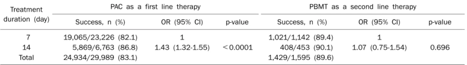
최근 대한헬리코박터 및 상부위장관 학회의 2001년부터 2010년까지 후향적 연구에 따르면 3제 요법의 제균율은 83.1%였다. 7일과 14일의 치료기간에 따른 제균율 차이를 보 면 7일 투여가 82.1%인 반면 14일 투여한 경우 제균율은 86.8%로 14일간 투여한 경우가 유의하게 제균율이 높았다 (Table 2). 2차 치료의 제균 결과는 89.6%로 우수한 제균 치 료 효과가 있었으며 이 경우 7일이나 14일 투여의 제균 치료 율은 89.4%와 90.1%로 차이가 없었다(Table 2).14 반면 2차 제균에 실패한 환자들에 대하여 실시한 3차 제균 치료에서
Table 2. Eradication of Helicobacter pylori according to Treatment Duration14 Treatment
duration (day)
PAC as a first line therapy PBMT as a second line therapy
Success, n (%) OR (95% CI) p-value Success, n (%) OR (95% CI) p-value
7 19,065/23,226 (82.1) 1 1,021/1,142 (89.4) 1
14 5,869/6,763 (86.8) 1.43 (1.32-1.55) <0.0001 408/453 (90.1) 1.07 (0.75-1.54) 0.696
Total 24,934/29,989 (83.1) 1,429/1,595 (89.6)
Total patient’s number 32,120.
Retrospective analysis from 2001 to 2010.
P, proton pump inhibitor; A, amoxicillin; C, clarithromycin; B, bismuth; M, metronidazole; T, tetracyclin.
Table 3. Eradication Rates of Helicobacter pylori according to Third Line Therapeutic Regimens14
Tertiary line therapy regimen Patient, n (%) Eradication rate, %
Total 141 (100) 73.8
PBMT 62 (44.0) 90.3
PAL 23 (16.3) 56.5
PAC 12 (8.5) 58.3
Others 44 (31.2) 63.6
Total number 32,120.
Retrospective analysis from 2001 to 2010.
P, proton pump inhibitor; B, bismuth; M, metronidazole; T, tetracyclin;
A, amoxicillin; L, levofloxacin; C, clarithromycin.
Table 4. Eradication Rates of Helicobacter pylori according to First Line Therapeutic Regimens14
First line therapy regimen Patient, n (%) Eradication rate, %
Total 32,120 (100) 83.2
PAC 30,565 (95.2) 83.2
PBMT 186 (0.6) 89.8
Sequential therapy 126 (0.4) 91.3 Concomitant therapy 303 (0.9) 89.8
Others 940 (2.9) 81.1
Total number 32,120.
Retrospective analysis from 2001 to 2010.
P, proton pump inhibitor; A, amoxicillin; C, clarithromycin; B, bismuth;
M, metronidazole; T, tetracyclin.
Table 5. Eradication Rates of Helicobacter pylori according to Second Line Therapeutic Regimens14
Second line therapy regimen Patient, n (%) Eradication rate, %
Total 2,056 (100) 83.5
PBMT 1,622 (78.9) 89.5
PAC 161 (7.8) 46.0
PTM 70 (3.4) 88.6
PLM 44 (2.1) 72.7
PAM 2 (0.1) 100
Others 157 (7.6) 60.5
Total number 32,120.
Retrospective analysis from 2001 to 2010.
P, proton pump inhibitor; B, bismuth; M, metronidazole; T, tetracyclin;
A, amoxicillin; C, clarithromycin; L, levofloxacin.
현재 국민건강보험공단에서 인정하고 있는 양성자 펌프억제 제, 아목시실린, 레보플로사실린의 제균 치료율은 56.5%로 매우 실망스런 결과를 보여주고 있다(Table 3).14 3차 제균 치 료 대상인 환자에게 2차 제균 치료제인 양성자 펌프억제제, 비스무스, 메트로니다졸과 테트라사이클린 4제 요법을 재투 여 하면 제균율이 90.3%로 나타나, 2차 제균에 실패한 환자에 게 3차 치료로 2차 제균 치료에 사용되었던 4제 요법을 재투 여할 것이 추천되고 있다(Table 3).14
기존의 치료제 외 순차치료(sequential therapy)의 1차 제 균 치료의 효과는 91.3%였고 동시치료(concomitant ther- apy)의 제균 치료 효과는 89.8%로 매우 우수한 효과를 보여 주고 있다(Table 4).14 이런 순차치료나 동시치료는 기존의 3 제 1차 치료제가 80% 이하의 제균 치료율을 보일 때 대체 치료제로 사용할 수 있을 것으로 보인다. 또한 2차 제균 치료 에서 1차 치료에 실패하였던 3제 요법을 재투여하는 경우로 클라리스로마이신이나 메트로니다졸을 변경하여 투여했을 때 의 제균율은 클라리스로마이신이 46%인 반면 메트로니다졸 을 포함한 3제 요법은 88.6%로, 클라리스로마이신을 포함한 1차 치료에 실패한 경우 메트로니다졸을 포함한 1차 제균 치 료도 하나의 대안이 될 수 있을 것이다(Table 5).14
5. 위암
헬리코박터 감염은 세계보건기구에 1급 발암요인으로 지
정되어 있으며 오랜 기간 지속되면 만성염증과 위축성 변화를 유발하고 위암을 발생시킨다.15,16 따라서 헬리코박터 감염과 위암의 발생은 연관성을 가지고 있다. 최근 한국에서 헬리코 박터 감염의 유병률은 점차적으로 감소하고 있고 새로 발생하 는 위암 환자는 2002년 이후 증가하다 2012년 유의하게 감소 하고 있다(Table 6).17 이는 헬리코박터 유병률 감소와 제균 치료, 위암의 전병변인 선종 등을 내시경치료로 제거하기 때 문이다. 위암 환자의 5년 생존율은 93년 이후 42.8%에서 2010년 67.0%로 급속히 호전되고 있다(Table 7).17 이는 1999년부터 실시된 국가암검진으로 인한 효과로 조기위암의
Table 6. Number of Newly Registered Gastric Cancer Patients in Korea17
2002 2003 2004 2005 2006 2007 2008 2009 2010 2011 2012
Patient (n) 23,220 23,926 23,630 26,380 26,443 26,805 28,378 30,005 30,592 31,832 30,847 Incidence rates (/100,000 person) 48.2 49.5 48.7 54.2 54.1 54.6 57.4 60.4 61.3 63.5 61.3
Table 7. Five-Year Survival Rates (%) of Five Major Cancers in Korea17
Rank Cancer Period
Increment
1993-1995 1996-2000 2001-2005 2006-2010
1 Thyroid cancer 94.2 94.9 98.3 99.8 5.6
2 Gastric cancer 42.8 46.6 57.7 67.0 24.2
3 Colon cancer 54.8 58.0 66.6 72.6 17.8
4 Lung cancer 11.3 12.7 16.1 19.7 8.4
5 Liver cancer 10.7 13.2 20.1 26.7 16.0
Table 8. TNM Stage and Types of Gastric Cancer Treatment at 10 Institutes in Korea18
Stage ESD Laparoscopic surgery
Open resection
Robotic surgery Total 1 233 (29.9) 242 (31.0) 116 (14.9) 35 (4.5) 626 (80.3) 2 1 (0.1) 43 (5.5) 31 (3.9) 9 (1.2) 84 (10.8)
3 0 20 (2.6) 45 (5.8) 3 (0.4) 68 (8.7)
4 0 1 (0.1) 1 (0.1) 0 2 (0.3)
234 (30) 306 (39.2) 193 (24.7) 47 (6.0) 780 (100) Values are presented as n (%).
Period, December 2011 to July 2012; total number, 780.
ESD, endoscopic mucosal dissection.
발견이 많아졌기 때문이다. 2011년에서 2012년까지 서울, 경 기 지역 7개 병원에 입원치료한 위암 환자를 분석한 경우 1기 환자가 80.3%, 2기 환자가 10.8%, 3기 환자가 8.7%였고 4기 환자는 0.3%에 불과하였다(Table 8).18 따라서 국가암검진 및 소화불량 증상으로 내시경 검사를 받아 위암이 조기에 발견되 기는 하지만, 위암 환자의 수가 줄어 들지는 않는다. 그러나 이와 같이 국가암검진과 내시경 검사를 통해 위암의 전병변이 발견되어 내시경 치료가 일반화된다면 향후 한국에서 위암의 발생도 감소할 것으로 생각된다.
결 론
한국에서 헬리코박터 유병률이 감소하면서 내시경으로 확 인된 소화성궤양 환자도 감소하고 있다. 이는 헬리코박터제균 치료로 인하여 소화성궤양 재발이 감소한 것이 원인으로 생각 된다. 그러나 약제 사용으로 인한 소화성궤양 환자는 점차 많 아지고 있으며 궤양 발생 연령도 높아지고 있다. 헬리코박터 유병률이 감소하는데도 위암 환자는 감소하지 않고 있지만, 헬리코박터 유병률의 감소와 일부 환자의 제균 치료, 위암 전
구병변의 내시경 치료의 증가 등으로 미래에는 감소할 것으로 생각된다.
REFERENCES
1. Moayyedi P. The health economics of Helicobacter pylori infection. Best Pract Res Clin Gastroenterol 2007;21:347-361.
2. Yim JY, Kim N, Choi SH, et al. Seroprevalence of Helicobacter py- lori in South Korea. Helicobacter 2007;12:333-340.
3. Kim JH, Kim HY, Kim NY, et al; Korea H. pylori Study Group, South Korea. Seroepidemiological study of Helicobacter pylori in- fection in asymptomatic people in South Korea. J Gastroenterol Hepatol 2001;16:969-975.
4. Lim SH, Kwon JW, Kim N, et al. Prevalence and risk factors of Helicobacter pylori infection in Korea: nationwide multicenter study over 13 years. BMC Gastroenterol 2013;13:104.
5. Bae SJ, Kim DS, Kim KM, Park CM, Kim HY; Health Insurance Review and Assessment Service (HIRA). Studies on the occur- enceof peptic ulcer disease and health care utilization patterns in Korean. A research report of HIRA (2010-05). Seoul: HIRA, 2010.
6. Kim JJ, Kim N, Park HK, et al. Clinical characteristics of patients diagnosed as peptic ulcer disease in the third referral center in 2007. Korean J Gastroenterol 2012;59:338-346.
7. Kwon JH, Choi MG, Lee SW, et al. Trends of gastrointestinal dis- eases at a single institution in Korea over the past two decades.
Gut Liver 2009;3:252-258.
8. Kim JI, Kim SG, Kim N, et al; Korean College of Helicobacter and Upper Gastrointestinal Research. Changing prevalence of upper gastrointestinal disease in 28 893 Koreans from 1995 to 2005.
Eur J Gastroenterol Hepatol 2009;21:787-793.
9. Jang HJ, Choi MH, Shin WG, et al. Has peptic ulcer disease changed during the past ten years in Korea? A prospective mul- ti-center study. Dig Dis Sci 2008;53:1527-1531.
10. Kim JI, Lee HJ, Kim JH, et al. Is there a difference between endo- scopic finding and Helicobacter pylori infection in patients with chronic gastritis? Korean J Helicobacter Up Gastrointest Res
2012;12:178-182.
11. Park HK, Kim N, Lee SW, et al; Korean College of Helicobacter and Upper Gastrointestinal Research. The distribution of endo- scopic gastritis in 25,536 heath check-up subjects in Korea.
Korean J Helicobacter Up Gastrointest Res 2012;12:237-243.
12. Sung KC, Shim SC, Kim SH, et al. Esophgogastroduodenoscopic findings in 9,137 healthy subjects examined for the secondary prevention. Korean J Gastrointest Endosc 1998;18:161-168.
13. Tytgat GN. Role of endoscopy and biopsy in the work up of dyspepsia. Gut 2002;50(Suppl 4):iv13-iv16.
14. Kim JG. Eradication rates of H. pylori in Korea over the last 10 years; retrospective nation-wide survey. In: Proceedings of 22nd Conference and Satellite Symposium of Korean College of Helicobacter and Upper Gatrointestinal Research; 2013 Dec 6-7; Seoul, Korea. Seoul: Korean College of Helicobacter and Upper Gastrointestinal Research, 2013.
15. Correa P. Human gastric carcinogenesis: a multistep and multi- factorial process--First American Cancer Society Award Lecture on Cancer Epidemiology and Prevention. Cancer Res 1992;52:
6735-6740.
16. Fox JG, Wang TC. Inflammation, atrophy, and gastric cancer. J Clin Invest 2007;117:60-69.
17. Ministry for Health, Welfare and Family Affairs. Annual report of cancer incidence (2007), cancer prevalence (2007) and survival (1993-2007) in Korea. Seoul: Ministry for Health, Welfare and Family Affairs, 2009.
18. Kim JH, Kim SS, Lee JH, et al. Analysis of medical costs of gastric cancer during the first year after diagnosis: a multicenter study.
In: Proceedings of Seoul International Symposium on Helico- bacter and Upper Gastrointestinal disease; 2014 Apr; Seoul, Korea. Seoul: Korean College of Helicobacter and Upper Gastro- intestinal Research, 2014.