帶下 처방 3종의 항염, 항소양, 항균 효능에 관한 실험 연구 : 銀花瀉肝湯, 少腹逐瘀湯, 完帶湯
1
열린부부한의원,
2상쾌한365한의원,
3동신대학교 한의과대학 한방부인과교실 이은규
1, 박찬욱
2, 김수현
3, 최유진
3, 박경미
3, 양승정
3, 조성희
3ABSTRACT
An Experimental Study on the Anti-inflammatory, Anti-pruritic and Anti-microbial Effects of the Three Herbal Prescription: Eunhwasagan-tang
(EST), Sobokchukeo-tang (SCT), Wandae-tang (WDT) Eun-Kyu Lee
1, Chan-Wook Park
2, Soo-Hyeon Kim
3, Yoo-Jin Choi
3,
Kyung-Mi Park
3, Seung-Jeong Yang
3, Seong-Hee Cho
31
Yeollin-bubu Korean Medical Clinic,
2Fresh-365 Korean Medical Clinic
3
Dept. of Korean Gynecology and Obstetrics, College of Korean Medicine, Dong-Shin University
Objectives: The purpose of this study was to investigate the in vitro anti-inflammatory, anti-pruritic and antimicrobial effects of the three herbal prescription (EST, SCT, WDT), which has been traditionally used for treating leukorrhea induced by various infections in the female genital tract.
Methods: In this experiment, the anti-inflammatory effects were evaluated by Nitric oxide (NO), Interlukine-1β (IL-1β), Interlukine-2 (IL-2), Interlukine-6 (IL-6), Tumor necrosis factor-α (TNF-α), Prostaglandin E
2(PGE
2), Leukotriene B
4(LTB
4) production amount and Inducible nitric oxide synthase (iNOS), Nuclear factor kappa B (NF-κB), Cyclooxygenase-2 (COX-2) gene expression levels in RAW264.7 cells. And the anti-pruritic effects were evaluated by Histamine, Acetylcholine (ACh), Acetylcholinesterase (AChE), Substance P production amount in Mast cell/9 (MC/9) and Pheochromocytoma 12 (PC12) cells. The anti-microbial effect was measured by inhibition zone diameter on Escherichia coli, Staphylococcus aureus, Pseudomonas aeruginosa, Candida albicans and Aspergillus niger.
Results: As a result of measuring anti-inflammatory efficacy, IL-1β, IL-2, IL-6, TNF-α, PGE
2, and LTB
4production amounts were significantly reduced in the EST, SCT, WDT extraction groups compared with the control group, and significantly decreased the amount of NF-κB, iNOS, and COX-2 gene expression and the amount of Phospho-Inhibitor kappa B alpha (p-IκB-α)/Inhibitor kappa B alpha (IκB-α) and NF-κB p65 protein expression. In addition, As a result of measuring the anti-pruritic effect, the amounts of histamine, ACh and Substance P were significantly decreased, and AChE production was slightly decreased, but it’s significance did not appear. Finally the anti-microbial effects of EST, SCT, WDT extraction groups against Pseudomonas aeruginosa, Candida albicans and Aspergillus niger was inhibited, however the growth of Escherichia coli and Staphylococcus aureus was not inhibited.
Conclusions: These data suggest that EST, SCT, WDT can be used to treat patients with leukorrhea.
Key Words: Eunhwasagan-tang (EST), Sobokchukeo-tang (SCT), Wandae-tang (WDT), Anti-inflammatory, Anti-pruritic, Anti-microbial
3 )
Corresponding author(Seong-Hee Cho) : Korean Hospital of Dong-Shin University, 141 Wolsan-ro, Namgu, Gwangju
Tel : 062-350-7213 E-mail : [email protected]
Ⅰ. 서 론
帶下는 여성 성기의 분비물을 통틀어 이르며, 여성 생식기의 병변 여부와 양 상을 알 수 있는 직관적인 증거로 부인 과 임상 진단 상 중요한 자료가 된다. 정 상 상태의 여성생식기 점막은 분비물로 적셔져 있으며 대개의 경우 질 밖으로 배출되지 않으나 분비물의 양이 과다하 거나, 性狀, 色調, 臭氣에 이상이 있고 기 타 음부 자극증상을 동반하는 경우 내생 식기의 염증과 연관되어 있는 경우가 많 다
1,2). 주로 질염, 자궁경관염, 난관염, 난 소염 등의 질환에서 나타나며
1), 원인균은 각종 세균, 트리코모나스(Trichomonas), 칸디다(Candida albican), 임균(Neisseria gonorrheae), 클라미디아(Chlamydia trachomatis) 등이 대부분이며 질염 및 자궁경관염의 치료에 준하여 균종에 따른 적절한 항생 제 및 항진균제를 투여한다
3).
한의학에서는 風冷, 寒濕, 濕熱, 濕痰, 食毒, 七情, 瘀血 등의 원인으로 脾腎虛 하여 任帶脈이 약해지고 견고하지 못하 게 되면 水濕이 생식기로 下注하여 帶下 가 발생한다고 보고 辨證에 따라 完帶 湯, 內補丸, 知柏地黃湯加味, 止帶方, 五 味消毒飮 등을 활용한다
1,4).
肝經濕熱로 인한 여성생식기의 염증성 질환에 널리 활용되고 있는 銀花瀉肝湯 은 龍膽瀉肝湯에 金銀花, 牧丹皮와 玄胡 索을 配伍하여 消炎, 散瘀, 抗菌, 鎭痛 작 용을 한층 강화시킨 처방으로서
5-7)항암 및 면역증강 효과
7), 소염⋅진통⋅해열⋅
진정⋅이뇨⋅항균⋅항산화 효과
8)등이 보 고되어 있다. 少腹逐瘀湯은 少腹 積塊疼 痛脹滿, 經血 接連不斷 粉紅兼白帶를 치 료할 목적으로 王淸任이 創方하였으며
9)항혈전 및 소염, 진통 효과
10)등이 보고 되어 있다. 完帶湯은 補中健脾 化濕止帶 하는 대표적인 固崩止帶方으로
11)이뇨, 소 염, 항균 효과
8), Gardnerella vaginalis에 대한 항균 효과 및 Clindamycin과의 병 용 효과
12)등이 보고되어 있다.
이에 銀花瀉肝湯, 少腹逐瘀湯, 完帶湯 의 현재까지 밝혀진 이러한 효능이 여성 생식기의 염증으로 유발된 帶下를 치료 하는데 적합하다고 생각되었으나 처방 별 항염, 항소양, 항균 효과에 대한 실험 적 비교 연구는 아직 접하지 못하였다.
본 연구자는 帶下 처방인 銀花瀉肝湯 (Eunhwasagan-tang, EST), 少腹逐瘀湯 (Sobokchukeo-tang, SCT), 完帶湯(Wandae- tang, WDT)의 항염, 항소양, 항균 효능 을 in vitro에서 알아보기 위하여 RAW264.7 세포, MC/9 세포, PC12 세포를 사용하였 다. 세포생존율을 측정하여 세포독성 여부 를 판별한 후 Nitric Oxide(NO), Cytokine, Prostaglandin E
2(PGE
2), Leukotriene B
4(LTB
4)의 생성량과 유전자 발현량을 측 정하고 Immunoblot 분석을 통해 항염 효 능을 평가하였고, Histamine, Acetylcholine (ACh), Acetylcholinesterase(AChE), Substance P 생성량을 측정하여 항소양 효능을 평 가하였으며, Escherichia coli, Staphylococcus aureus, Pseudomonas aeruginosa, Candida albicans, Aspergillus niger의 생장억제환 크기를 측정하여 항균 효능을 평가하여 유효한 결과를 얻었기에 보고하는 바이다.
Ⅱ. 재료 및 방법
1. 재 료 1) 약 재
본 실험에 사용한 EST
5), SCT
9), WDT
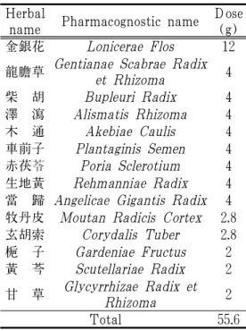
11)의 구성 약재들은 ㈜옴니허브에서 구입 하여 대전대학교 난치성 면역질환의 동서 생명의학연구 지역혁신센터에서 정선 후, 사용하였고, 각 1첩의 내용 및 분량은 다 음과 같다(Table 1, 2, 3).
Herbal
name Pharmacognostic name Dose (g) 金銀花 Lonicerae Flos 12 龍膽草 Gentianae Scabrae Radix
et Rhizoma 4 柴 胡 Bupleuri Radix 4 澤 瀉 Alismatis Rhizoma 4 木 通 Akebiae Caulis 4 車前子 Plantaginis Semen 4 赤茯苓 Poria Sclerotium 4 生地黃 Rehmanniae Radix 4 當 歸 Angelicae Gigantis Radix 4 牧丹皮 Moutan Radicis Cortex 2.8 玄胡索 Corydalis Tuber 2.8 梔 子 Gardeniae Fructus 2 黃 芩 Scutellariae Radix 2 甘 草 Glycyrrhizae Radix et
Rhizoma 2
Total 55.6
Table 1. The Composition of Eunhwasagan -tang
Herbal
name Pharmacognostic name Dose (g) 當 歸 Angelicae Gigantis Radix 12 蒲 黃 Typhae Pollen 12 赤芍藥 Paeoniae Radix 8 五靈脂 Trogopterorum Faeces 8
沒 藥 Myrrha 4
肉 桂 Cinnamomi Cortex 4 川 芎 Cnidii Rhizoma 4 玄胡索 Corydalis Tuber 4 小茴香 Foeniculi Fructus 3 乾 薑 Zingiberis Rhizoma 0.8
Total 55.8
Table 2. The Composition of Sobokchukeo -tang
Herbal
name Pharmacognostic name Dose (g) 白 朮 Atractylodis Rhizoma Alba 30 山 藥 Dioscoreae Rhizoma 30 白芍藥 Paeoniae Radix 15 車前子 Bupleuri Radix 9 蒼 朮 Atractylodis Rhizoma 9 人 蔘 Ginseng Radix 6 甘 草 Glycyrrhizae Radix et Rhizoma 3 柴 胡 Bupleuri Radix 1.8 陳 皮 Citri Unshius Pericarpium 1.5 荊芥穗 Schizonepetae Spica 1.5
Total 55.6
Table 3. The Composition of Wandae -tang
2) 시 약
사용된 시약은 dulbecco's Modified Eagle's Medium(DMEM : Gibco BRL, U.S.A.), fetal bovine serum(FBS : Gibco BRL, U.S.A.), horse serum(Gibco BRL, U.S.A.), penicillin-streptomycin(Sigma, U.S.A.), 2-mercaptoethanol(Gibco BRL, U.S.A.), L-glutamine(Gibco BRL, U.S.A.), T-stim (Corning, U.S.A.), trypan blue(Sigma, U.S.A.), EZ-Cytox(Daeilab, Korea), lipopolysaccharide (LPS : Sigma, U.S.A.), phorbol 12-myristate 13-acetate(PMA : Sigma, U.S.A.), ionomycin (Sigma, U.S.A.), nerve growth factor(NGF : Sigma, U.S.A.), dulbecco's phosphate buffered saline(D-PBS : Welgene, Korea), 2′,7′-Dichlorofluorescein diacetate(DCF-DA : Sigma, U.S.A.), nitric oxide detection kit (Intron Biotechnology, Korea), mouse cytokine milliplex map immunoassay kit (Millipore, U.S.A.), PGE
2Parameter Assay Kit(R&D systems Co., U.S.A.), LTB
4Parameter Assay Kit(R&D systems Co.,
U.S.A.), Total RNA prep kit(Intronbio,
Korea), AccuPower CycleScript RT PreMix (Bioneer, Korea), SYBR Green(Qiagen, Germany), DEPC-DW(Bioneer Co., Korea), lysis buffer(Intronbio, Korea), BCA protein assay kit(Thermo Fisher Scientific, U.S.A.), tris-buffered saline with Tween20(TBST : Intronbio, Korea), IκB-α Rabbit mAb (Cell Signaling Technology, U.S.A.), phospho- IκBα Rabbit mAb(Cell Signaling Technology, U.S.A.), anti-NF-κB P65 antibody(abcam, U.K.), anti-β-actin antibody(abcam, U.K.), goat anti-rabbit IgG(abcam, U.K.), western ECL blotting substrates(BIO-RAD, U.S.A.), histamine ELISA Kit(MyBioSource, U.S.A.), acetylcholine ELISA Kit(MyBioSource, U.S.A.), acetylcholinesterase ELISA Kit (MyBioSource, U.S.A.), substance P ELISA Kit(MyBioSource, U.S.A.), nutrient agar (NA : Difco, USA), yeast malt(YM : Difco, USA), bacto agar(Difco, USA) 등을 사용하였다.
3) 기 기
사용된 기기는 rotary vacuum evaporator (Büchi B-480, Switzerland), freeze dryer (EYELA FDU-540, Japan), HPLC (Shimadzu, Co., Japan), CO
2incubator (Forma scientific Co., U.S.A.), clean bench (Vision scientific, Korea), autoclave(Sanyo, Japan), vortex mixer(Vision scientific, Korea), centrifuge(Hanil, Korea), deep-freezer (Sanyo, Japan), ice-maker(Vision scientific, Korea), flow cytometer(Becton Dickinson, U.S.A.), plate shaker(Lab-Line, U.S.A.), luminex(Millipore, U.S.A.), micro plate reader(Molecular Devices, U.S.A.), flow cytometer(Becton Dickinson, U.S.A.), Nanodrop(Thermo Fisher Scientific, U.S.A.),
Alpha Cycler 1 PCRmax(PCRmax, U.K.) real time PCR(Qiagen, Germany) Electrophoresis Chambers(BIO-RAD, U.S.A.), Fusion FX (Vilber Lourmat, France) 등을 사용하 였다.
2. 방 법 1) 시료 추출
EST, SCT, WDT의 각 1첩 분량에 1000 ml의 증류수를 넣어 3시간 동안 환 류추출하였으며, 여과액을 rotary vacuum evaporator로 감압 농축 후, freeze dryer 로 농축액을 동결 건조하였다. EST는 10.4 g(수율 18.7%), SCT는 11.1 g(수율 19.9%), WDT는 24.9 g(수율 23.3%)의 분말을 획득하여 초저온 냉동고(-80℃) 에서 보관하면서 필요에 따라 증류수에 희석하여 농도를 조절하여 사용하였다.
2) 세포 배양
RAW264.7 세포는 10% fetal bovine serum(FBS)와 1% penicillin-streptomycin 으로 구성된 DMEM 배지를 사용하였고 MC/9 세포는 10% FBS, 1% penicillin- streptomycin, 0.055 mM 2-mercaptoethanol, 2 mM L-glutamine, 10% T-Stim으로 구 성된 DMEM 배지를 사용하였으며, PC12 세포는 10% horse serum과 5% FBS로 구성된 DMEM 배지를 사용하였다. 모 든 세포들은 세포배양기에서 37℃, 5%
CO
2환경이 유지된 상태에서 배양하였 으며, 2-3일 주기로 계대 배양하였다.
3) 세포생존율 측정
RAW264.7 세포는 96 well plate에 1.5×10
5cells/well로 분주하였고 MC/9 세포는 48
well plate에 2×10
5cells/well로 분주하였
으며, PC12 세포는 collagen으로 코팅된
48 well plate에 2×10
5cells/well로 분주
하여 24시간 동안 배양하였다. 24시간 후, 각각의 시료를 50, 100 μg/ml의 농도 로 처리하여 RAW264.7 세포는 24시간, MC/9 세포와 PC12 세포는 48시간 동안 배양하였다. 모든 배양이 끝난 후, 배양 액 100 μl당 EZ-Cytox 용액 10 μl를 첨 가하여 30분간 세포배양기에서 반응시켰 다. 반응 후, 450 nm에서 흡광도 변화를 측정하여 백분율로 대조군에 대한 세포 생존율을 나타내었다.
4) 항염증 효능평가 (1) NO 생성량 측정
96 well plate에 RAW264.7 세포를 1.5×10
5cells/well로 분주하여 24시간 동안 배양 하였다. 배양 후, 50, 100 μg/ml 농도의 시료와 1 μg/ml의 LPS를 함께 처리하여 다시 24시간 동안 배양하였다. 배양 후, 50 μl의 N1 buffer를 각 well에 처리하여 10분간 상온에서 반응시킨 후, 50 μl의 N2 buffer를 각 well에 처리하고 10분간 반응시켰다. 반응 후, 540 nm에서 흡광 도 변화를 측정하여 백분율로 대조군에 대한 NO 생성량을 나타내었다.
(2) Cytokine 생성량 측정
12 well plate에 RAW264.7 세포를 2×10
5cells/well로 분주하여 24시간 동안 배양 하였다. 배양 후, 50, 100 μg/ml 농도의 시료와 1 μg/ml의 LPS를 함께 처리하여 다시 24시간 동안 배양하였다. 배양액을 5분간 1200 rpm에서 원심분리하여 얻은 상등액과 standard를 96 well plate에 25 μl씩 분주하고 assay buffer, matrix buffer, antibody-immobilized beads를 각각 25 μl 씩 가하여 혼합하여 2시간 동안 실온에 서 반응시킨 후 washing 완충 용액을 사 용하여 2회 세척하였다. 세척 후, detection antibody를 25 μl 가하여 1시간 동안 실
온에서 반응시키고 추가로 Streptavidin- Phycoerythrin을 25 μl 가하여 30분 동안 실온에서 반응시킨 뒤 washing 완충 용 액을 사용하여 2회 세척하였다. 세척 후, PBS를 150 μl 가하여 5분간 shaking한 후, Luminex를 이용하여 측정하여 절대 값으로 나타내었다.
(3) PGE
2, LTB
4생성량 측정 12 well plate에 RAW264.7 세포를 2×10
5cells/well로 분주하여 24시간 동안 배양 하였다. 배양 후, 50, 100 μg/ml 농도의 시료와 1 μg/ml LPS를 함께 처리하여 다시 24시간 동안 배양하였다. 배양액을 1200 rpm에서 5분간 원심분리하여 얻은 상등액과 standard를 96 well plate에 100 μl씩 분주하고 37℃에서 90분간 반 응시켰다. 반응 후, washing buffer를 이 용하여 3회 세척한 후, detection antibody 를 100 μl 가하여 다시 37℃에서 60분간 반응시키고 세척하였다. 세척 후, HRP conjugate를 100 μl씩 가하여 37℃에서 30 분간 반응시키고 세척한 뒤 다시 substrate reagent를 90 μl씩 가하여 37℃에서 15분 간 반응시키고 stop solution을 50 μl 추 가하여 ELISA reader기를 통해 450 nm 에서 흡광도를 측정하였으며, standard curve를 기준으로 하여 절대 값으로 나 타내었다.
(4) 유전자 발현량 측정
① RNA 추출
6 well plate에 RAW264.7 세포를 1×10
6cells/well로 분주하여 24시간 동안 배양
하였다. 배양 후, 50, 100 μg/ml 농도의
시료와 1 μg/ml LPS를 함께 처리하여 다
시 24시간 동안 배양하였다. 이후, 1200 rpm
에서 5분 동안 원심분리하여 얻은 RAW264.7
세포에 easy blue 1 ml와 chloroform 200
μl를 넣고 vortexing 해준 후, 13000 rpm, 4℃에서 10분 동안 원심분리 해준다. 상 층액 400 μl를 binding buffer 400 μl와 1분 동안 실온에서 반응시킨 뒤 반응액 700 μl 를 column에 주입하여 30초 동안 13000 rpm에서 원심분리 한다. Column에 700 μl의 washing buffer A를 넣고 30초 동안 13000 rpm에서 원심분리한 후, 700 μl의 washing buffer B를 넣고 동일하게 원심 분리 한다. Column 하단을 Ep tube로 교 체한 뒤, column에 50 μl의 elution buffer 를 넣고 1분 동안 반응시킨 뒤 1분 동안 13000 rpm에서 원심분리하여 추출된 total RNA를 모은다.
② cDNA 합성
역전사(reverse transcription) 반응은 RT premix kit의 mixture(reaction buffer, dNTPs mixture, RNase inhibitor, stabilizer, oligo dT15 primer)를 사용하여 total RNA가 1 μg이 되도록 diethyl pyrocarbonate (DEPC) 처리된 증류수에 최종 부피가 20 μl가 되도록 첨가하였다. 이 반응 혼 합액을 잘 섞은 후 45℃에서 60분간 반 응시켜서 first-strand cDNA를 합성한 뒤, 95℃에서 5분간 방치하여 M-MLV RT를 불활성화 시킨 후, 합성이 완료된 cDNA를 polymerase chain reaction(PCR) 에 사용하였다.
③ 유전자 증폭
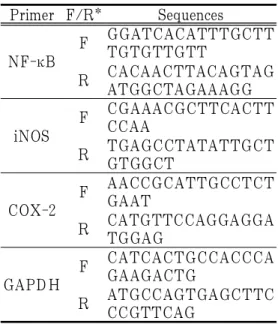
합성이 끝난 cDNA를 증폭시키기 위하 여 real-time PCR을 진행하였으며, real-time 전용 tube에 cDNA 1 μl, 각 primer 2 μl, SYBR Green 10 μl, DEPC-DW 5 μl씩 을 넣었다. 이후 94℃에서 5분 동안 반응 한 다음 94℃에서 15초, 60℃에서 30초, 72℃에서 30초를 총 40회 반복하여 진행 하였다. 유전자 발현량은 대조군에 비하
여 계산하였으며, 사용된 primer의 sequence 는 Table 4와 같다.
Primer F/R* Sequences
NF-κB
F GGATCACATTTGCTT TGTGTTGTT
R CACAACTTACAGTAG ATGGCTAGAAAGG
iNOS
F CGAAACGCTTCACTT CCAA
R TGAGCCTATATTGCT GTGGCT
COX-2
F AACCGCATTGCCTCT GAAT
R CATGTTCCAGGAGGA TGGAG
GAPDH
F CATCACTGCCACCCA GAAGACTG
R ATGCCAGTGAGCTTC CCGTTCAG
* F : forward, R : reverse
Table 4. The Sequences of Primers
(5) Immunoblot 분석
① 단백질 분리 및 정량
6 well plate에 RAW264.7 세포를 1×10
6cells/well로 분주하여 24시간 동안 배양 하였다. 배양 후, 50, 100 μg/ml 농도의 시료와 1 μg/ml LPS를 함께 처리하여 다시 24시간 동안 배양하였다. 이후, 1200 rpm에서 5분 동안 원심분리하여 얻은 RAW264.7 세포에 lysis buffer(10 mM Tris-HCl [pH 7.6], 140 mM NaCl, 1 mM phenylmethylsulphonyl fluoride, 1%
Nonidet P-40, 0.5% deoxycholate, 2% β
-mercaptoethanol, 10 μg/ml pepstatin A
and 10 μg/ml aprotinin)를 넣고 4℃에
서 15분간 방치하였다. 방치 후, 13000
rpm, 4℃에서 10분 동안 원심분리 하여
상층액을 얻었으며, BCA protein assay
kit를 이용하여 상층액의 단백질을 정량
하였다.
② 전기영동 및 immunoblot
정량한 단백질을 20 μg씩 10% SDS- polyacrylamide gel에 전기영동하여 단백 질을 분리시킨 후, PVDF membrane에 분리한 단백질을 transfer하였으며, 비 특 이적인 단백질을 blocking하기 위해 5%
skim milk에 담가 4℃에서 90분 동안 방 치하였다. 이후, membrane을 TBST로 세 척하고 희석한 IκB-α(1:1000), p-IκB-α (1:1000), NF-κB P65(1:1000), β-actin (1:1000) antibody에 담가 상온에서 2시 간 동안 반응시켰다. 다시 TBST로 세척 하고 희석한 goat anti-rabbit IgG(1:1000) antibody에 담가 상온에서 1시간 동안 반응시켰다. 이후, western ECL blotting substrates를 처리하고 Fusion FX을 통 해 단백질 발현량을 측정하였다.
5) 항소양증 효능평가 (1) Histamine 생성량 측정
12 well plate에 MC/9 세포를 2×10
5cells/well로 분주하여 24시간 동안 배양 하였다. 배양 후, 50, 100 μg/ml 농도의 시료와 50 ng/ml PMA, 0.5 uM ionomycin 을 함께 처리하여 다시 48시간 동안 배 양하였다. 배양액을 5분간 1200 rpm에서 원심분리하여 얻은 상등액과 standard를 96 well plate에 100 μl씩 넣고 37℃에서 120분간 반응시켰다. 반응 후, washing buffer를 사용하여 3회 세척을 진행하고 Biotin-conjugate를 100 μl씩 넣어 다시 37℃에서 60분간 반응시켰다. 다시 세척 을 진행하고 Streptavidin-HRP를 100 μl 씩 넣어 37℃에서 30분간 반응시켰다. 마 지막 세척을 진행하고 substrate solution 을 100 μl씩 넣어 37℃에서 15분간 반응 시켰다. 반응 후, 50 μl의 stop solution을
추가하여 ELISA reader기를 통해 450 nm 에서 흡광도를 측정하였으며, standard curve를 기준으로 절대 값으로 나타내었다.
(2) Acetylcholine, acetylcholinesterase, substance P 생성량 측정
Collagen으로 코팅된 12 well plate에 PC12 세포를 2×10
5cells/well로 분주하 여 24시간 동안 배양하였다. 배양 후, 50, 100 μg/ml 농도의 시료와 50 ng/ml NGF 를 함께 처리하여 다시 48시간 동안 배양 하였다. 이후, 배양액을 1200 rpm에서 5분 간 원심분리하여 얻은 상등액과 standard 를 96 well plate에 100 μl씩 넣고 conjugate 를 50 μl씩 추가하여 37℃에서 60분간 반 응시켰다. 반응 후, washing buffer를 사용 하여 5회 세척 작업을 진행하고 substrate A와 B를 각각 50 μl씩 넣어 37℃에서 15 분간 반응시켰다. 마지막으로 stop solution 을 50 μl 추가하여 ELISA reader기를 통 해 450 nm에서 흡광도를 측정하였으며, standard curve를 기준으로 절대 값으로 나타내었다.
6) 항균 효능평가
식약처 공시 균주인 대장균 Escherichia
coli(E. coli), 그람 양성균 Staphylococcus
aureus(S. aureus), 그람 음성균 Pseudomonas
aeruginosa(P. aeruginosa), 진균인 Candida
albicans(C. albicans), Aspergillus niger
(A. niger) 5종류로 항균 효능을 측정하
였다. E. coli, S.aureus, P. aeruginosa는
NA 배지를 사용하였으며, C. albicans,
A. niger는 YM 배지를 사용하여 배양하
였으며, 배양한 균을 각 고체배지에 10
7CFU/ml로 도말하였다. Paper disc 위에
100 μg/ml 농도의 시료를 30 μl씩 접종
하여 건조한 후 각 균주가 도말된 배지
위에 올려놓고 37℃의 배양기에서 24시
간 배양하여 생장억제환의 크기를 측정 하였다.
3. 통계처리
통계적 분석은 SPSS 21.0 for windows (SPSS Inc., USA)를 이용하여 평균±표 준편차로 나타내었으며, ANOVA를 사용 하여 다중 비교하였고 Duncan test를 통 해 p<0.05, p<0.01 및 p<0.001 수준에서 유 의성을 검정하였다.
Ⅲ. 실험 결과
1. 세포생존율 1) RAW264.7 세포
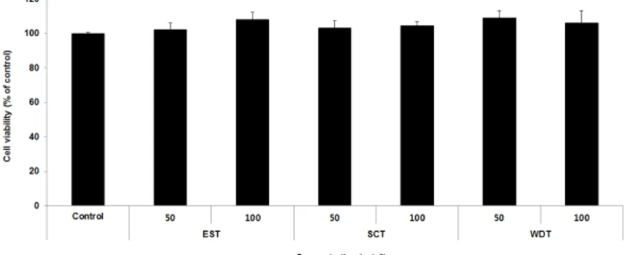
RAW264.7 세포에서 세포생존율을 측정 한 결과, 3가지 처방은 모든 농도에서 세포 에 대한 독성이 나타나지 않았다(Fig. 1).
2) MC/9 세포
MC/9 세포에서 세포생존율을 측정한 결과, 3가지 처방은 모든 농도에서 세포에 대한 독성이 나타나지 않았다(Fig. 2).
Fig. 1. Cell viability of RAW264.7 cells treated with samples.
RAW264.7 cells were treated by 50 and 100 μg/ml of each samples for 24 h. Treated cells were exposed by EZ-Cytox for 30 min and then absorbance were measured at 450 nm using microplate reader.
Cell viability were calculated as percentage relative to the control. The result were presented by the mean±S.D from three independent experiments.
Fig. 2. Cell viability of MC/9 cells treated with samples.
MC/9 cells were treated by 50 and 100 μg/ml of each samples for 48 h. Treated cells were exposed by EZ-Cytox for 30 min and then absorbance were measured at 450 nm using microplate reader.
Cell viability were calculated as percentage relative to the control. The result were presented by the mean±S.D from three independent experiments.
3) PC12 세포
PC12 세포에서 세포생존율을 측정한 결
과, 3가지 처방은 모든 농도에서 세포에 대한 독성이 나타나지 않았다(Fig. 3).
Fig. 3. Cell viability of PC12 cells treated with samples.
PC12 cells were treated by 50 and 100 μg/ml of each samples for 48 h. Treated cells were exposed by EZ-Cytox for 30 min and then absorbance were measured at 450 nm using microplate reader. Cell viability were calculated as percentage relative to the control. The result were presented by the mean±S.D from three independent experiments.
2. 항염증 효능평가 1) NO 생성량
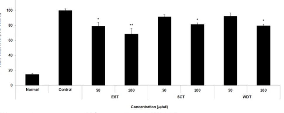
NO 생성량을 측정한 결과, EST는 모 든 농도에서 대조군에 비해 유의성 있는
(* : p<0.05, ** ; p<0.01) 감소가 나타났 으며, SCT와 WDT는 100 μg/ml 농도에 서 대조군에 비해 유의한(* : p<0.05) 감 소가 나타났다(Fig. 4).
Fig. 4. Effects of samples on NO level in RAW264.7 cells.
RAW264.7 cells were treated by 50 and 100 μg/ml of each samples with 1 μg/ml LPS for 24 h.
NO level were calculated as percentage relative to the control. The result were presented by the mean±S.D from three independent experiments (Significance of results, * : p<0.05, ** ; p<0.01 compared to control).
2) Cytokine 생성량 (1) IL-1β
IL-1β 생성량을 측정한 결과, SCT 50
μg/ml를 제외한 모든 농도에서 대조군 에 비해 유의한(* : p<0.05, ** ; p<0.01,
*** : p<0.001) 감소가 나타났다(Fig. 5).
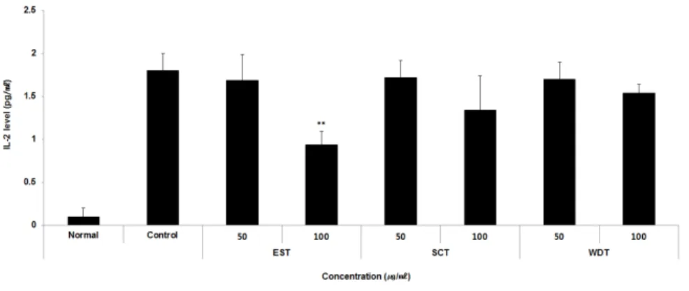
(2) IL-2
IL-2 생성량을 측정한 결과, EST 100
μg/ml 농도에서만 대조군에 비해 유의한 (** ; p<0.01) 감소가 나타났다(Fig. 6).
Fig. 5. Effects of samples on IL-1β level in RAW264.7 cells.
RAW264.7 cells were treated by 50 and 100 μg/ml of each samples with 1 μg/ml LPS for 24 h.
Treated cells were measured by mouse cytokine milliplex map immunoassay kit. The result were presented by the mean±S.D from three independent experiments (Significance of results, * : p<0.05,
** ; p<0.01, *** : p<0.001 compared to control).
Fig. 6. Effects of samples on IL-2 level in RAW264.7 cells.
RAW264.7 cells were treated by 50 and 100 μg/ml of each samples with 1 μg/ml LPS for 24 h.
Treated cells were measured by mouse cytokine milliplex map immunoassay kit. The result were presented by the mean±S.D from three independent experiments (Significance of results, ** ; p<0.01 compared to control).
(3) IL-6
IL-6 생성량을 측정한 결과, EST 50, 100 μg/ml 농도에서만 대조군에 비해 유 의한(** ; p<0.01, *** : p<0.001) 감소가 나타났다(Fig. 7).
(4) TNF-α
TNF-α 생성량을 측정한 결과, SCT 50 μg/ml를 제외한 모든 농도에서 대조
군에 비해 유의한(* : p<0.05, ** ; p<0.01,
*** : p<0.001) 감소가 나타났다(Fig. 8).
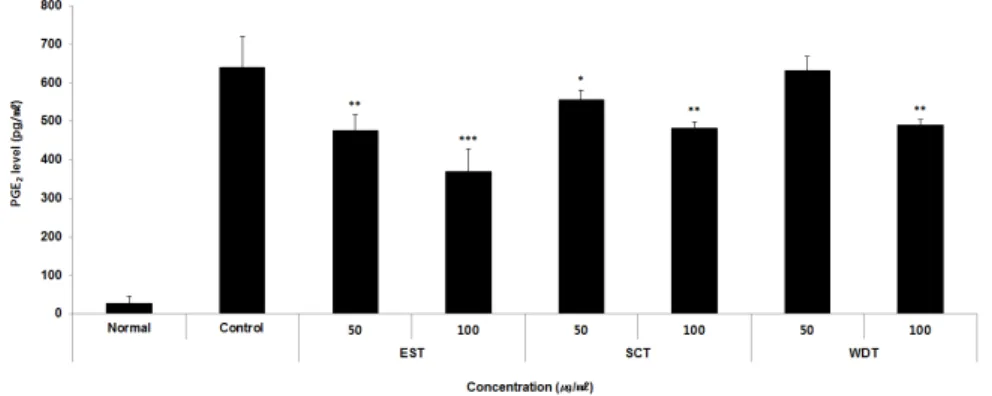
(5) PGE
2PGE
2생성량을 측정한 결과, WDT 50 μg/ml를 제외한 모든 농도에서 대조군 에 비해 유의한(* : p<0.05, ** ; p<0.01,
*** : p<0.001) 감소가 나타났다(Fig. 9).
Fig. 7. Effects of samples on IL-6 level in RAW264.7 cells.
RAW264.7 cells were treated by 50 and 100 μg/ml of each samples with 1 μg/ml LPS for 24 h.
Treated cells were measured by mouse cytokine milliplex map immunoassay kit. The result were presented by the mean±S.D from three independent experiments (Significance of results, ** ; p<0.01, *** : p<0.001 compared to control).
Fig. 8. Effects of samples on TNF-α level in RAW264.7 cells.
RAW264.7 cells were treated by 50 and 100 μg/ml of each samples with 1 μg/ml LPS for 24 h.
Treated cells were measured by mouse cytokine milliplex map immunoassay kit. The result were presented by the mean±S.D from three independent experiments (Significance of results, * : p<0.05, ** ; p<0.01, *** : p<0.001 compared to control).
Fig. 9. Effects of samples on PGE
2level in RAW264.7 cells.
RAW264.7 cells were treated by 50 and 100 μg/ml of each samples with 1 μg/ml LPS for 24 h.
Treated cells were measured by PGE2 Parameter Assay Kit. The result were presented by the mean±S.D from three independent experiments (Significance of results, * : p<0.05, ** ; p<0.01,
*** : p<0.001 compared to control).
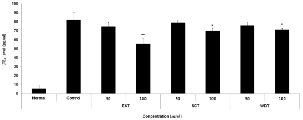
(6) LTB
4LTB
4생성량을 측정한 결과, 3가지 처 방은 100 μg/ml 농도에서 대조군에 비해
유의한(* : p<0.05, ** ; p<0.01) 감소가 나타났다(Fig. 10).
Fig. 10. Effects of samples on LTB
4level in RAW264.7 cells.
RAW264.7 cells were treated by 50 and 100 μg/ml of each samples with 1 μg/ml LPS for 24 h.
Treated cells were measured by LTB4 Parameter Assay Kit. The result were presented by the mean±S.D from three independent experiments (Significance of results, * : p<0.05, ** ; p<0.01 compared to control).
3) 유전자 발현량 (1) NF-κB
NF-κB 유전자 발현량을 측정한 결과, 3가지 처방은 모든 농도에서 대조군에 비해 유의한(** : p<0.01, *** : p<0.001) 감소가 나타났다(Fig. 11).
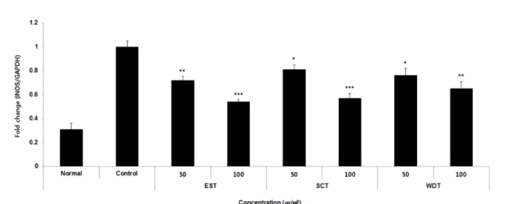
(2) iNOS
iNOS 유전자 발현량을 측정한 결과, 3 가지 처방은 모든 농도에서 대조군에 비 해 유의한(* : p<0.05, ** : p<0.01, *** : p<0.001) 감소가 나타났다(Fig. 12).
Fig. 11. Effects of samples on NF-κB mRNA expression level in RAW264.7 cells.
RAW264.7 cells were treated by 50 and 100 μg/ml of each samples with 1 μg/ml LPS for 24 h.
The mRNA expression level were measured by polymerase chain reaction. The result were presented by the mean±S.D from three independent experiments (Significance of results, ** : p<0.01, *** : p<0.001 compared to control).
Fig. 12. Effects of samples on iNOS mRNA expression level in RAW264.7 cells.
RAW264.7 cells were treated by 50 and 100 μg/ml of each samples with 1 μg/ml LPS for 24 h.
The mRNA expression level were measured by polymerase chain reaction. The result were presented by the mean±S.D from three independent experiments (Significance of results, * : p<0.05, ** : p<0.01, *** : p<0.001 compared to control).
(3) COX-2
COX-2 유전자 발현량을 측정한 결과, EST는 모든 농도에서 대조군에 비해 유의 성 있는(*** : p<0.001) 감소가 나타났으
며, SCT와 WDT는 100 μg/ml 농도에서 대조군에 비해 유의한(* : p<0.05, *** : p<0.001) 감소가 나타났다(Fig. 13).
Fig. 13. Effects of samples on COX-2 mRNA expression level in RAW264.7 cells.
RAW264.7 cells were treated by 50 and 100 μg/ml of each samples with 1 μg/ml LPS for 24 h.
The mRNA expression level were measured by polymerase chain reaction. The result were presented by the mean±S.D from three independent experiments (Significance of results, * : p<0.05, *** : p<0.001 compared to control).
4) Immunoblot (1) p-IκB-α/IκB-α
p-IκB-α/IκB-α 단백질 발현량을 측정 한 결과, 3가지 처방은 모든 농도에서 대 조군에 비해 유의한(*** : p<0.001) 감 소가 나타났다(Fig. 14).
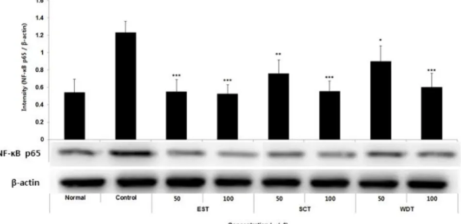
(2) NF-κB p65
NF-κB p65 단백질 발현량을 측정한 결과, 3가지 처방은 모든 농도에서 대조 군에 비해 유의한(* : p<0.05, ** : p<0.01,
*** : p<0.001) 감소가 나타났다(Fig. 15).
Fig. 14. Effect of samples on p-IκB-α/IκB-α protein expression level in RAW264.7 cells.
RAW264.7 cells were treated by 50 and 100 μg/ml of each samples with 1 μg/ml LPS for 24 h. The protein expression level were measured by immunoblot. The result were presented by the mean±S.D from three independent experiments (Significance of results, *** : p<0.001 compared to control).
Fig. 15. Effects of samples on NF-κB protein expression level in RAW264.7 cells.
RAW264.7 cells were treated by 50 and 100 μg/ml of each samples with 1 μg/ml LPS for 24 h.
The protein expression level were measured by immunoblot. The result were presented by the mean±S.D from three independent experiments (Significance of results, * : p<0.05, ** : p<0.01,
*** : p<0.001 compared to control).
3. 항소양증 효능평가 1) Histamine
Histamine 생성량을 측정한 결과, EST 50, 100 μg/ml 농도에서만 대조군에 비 해 유의한(* ; p<0.05, *** : p<0.001) 감 소가 나타났다(Fig. 16).
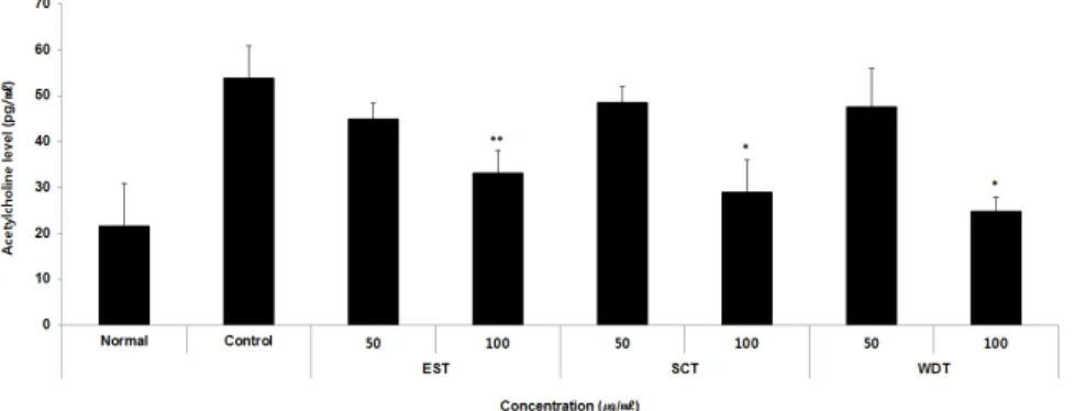
2) Acetylcholine
Acetylcholine 생성량을 측정한 결과, 3가
지 처방은 100 μg/ml 농도에서만 대조군
에 비해 유의한(* : p<0.05, ** ; p<0.01)
감소가 나타났다(Fig. 17).
Fig. 16. Effects of samples on histamine level in MC/9 cells.
MC/9 cells were treated by 50 and 100 μg/ml of each samples with 50 ng/ml PMA and 0.5 uM ionomycin for 48 h. Treated cells were measured by histamine ELISA kit. The result were presented by the mean±S.D from three independent experiments (Significance of results, * ; p<0.05, *** : p<0.001 compared to control).
Fig. 17. Effects of samples on acetylcholine level in PC12 cells.
PC12 cells were treated by 50 and 100 μg/ml of each samples with 50 ng/ml NGF for 48 h.
Treated cells were measured by acetylcholine ELISA kit. The result were presented by the mean±S.D from three independent experiments (Significance of results, * : p<0.05, ** ; p<0.01 compared to control).
3) Acetylcholinesterase
Acetylcholinesterase 생성량을 측정한 결과, 3가지 처방 모두 대조군에 비해 약간 감소하였으나, 유의성은 없었다(Fig. 18).
4) Substance P
Substance P 생성량을 측정한 결과,
WDT 50 μg/ml를 제외한 모든 농도에서
대조군에 비해 유의한(* : p<0.05, ** ;
p<0.01, *** : p<0.001) 감소가 나타났다
(Fig. 19).
Fig. 18. Effects of samples on acetylcholinesterase level in PC12 cells.
PC12 cells were treated by 50 and 100 μg/ml of each samples with 50 ng/ml NGF for 48 h.
Treated cells were measured by acetylcholinesterase ELISA kit. The result were presented by the mean±S.D from three independent experiments.
Fig. 19. Effects of samples on substance P level in PC12 cells.
PC12 cells were treated by 50 and 100 μg/ml of each samples with 50 ng/ml NGF for 48 h.
Treated cells were measured by substance P ELISA kit. The result were presented by the mean±S.D from three independent experiments (Significance of results, * : p<0.05, ** ; p<0.01,
*** : p<0.001 compared to control).
4. 항균 효능평가
생장억제환 크기를 측정한 결과, 3가지 처방 모두 Escherichia coli와 Staphylococcus aureus의 생장은 억제하지 못하였으나
Pseudomonas aeruginosa, Candida albicans,
Aspergillus niger의 생장을 억제하는 효
능이 EST, WDT, SCT 순으로 나타났다
(Fig. 20).
Fig. 20. Effects of samples on clear zone diameter of bacteria.
100 μg/ml of each samples were loaded into the solid medium inoculated with bacteria and the clear zone diameter was measured after 24 hours. (mean±standard deviation, n=four independent experiments).