Kor. J. Pharmacogn.
47(1) : 12∼ 17 (2016)
12
떡윤노리나무로부터 분리된 Lyoniside의 함량분석과 항염증 효과
우경완·성태경·이혜미·장지훈·이기호·조현우·조정희·안병관* 한약진흥재단 한약자원본부 한약재연구팀
Quantitative Analysis and Anti-inflammatory of Lyoniside from the Pourthiaea villosa var. brunnea
Kyeong Wan Woo, Tae Kyoung Seong, Hye Mi Lee, Ji Hun Jang, Ki Ho Lee, Hyun Woo Cho, Jung Hee Cho, and Byeongkwan An*
Traditional Korean Medicines Research Team, Naional Development Institute of Korea Medicine, 288, Woodlandgil, Anyangmyeon, Jangheunggun, Jeollanamdo59338, Korea
Abstract − In this study, the purification of the MeOH extract from the stems and leaves of Pourthiaea villosa var. brunnea using column chromatography furnished a main compound, lyoniside. The structure was elucidated on the basis of 1H and 13C NMR spectroscopic data. Quantitative analysis of lyoniside was conducted by HPLC method and the highest content of lyoniside was found in 50% MeOH reflux extraction. To investigate the anti-inflammatory effect of the lyoniside, we measured nitric oxide and pro-inflammatory cytokines such as TNF-α, IL-1β, IL-6 levels in lipopolysaccharide-induced murine mac- rophage cell line RAW 264.7. As a results, lyoniside decreased the level of nitric oxide and IL-6 in concentration dose depen- dent manner in RAW 264.7 cells.
Key words − Pourthiaea villosa var. brunnea, Lyoniside, Quantitative analysis, Anti-inflammation, Cytokine
떡윤노리나무(Pourthiaea villosa var. brunnea)는 장미과 (Rosaceae)에 속하는 낙엽활엽관목으로 잎은 호생하며 길이 3-8 cm로 도란형이고, 윤노리나무에 비해 잎이 두껍고 잎자 루가 짧으며, 지름 3-5 cm의 흰꽃이 산방화서로 핀다. 열매 는 타원형이며 길이 12 mm의 열매가 9월에 적색으로 익는 다.1)국내에는 Pourthiaea 속에 윤노리나무(P. villosa), 좀윤 노리나무(P. villosa var. coreana), 민윤노리나무(P. villosa var. laevis), 털윤노리나무(P. villosa var. zollingeri) 등이 자 생하고 있으며, 이중 P. villosa 가지의 MeOH추출물에서 항 산화 및 항염증 활성 연구와2) P. villosa 과실에서 flavonoid 와 phenol성 성분을 발표하였고,3) 이 외의 식물에서는 성분 및 생리활성에 관한 연구가 보고되지 않은 실정이다. 따라 서 본 연구실에서는 국내 자생하는 토종자원의 한약재 기 반구축사업의 일환으로 떡윤노리나무에 대한 지표성분 분 리 및 함량분석 그리고 항염증활성 연구를 진행하였다.
재료 및 방법
실험재료 − 본 실험에 사용한 떡윤노리나무는 2013년 5 월에 제주도에서 채취하였으며, 목포대학교 한약자원학과 김휘 교수에게 의뢰하여 정확히 감정하였으며, 음건세절하 여 실험에 사용하였다. 표본은 한약진흥재단 한약재연구팀 에 보관하였다.
기기 및 시약 − 추출 및 분획용 시약, TLC 및 column용 시약 등은 특급시약을 사용하였다. Thin layer chromatography 용 plate는 precoated silica gel 60 F254 plate(layer thickness 0.25 mm, 20×20 cm. Art. 5715, Merck)와 precoated RP-18 F254S plate(layer thickness 0.25 mm, 20×20 cm. Art. 5423, Merck)를 사용하였으며, UV의 검색은 254와 365 nm에 측 정하였다. Column chromatography의 충진제는 YMC gel ODS-A(50 µm)를 사용하였다. Preparative HPLC는 Agilent A1200 series(Agilent, U.S.A)을 column은 Luna 5 µ C18(2) 100A column(250×30.00 mm, Phenomenex)을 사용하였다.
단일 화합물의 구조를 규명하기 위해 이용한 NMR(Nuclear magnetic resonance) spectrum은 Varian 600 spectrometer
*교신저자(E-mail):[email protected] (Tel): +82-63-860-2811
(1H: 600 MHz, 13C: 150 MHz)를 사용하여 측정하였고, NMR 용매는 Sigma. Inc(USA)의 DMSO-d6를 사용하였다.
항염증 효능에 이용한 시약은 Dulbecco's modified eagle's medium(DMEM)과 fetal bovine serum(FBS), penicillin, streptomycin은 Gibco/BRL(Eggenstein, Germany)에서 구입하 였고, 3-(4,5-dimethylthiazol-2-yl)-5-(3-carboxymethoxyphenyl)- 2-(4-sulfophenyl)-2H-tetrazolium(MTS, CellTiter 96AQueous One Solution Cell Proliferation Assay), Griess reagent system은 Promega(Madison, USA)에서 구입하였다. Lipo- polysaccaride(LPS), dimethyl sulfoxide(DMSO)는 Sigma Chemical Co.(St. Lousi, MO, USA)에서 구입하였고, PEG2 ELISA kit 는 Enzo Life Sciences Co.(Farmingdale, NY, USA)에서 구입하였고, TNF-α, IL-1β, IL-6의 enzyme- linked immunosorbent assay(ELISA) Kit는 R&D systems (DuoSet ELISA Development Systems, MN, USA)에서 구 입하였다.
추출 및 분획 − 신선한 떡윤노리나무의 가는줄기 및 잎 (5 kg)를 100% MeOH로 3시간, 3회 반복 환류냉각 추출하 고 여과, 감압 농축하여 156 g의 MeOH extract를 얻었다.
MeOH extract(156 g)을 증류수에 녹여 n-Hexane, n-BuOH 로 차례로 용매 분획하여 각각 19 g, 76 g을 그리고 H2O 분 획 55 g을 얻었다.
지표물질 분리 − BuOH 분획물(76 g)을 ODS gel(50 μm, 350 g, YMC, Japan)이 충진 된 column(8 cm×100 cm)에 Acetonitrile-Water(5:95→60:40 in 0.02% formic acid)을 용 매 조건으로 하여 column chromatography를 실시하였고, 7 개의 소분획(PVB1~7)을 얻었다. 그 중 PVB3 소분획물 2.3 g을 preparative-HPLC(Agilent, infinity 1260, USA)를 사용 하여 화합물 1(40 mg)을 분리하였다(Fig. 1).
Lyoniside (1) − Yellowish gum; 1H NMR (600 MHz, DMSO-d6) δ 6.53 (1H, s, H-2), 6.33 (2H, s, H-2', 6'), 4.25 (1H, d, J=6.5 Hz, H-7'), 4.11 (1H, d, J=7.5 Hz, H- 1''), 3.76 (3H, s, 3-OCH3), 3.68 (1H, m, H-5''a), 3.65 (1H, m, H-9'a), 3.63 (6H, s, 3',5'-OCH3), 3.49 (1H, dt, J=9.0, 4.0 Hz, H-9a), 3.34 (1H, m, H-9b), 3.26 (1H, m, H-4''), 3.25 (1H, m, H-9'b), 3.23 (3H, s, 5-OCH3), 3.10 (1H, td, J=9.0, 3.5 Hz, H-3''), 3.02 (1H, m, H-5''b), 3.00 (1H, m, H-2''), 2.62 (1H, dd, J=15.0, 4.0 Hz, H-7), 2.52 (1H, m,
H-7), 1.90 (1H, m, H-8'), 1.50 (1H, m, H-8); 13C NMR (150 MHz, DMSO-d6) δ 147.9 (C-3', 5'), 147.3 (C-3), 146.9 (C-5), 138.0 (C-1'), 137.6 (C-4), 133.6 (C-4'), 128.7 (C-1), 125.3 (C-6), 107.0 (C-2), 106.2 (C-2', 6'), 104.4 (C- 1''), 77.2 (C-3''), 73.7 (C-2''), 70.0 (C-4''), 69.2 (C-9'), 66.1 (C-5''), 64.0 (C-9), 59.0 (5-OCH3), 56.4 (3',5'-OCH3), 56.0 (3-OCH3), 45.0 (C-8'), 41.4 (C-7'), 39.2 (C-8), 33.0 (C-7);
LC ESI IT-TOF MS: m/z 551 [M-H]−.
표준용액 조제 − 떡윤노리나무로부터 분리, 정제한 lyoniside을 1.03 mg을 취해 MeOH를 10 mL가하여 stock solution을 조제하였다. 이 stock solution을 희석하여 103, 51.5, 25.75, 12.88, 6.44 ppm 농도의 표준용액을 조제하였다.
검액 조제 − 용매에 따른 추출효율 분석으로 시료를 가루 로 하여 약 1.0 g을 정확히 칭량하고 MeOH, 50% MeOH, EtOH을 각각 10 mL씩 첨가하여 30 분간 초음파 추출한 다 음 여과 후 각각의 추출용매를 넣어 정확하게 10 mL가 되 게 하여 검액으로 사용하였다. 추출조건에 따른 함량 분석 으로는 시료를 가루로 하여 약 0.5 g을 정확히 칭량하고 추 출효율이 가장 좋았던 50% MeOH 25 mL를 첨가하여 초음 파 30분, 30분 2회, 60분과 환류 30분, 30분 2회, 60분 추 출한 다음 여과 후 정확하게 25 mL가 되게 하여 검액으로 사용하였다.
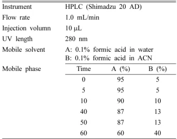
HPLC 분석 조건 − 분리된 lyoniside의 분석 조건을 Table I과 같이 설정한 다음, 이 조건에 따라 함량분석을 실시하였다.
검량선 및 직선성 (Linearity) − 지표물질 lyoniside의 표 준용액을 MeOH로 희석하여 5개의 농도(103, 51.5, 25.75, 12.88, 6.44 µg/mL)가 되도록 만들어 실험하였다. 검량선은 y=ax+b(y: peak 면적, x: 시료농도, a: 직선의 기울기, b: y 절편)의 형태로 작성하였다.
세포 배양 − 한국세포주은행(KCLB, Seoul, Korea)으로부 터 분양 받은 마우스 대식세포주인 RAW 264.7 cell은 Table I. HPLC condition of lyoniside
Instrument HPLC (Shimadzu 20 AD) Flow rate 1.0 mL/min
Injection volumn 10μL UV length 280 nm
Mobile solvent A: 0.1% formic acid in water B: 0.1% formic acid in ACN Mobile phase Time A (%) B (%)
0 95 5
5 95 5
10 90 10
40 87 13
50 87 13
60 60 40
Fig. 1. Chemical structures of compound 1.
Penicillin/streptomycin 100 unit/mL과 10% FBS가 함유된 DMEM 배지를 사용하여 37oC, 5% CO2 incubator에서 배 양하였다.
세포 독성 − Lyoniside의 RAW 264.7 cell에 대한 세포 독성 효과를 측정하기 위해 MTS assay를 실시하였다. 96 well plate에 3×10⁵ cells/mL로 분주하여 배양하였고 10%
FBS DMEM 조건에서 시료를 농도별로 전처리하고 여기에 LPS(500 ng/mL)를 각각 처리한 다음 RAW 264.7 cell를 24 시간 배양하였다. 그 후의 각각 세포 배양액 용량의 1/10 배 의 MTS 용해액을 첨가한 후 37oC에서 2 시간 배양한 후 ELISA microplate reader(Infinite 200 pro, TECAN, Austria) 를 이용하여 490 nm에서 흡광도를 측정하였다.
Nitrite Assay − LPS로 활성화된 RAW 264.7 cell에서 lyoniside의 NO 생성 억제를 측정하기 위해 시료를 여러 농 도로 처리한 실험군과 대조군을 LPS(500 ng/mL)와 함께 24 시간 세포 배양 후 griess reagent system(G2930, Promega, USA)를 이용하여 NO를 측정하였다. 96 well plate에 세포 배양 상등액과 griess reagent를 1:1로 혼합하여 넣고 10분 동안 반응 시킨 후 ELISA microplate reader(Infinite 200 pro, TECAN, Austria)를 이용하여 540 nm에서 흡광도를 측 정하였다.
Cytokines (TNF-α, IL-1β, IL-6) Assay − LPS(500 ng/
mL)로 RAW 264.7 cell을 자극하기 전 lyoniside를 30분 동 안 전처리 하였다. Pro-inflammatory cytokine의 염증매개물 질의 생성에 미치는 약물의 효과를 검증하기 위해서 LPS로 자극한 후 24 시간 뒤 이들 염증매개와 관련된 cytokine을 세포 상층액에서 명시된 ELISA kit의 protocol 대로 ELISA 법으로 정량하였다.
통계처리 − 본 실험에서 얻은 결과에 대해서는 평균치±
표준오차(mean±S.E.)로 나타내었으며, 대조군과 각 실험군 과의 평균의 차이는 Student’s t-test로 분석하여 p-value 값 이 0.05 미만일 때 통계적으로 유의한 차이가 있는 것으로 판정하였다.
결과 및 고찰
화합물의 구조 − 화합물 1은 무색의 결정으로 분리되었 으며, 1H-NMR spectrum에서 3개의 benzene ring proton δ 6.53 (1H, s, H-2), 6.33 (2H, s, H-2', 6') peak와 2개의 oxygenated methylene group δ 3.65 (1H, m, H-9'a), 3.49 (1H, dt, J=9.1, 4.1 Hz, H-9a), 3.34 (1H, m, H-9b), 3.25 (1H, m, H-9'b) peak가 관찰되었으며, 3개의 methine proton δ 4.25 (1H, d, J=6.6 Hz, H-7'), 1.90 (1H, m, H-8'), 1.50 (1H, m, H-8) peak와 1개의 methylene proton δ 2.62 (1H, dd, J=15.2, 4.4 Hz, H-7), 2.52 (1H, m, H-7) peak, 4개의 methoxy proton δ 3.76(3H, s, 3-OCH3), 3.63 (6H, s, 3',5'- OCH3), 3.23 (3H, s, 5-OCH3) peak이 관찰되었다. 13C-NMR spectrum을 관찰한 결과 12개의 aromatic carbon, 2개의 oxygenated carbon, 4개의 methoxy carbon을 포함한 총 21 개의 peak을 확인하였고, lignan 계열인 것을 예측하였다4). 추가적으로 1H-NMR spectrum에서 arabinose의 anomeric proton δ 4.11 (1H, d, J=7.5 Hz, H-1'')과 13C-NMR spectrum 에서 δ 104.4 (C-1''), 77.2 (C-3''), 73.7 (C-2''), 70.0 (C-4''), 66.1 (C-5'')을 관찰하였다.5) 이상의 결과를 바탕으로 기존의 문헌과 비교하여 화합물 1은 lyoniside로 구조 결정하였다.6)
함량분석 − 떡윤노리나무로 분리한 화합물 lyoniside의 분
Fig. 2. HPLC chromatogram of P. villosa var. brunnea extract (PVE) and lyoniside.
석법 확립을 위해 Table I과 같은 조건으로 진행하여 43.0 min 에 관찰하였다(Fig. 2). 분석법을 바탕으로 추출용매(50%
MeOH, 100% MeOH, EtOH)에 따른 지표성분 함량 비교시 50% MeOH 환류 추출에서 가장 높은 효율을 확인하였다 (Table II). 50% MeOH을 추출용매로 하여 추출조건별 실험 결과 환류 추출 30분에서 가장 높은 효율을 확인하였다(Table III). 지표물질의 검량선 결과 Fig. 3과 같고, 상관계수(R2)은 0.9999로 높은 직선성을 보였다. 이와 같은 방법으로 떡윤 노리나무 50% MeOH 추출물에 함유하는 lyoniside의 함량 을 측정한 결과 0.073%으로 확인되었다(Table IV).
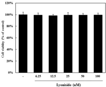
항염증활성 − 떡윤노리나무로 분리한 화합물 lyoniside의 RAW 264.7에서의 항염증활성을 알아보기 위하여, lyoniside 의 세포독성을 MTS 분석법을 이용하여 세포생존율을 측정 하였다. Lyoniside를 6.25, 12.5, 25, 50, 100 µM의 농도로 처리하였을 때 세포독성이 관찰되지 않았고(Fig. 4), 독성이 없는 농도(6.25, 12.5, 25, 50, 100 µM)에서 생성되는 NO 양
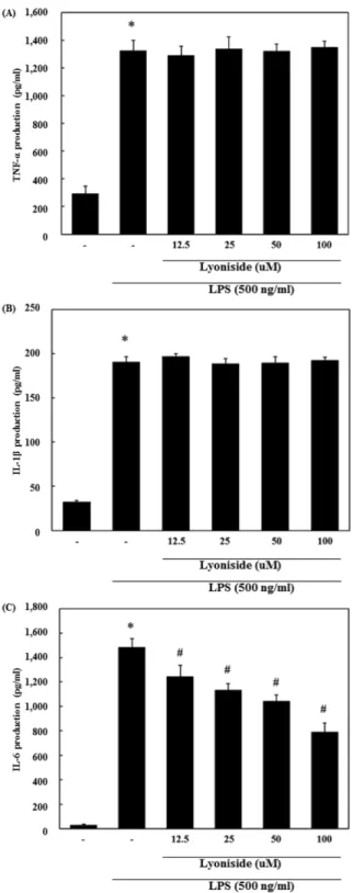
을 측정한 결과, 농도별로 유의성있게 NO생성을 억제하는 것을 확인하였다(Fig. 5). Lyoniside가 RAW 264.7 cell에서 LPS로 유도 되는 각종 전염증성 및 염증성 cytokine들의 발 현에 대한 영향을 조사하기 위하여 전염증성 및 염증성 cytokine의 생성을 조사하였다. 세포 상층액을 ELISA 방법 으로 측정한 결과 lyoniside의 12.5~100 µM 농도에서 IL-6 를 농도 의존적으로 유의성 있게 억제하였고, IL-1β, TNF- α는 LPS만 처리한 군과 비교하여 차이가 없었다(Fig. 6). 대 Table II. The contents of lyoniside by extract solvents
Solvents
Content (%)
MeOH 50% MeOH EtOH
0.015 0.054 0.003
Table III. The contents of lyoniside by extraction time and methods
Extraction method Sonication Reflux
Time (min) 30 30*2 60 30 30*2 60
Content (%) 0.061 0.065 0.065 0.071 0.070 0.069
0.062 0.068 0.068 0.074 0.070 0.068
0.065 0.065 0.070 0.073 0.070 0.069
SD 0.002 0.001 0.002 0.001 0.000 0.001
Average 0.063 0.066 0.068 0.073 0.070 0.069
Table IV. Calibration data for analysis of lyoniside
Compound
Regression equation
y=ax+b Contents
(mg/g) r2 LOD
(μg/mL) LOQ
(μg/mL) Slope(a) Intercept(b)
Lyoniside 3763.66 -2207.37 0.73±0.01 0.9999 0.70 2.13
Fig. 3. Calibration curve of lyoniside.
Fig. 4. Effect of lyoniside on cell viability in RAW 264.7 cells. RAW 264.7 cells were incubated for 24 hr in the pres- ence or absence of lyoniside at indicated dose. Cell viability was evaluated by MTS assay as described in materials and methods. Statistical significance: *p<0.05, when compared to the normal group. Significant differences between treated groups were determined using the Student’s t-test. Data rep- resent the mean±S.E. of triplicate determinations from three separate experiments.
식세포는 외부인자에 대한 방어기전으로 매우 중요한 역할 을 하며 NO, prostaglandins(PGs) 그리고 pro-inflammatory cytokines 등 다양한 염증 매개물질을 조절한다7). 이 중 NO 의 형성은 아미노산의 한 종류인 아르기닌의 deamination 반응에 의해 생성되며, 과하게 유도되었을 경우 각종 염증 질환을 발생시키게 된다8,9). 이런 이론들을 바탕으로 NO의 형성에 lyoniside가 저해하는지 확인한 결과 감소되는 경향 을 확인할 수 있었다. 염증매개물질인 TNF-α와 IL-6, IL-1β 는 전염증성 매개인자로서 세포 대 세포로 신호전달을 하 여 염증과 관련된 일련의 반응을 촉진시키는 cytokine으로 잘 알려져 있다10). Lyoniside는 LPS에 의해 유도된 IL-6의 생성을 농도 의존적으로 유의성 있게 저해하였다. 본 항염 증실험 결과를 요약하면 lyoniside는 대식세포에서 IL-6를 저해하여 LPS에 의해 유도된 염증반응을 감소시켜 NO생 성을 감소시키는 것을 확인할 수 있었다. 하지만 lyoniside 가 IL-6 만을 어떠한 원리로 저해하는지에 대한 유전자 전 사 및 번역단계에서 추가적인 실험을 할 필요가 있다.
결 론
본 연구에서는 떡윤노리나무의 가는 줄기 및 잎으로부터 각종 column chromatography법을 이용하여 주성분인 Fig. 5. Effect of lyoniside on LPS-induced NO production in RAW 264.7 cells. RAW 264.7 cells were pretreated with the indicated concentration of lyoniside for 30 minutes before being incubated with LPS (500 ng/mL) for 24 hr. The culture supernatant was subsequently isolated and analyzed for LPS treated group. Statistical significance: *p<0.05, when compared to the normal group; #p<0.05, when compared to the LPS alone treated group. Significant differences between treated groups were determined using the Student's t-test. Values shown are the mean±S.E. of triplicate determinations from three separate experiments. NOR: normal, LPS: LPS treated.
Fig. 6. Effect of lyoniside on LPS-induced TNF-α (A), IL-1β (B), IL-6 (C) production in RAW 264.7 cells. Cells were pre- treated with the indicated concentrations of lyoniside for 30 minutes before being incubated with LPS (500 ng/mL) for 24 hr. Production of TNF-α, IL-1β, IL-6 was measured by ELISA. Data are mean±S.E. values of triplicate determinations from three separate experiments. *p<0.05, when compared to the normal group; #p<0.05, when compared to the LPS alone treated group.
lyoniside(1)를 분리하였고, 이를 지표성분으로 하여 함량분 석을 실시하였다. 50% MeOH 환류 30분 추출시 0.073%으 로 가장 많은 양을 함유하는 것으로 검출되었다. Lyoniside 의 항염증 활성 측정시 RAW 264.7 세포에서 lyoniside의 독성이 없었고, LPS에 의해 유도되는 NO생성이 감소하는 경향을 보였으며, IL-6 단백질을 저해하는 것을 확인할 수 있었다.
사 사
이 연구는 2014년도 보건복지부 한의약 산업 육성을 위 한 기반구축(한국 토종자원의 한약재 기반구축사업) 결과의 일부이며 지원에 감사드립니다.
인용문헌
1.이창복 (2003) 원색 대한식물도감(상), 577, 향문사, 서울.
2. Kim, H. H., Kwon, J. H., Park, K. H., Kim, M. H., Oh, M.
H., Choe, K. I., Park, S. H., Jin, H. Y., Kim, S. S. and Lee, M. W. (2012) Screening of antioxidative activities and anti- inflammatory activities in local native plants. Kor. J. Phar- macogn. 43: 85-93.
3. Lee, H. J., Ahn, D., Lee, E. B., Lee, T. G. and Kim, D. K.
(2013) Phenolic components from the fruits of Pourthiaea villosa. Kor. J. Pharmacogn. 44: 16-21.
4. Suh, W. S., Kim, K. H., Kim, H. K., Choi, S. U. and Lee, K.
R. (2015) Three new lignan derivatives from Lindera glauca (Siebold et Zucc.) Blume. Helv. Chim. Acta. 98: 1087-1094.
5. Su, D., Tang, W., Hu, Y., Liu, Y., Yu, S., Ma, S., Qu, J. and Yu, D. (2008) Lignan glycosides from Neoalsomitra inte- grifoliola. J. Nat. Prod. 71: 784-788.
6. Zheng, M. S., Li, G., Li, Y., Seo, C. S., Lee, Y. K., Jung, J.
S., Song, D. K., Bae, H. B., Kwak, S. H., Chang, H. W., Kim, J. R. and Son, J. K. (2011) Protective constituents against sep- sis in mice from the root barks of Ulmus davidiana var.
japonica. Arch. Pharmacal. Res. 34: 1443-1450.
7. Iontcheva, I., Amar, S., Zawawi, K. H., Kantarci, A. and Van Dyke, T. E. (2004). Role for moesin in lipopolysaccharid- estimulated signal transduction. Infect Immun. 72: 2312- 2320.
8. McCartney-Francis, N., Allen, J. B., Mizel, D. E., Albina, J.
E., Xie, Q. W., Nathan, C. F. and Wahl, S. M. (1993). Sup- pression of arthritis by an inhibitor of nitric oxide synthase. J.
Exp. Med. 178: 749-754.
9. Weisz, A., Cicatiello, I. and Esumi, H. (1996). Regulation of the mouse inducible-type nitric oxide synthase gene promoter by interferon-gamma, bacterical lipopolysaccharide and NG- monomethyl-L-arginene. Biochem. J. 316: 209-215.
10. Feldmann, M., Brennan, F. M. and Maini, R. N. (1996). Role of cytokines in rheumatoid arthritis. Annu. Rev. Immunol. 14:
397-440.
(2015. 11. 27 접수; 2016. 1. 25 심사; 2016. 3. 3 게재확정)