Effects of Root Extracts from
Angelica gigasand
Angelica acutilobaon Inflammatory Mediators in Mouse Macrophages
Taesook Yoon, Myeoung Sook Cheon, Do Yeon Lee, Byeong Cheol Moon, Hye-Won Lee, Byung Kil Choo and Ho Kyoung Kim*
Department of Herbal Resources Research, Korea Institute of Oriental Medicine, Daejeon, 305-811, Korea Received October 18, 2007; Accepted December 12, 2007
Root extracts of Angelica gigas and A. acutiloba have been used traditionally for the treatment of gynecological diseases, as well as anemia, blood stasis, and inflammatory pain, as blood tonics in Oriental medicine. In the present study, we investigated the effects of A. gigas and A. acutiloba on inflammatory mediators in mouse macrophages and compared their activities. Many studies suggest that prostaglandin E2 (PGE2) biosynthesis and nitric oxide (NO) production play important roles in the processes of both inflammation and carcinogenesis. Ethanolic extracts from the roots of both species exhibited significant inhibitory effects on PGE2 generation in lipopolysaccharide- stimulated RAW 264.7 cells. In particular, the extract from A. gigas was more effective than that from A. acutiloba. Although neither inhibited NO generation, the extract from A. acutiloba
stimulated NO generation. Our results suggest that the roots of A. gigas might possess more anti- inflammatory and/or cancer chemopreventative activity than that of A. acutiloba due to the suppression of cyclooxygenase-2 (COX2)-mediated PGE2 production. In addition, A. acutiloba
might exert anti-tumor activity through an increase in macrophage-produced NO.
Key words: Angelica acutiloba, Angelica gigas, anti-inflammatory, anti-tumor, cancer chemopreventative, cyclooxygenase-2, inducible nitric oxide synthase, nitric oxide, prostaglandin E2
Root extracts of Angelica gigas Nakai and A. acutiloba Kitagawa (Umbelliferae) have been prescribed for the treatment of gynecological diseases, as well as anemia, blood stasis, and inflammatory pain, as blood tonics in traditional Oriental medicine [Sarker and Nahar, 2004].
The roots of A. gigas, Angelicae Gigantis Radix, are mainly used in Korea, whereas the roots of A. acutiloba, Angelicae Acutilobae Radix, are used in Japan. Both species have been substituted indiscriminately for Angelicae Radix in both countries for many years. The roots of A. gigas have been known to possess more effective anti-inflammatory, analgesic, and antithrombotic activities than that of A. acutiloba, whereas the latter is believed to have more hematopoietic effects than the former. However, there have been no guidelines for alternative clinical applications.
Previous studies have focused on revealing their activities and active components, reporting that Angelicae Radix possesses anti-microbial [Lee et al., 2003b], anti- oxidative [Choi et al., 2003], anti-inflammatory [Choi et
al., 2005], analgesic [Choi et al., 2003; Tanaka et al., 1977], anti-cancer [Lee et al., 2003a; Yamada et al., 1990; Yim et al., 2005], immunomodulatory [Kumazawa et al., 1982; Kumazawa et al., 1985], hematopoietic [Hatano et al., 2004], and antithrombotic [Lee et al., 2003c] properties. Active components isolated from these plants primarily include coumarins, acetylene compounds, chalcones, sesquiterpenes, and polysaccharides [Sarker and Nahar, 2004]. Comparative studies on the activities of both species have been performed to understand the differences in their efficacies [Ham et al., 1996; Kang et al., 2003; Song et al., 2004]. Nevertheless, these differences remain undetermined. The present study evaluated the differences in the anti-inflammatory activities of A. gigas and A. acutiloba in mouse macrophages to understand their traditional medicinal uses, alternative medicinal uses in modern society, and potential for drug development.
Many studies have suggested that prostaglandins (PGs) and nitric oxide (NO) are involved in various patho- physiological processes, including inflammation and carcinogenesis, and that inducible isoforms of cyclo- oxygenase (COX) and nitric oxide synthase (NOS) are primarily responsible for their production [Schmidt and Walter, 1994; Simon, 1999]. Two isoforms of COX,
*Corresponding author
Phone: 82-42-868-9502, Fax: 82-42-863-9434 E-mail: [email protected]
designated COX-1 and COX-2, have been identified and shown to catalyze the biosynthesis of PGs from arachidonic acid [Vane et al., 1998]. COX-1 is constitutively expressed in most tissues and appears to be responsible for normal, physiological, housekeeping roles. In contrast, COX-2 is undetectable in most normal tissues, but is induced by pro-inflammatory cytokines, growth factors, oncogenes, carcinogens, and tumor promoters, implying a role for COX-2 in both inflammation and control of cell growth [Oshima et al., 1996; Subbaramaiah et al., 1996]. Thus, compounds that inhibit the activity or expression of COX-2 might be important targets for anti-inflammation and cancer chemoprevention.
NOS is an important enzyme involved in the regulation of inflammation, vascular tone, neurotransmission, tumor cells, and homeostasis of the human body. NO is generated via the oxidation of the terminal guanidine nitrogen atom of L-arginine by NOS and is released during a variety of pathophysiological responses, including circulatory shock, inflammation, and carcinogenesis [Mordan et al., 1993; Ohshima and Bartsch, 1994]. NO has also been proposed to be an important mediator of tumor growth. For example, endogenously formed NO appears to cause the neoplastic transformation of mouse fibroblasts [Mordan et al., 1993]. The overexpression of NOS has also been detected in several human tumors [Gallo et al., 1998; Radomski et al., 1991; Thomsen et
al., 1995]. Thus, selective inhibitors of either COX-2 or NOS enzyme activity can act as selective agents for the suppression of genes that may be overexpressed during the inflammatory or carcinogenic process.
Indeed, selective COX-2 inhibitors, such as celecoxib, NS-398, and sulindac, have been reported to prevent cancer and treat inflammation. NO also is reported to participate in the cytolytic function of macrophages [Palmer et al., 1988]. Macrophages play a significant role in host defense through the growth inhibition of a wide variety of tumor cells and microorganisms. Administration of NOS inhibitors to mice has promoted the growth of several transplantable tumors [Farias-Eisner et al., 1994;
Yim et al., 1993]. In addition, melanoma cells transfected with NOS cDNA were not able to proliferate or metastasize well [Xie et al., 1995]. Recently, the anti-tumor activities of other macrophage-produced cytokines (IL-1β and TNF-α) have been well characterized [Gorelik et al., 1996; Hakim et al., 1996; Lasek et al., 1997; Yip et al., 1995]. Therefore, the selective activators of the NOS enzyme can exhibit anti-tumor effects. In the present study, we investigated the effects of root extracts of A. gigas and
A. acutiloba against PGE2 and NO production and compare these effects with their anti-inflammatory, cancer chemo- preventive, and anti-cancer activities.
Materials and Methods
Chemicals. Dulbecco’s modified Eagle medium (DMEM), fetal bovine serum (FBS), and penicillin and streptomycin solutions were purchased from Invitrogen Co. (Grand Island, NY, USA). Lipopolysaccharide (LPS, Escherichia coli 0111: B4), 3-(4,5-dimethylthiazol-2-yl)- 2,5-diphenyltetrazolium bromide (MTT), acetylsalicylic acid (aspirin, ASA), NG-monomethyl-L-arginine (L- NMMA), and other chemicals were from Sigma (St.
Louis, MO, USA).
Preparation of samples. Roots from A. gigas and A.
acutiloba were obtained from the Oriental Drug Store (Omniherb Co.) in Yeongcheon, Korea. Their botanical identifications were authenticated by their microscopic characteristics. The voucher specimens have been deposited at the Herbarium of the Department of Herbal Resources Research, Korea Institute of Oriental Medicine, Daejeon, Korea. The dried herbs were decocted with 70%
ethanol for 4 h. The decoctions were filtered and freeze- dried. The yields of the extractions from A. gigas and A.
acutiloba were 8.8% and 13.6%, respectively. Twenty milligrams of the freeze-dried powder were dissolved in 1 mL of 10% dimethyl sulfoxide (DMSO) and then filtered through a 0.2-mm syringe filter.
Cell culture. Mouse macrophage RAW 264.7 cells were obtained from the American Type Culture Collection (ATCC, Rockville, MD, USA). Cells were cultured in DMEM supplemented with 10% heat-inactivated FBS, 100 units/mL penicillin, and 100µg/mL streptomycin, and then incubated at 37oC in 5% CO2 in a humidified atmosphere.
Cell viability assay. To investigate the cytotoxicity of the test samples, cell viability was determined by the mitochondrial-dependent reduction of MTT to formazan.
RAW 264.7 cells (5 × 10⁵ cells/mL) were cultured in 96- well plates for 24 h after treatment with each concentration of sample. MTT solution (500µg/mL final concentration) was added to each well and further incubated for 1 h at 37oC. Media were discarded and DMSO was added to each well to dissolve the generated formazan. The absorbance was measured at 570 nm using a spectaMAX 340 reader (Molecular Devices, Silicon Valley, CA, USA) and the percent survival was determined by comparison with a control group.
Measurements of PGE2 production. RAW264.7 macrophages in 10% FBS-DMEM media were plated in 96-well plates (5 × 10⁵ cells/mL) and then incubated for 24 h. The medium was replaced with new medium, and the cells were then incubated in medium containing 1µg/
mL LPS and the test samples. After an additional 20-h incubation, the medium was removed and analyzed using
a commercially available PGE2 enzyme immunoassay (R&D Systems Inc., Minneapolis, MN, USA) according to the manufacturer’s instructions. Aspirin (250µM) was used as the reference drug. One hundred percent activity was defined as the difference between PGE2 accumulation in the absence (blank) and in the presence (control) of LPS for 20 h in triplicate. The percent inhibition was calculated as [1− (PGE2 level of sample−PGE2 level of blank/PGE2 level of control−PGE2 level of blank)] × 100%.
Measurement of NO generation. RAW264.7 macro- phages in 10% FBS-DMEM without phenol red media were plated in 96-well plates (5 × 10⁵ cells/mL) and then incubated for 24 h. The medium was replaced with new medium and the cells were then incubated in medium containing 1µg/mL LPS and the test samples. After an additional 20-h incubation, the medium was removed and analyzed by a commercially available NO colorimetric assay (R&D Systems Inc., Minneapolis, MN, USA) based on the Griess reaction [Green et al., 1982] according to the manufacturer’s instructions. L-NMMA (100 mM), a non-selective inhibitor of NOS [Leiper and Vallance, 1999], was used as the reference drug. One hundred percent activity was defined as the difference between NO formation in the absence (blank) and in the presence (control) of LPS for 20 h in triplicate. The percent inhibition was calculated as [1−(NO level of sample− NO level of blank/NO level of control−NO level of blank)] × 100%.
Statistical analysis. All experiments were repeated at least three times. The results were presented as the mean ± S.D. Statistical significance between the groups was analyzed by Student’s t-test. A p value less than 0.05 was considered to be statistically significant.
Results and Discussion
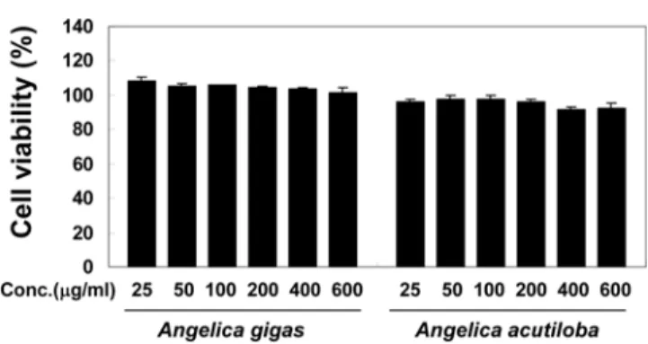
The MTT assay was performed to investigate the potential effects of root extracts from A. gigas and A.
acutiloba on the viability of mouse macrophage RAW 264.7 cells. Compared with untreated cells (100% viable), no significant decrease in cell viability was observed at sample concentrations from 25 to 600µg/mL (Fig. 1).
The percentage of viable cells was greater than 90% for each group. The results of the cell viability assay showed that the extracts from the roots of A. gigas and A.
acutiloba are not cytotoxic at concentrations below 600
µg/mL. Thus, we decided to use a test concentration of 400µg/mL in activity assays.
Since both PGE2 and NO have been previously implicated as important mediators in disease pathogenesis, including inflammation, cancer, multiple sclerosis, Parkinson’s
syndrome, and Alzheimer’s disease, we evaluated the inhibitory activities of root extracts from A. gigas and A.
acutiloba on inflammatory PGE2 production. We measured the level of PGE2 in LPS-stimulated RAW 264.7 cells.
The treatment of RAW 264.7 cells with LPS (1µg/mL) increased the production of PGE2 from endogenous arachidonic acid dramatically up to 343 pg/mL from a Fig. 1. The effects of root extracts from A. gigas and A.
acutiloba on cell viability. RAW 264.7 cell viability was evaluated by MTT colorimetric assay after cells were treated for 24 h with extracts of each concentration (25, 50, 100, 200, 400, and 600µg/mL). The percent of viable cells was greater than 90% for each group and no signifi- cant decrease in cell viability was observed at concentra- tions below 600µg/mL. Data represent the mean ± S.D. of triplicate tests.
Fig. 2. The effects of root extracts from A. gigas and A.
acutiloba on LPS-induced PGE2 production. After RAW 264.7 macrophages were stimulated with 1µg/mL LPS and 400µg/mL test sample for 20 h, PGE2 production was measured by PGE2 enzyme immunoassay. Blank and con- trol samples indicate the unstimulated and LPS-stimulated cells, respectively. Aspirin (250µM) was used as the refer- ence drug. Root extracts from both species were found to inhibit COX-2-mediated PGE2 production significantly and the extract of A. gigas suppressed PGE2 synthesis more effectively than that of A. acutiloba. Data represent the mean ± S.D. of three independent experiments, each per- formed in triplicate. Symbols * and ** represent the statis- tically significant p< 0.05 and p< 0.01 from the control determined by Student's t-test.
basal level of 51 pg/mL (no LPS). In this assay system, aspirin (250µM) was used as the reference drug. As shown in Fig. 2, root extracts from both species inhibited the LPS-induced PGE2 production significantly, and the root extract of A. gigas suppressed PGE2 synthesis more effectively than did A. acutiloba. The inhibitory activity of the root extract of A. gigas (97% inhibition at a test concentration of 400µg/mL) was approximately two- fold more potent than that of A. acutiloba (58% inhibition at a test concentration of 400µg/mL). The activity of A.
gigas against inflammatory PGE2 production was sufficient to reduce PGE2 levels to basal levels similar to aspirin (96% inhibition at 250µM). These results suggest that the pharmacological effects of root extracts from both species are involved in suppressing COX-2-mediated PGE2 generation and that A. gigas is more effective than is A. acutiloba.
The level of nitrite, a metabolite of NO that is used as an indicator for NO generation, was monitored in cultured LPS-stimulated RAW 264.7 cells to evaluate the effects of the root extracts from A. gigas and A. acutiloba on inflammatory NO formation. LPS (1µg/mL) markedly increased the production of NO from a basal level of 11
µM to 25µM after a 20-h incubation. In this assay system, L-NMMA (100µM), a non-selective inhibitor of NOS [Leiper and Vallance, 1999], was used as the
reference drug. When the cells were simultaneously treated with root extracts from A. gigas and A. acutiloba, no significant inhibitory effect on NO production was observed. However, the extract from A. acutiloba
stimulated NO generation (Fig. 3). These results indicate that extract anti-inflammatory activities are not involved in the suppression of iNOS-mediated NO formation and that A. acutiloba may exert anti-tumor activity as a result of the stimulation of macrophage-produced NO production.
In conclusion, our study suggests that the inhibitory effects on PGE2 production by root extracts from both A.
species tested are possibly, in part, related to their anti- inflammatory activities, and that A. gigas may be more efficacious in the treatment of inflammatory disease than
A. acutiloba. Also, A. acutiloba may have a greater potential for cancer treatment than A. gigas via the enhancement of macrophage-induced NO generation.
However, the mechanisms of action in these cases remain to be clarified. It is well known that NF-κB and AP-1 are major transcription factors that induce COX-2 and iNOS gene expression after LPS treatment [Aktan, 2004; Xie et
al., 1994]. To clarify the differences between the possible underlying molecular mechanisms that are responsible for the distinct pharmacological activities of both A. gigas and A. acutiloba, we will perform further studies that investigate the signal transduction pathways involved in both NF-κB and AP-1.
References
Aktan F (2004) iNOS-mediated nitric oxide production and its regulation. Life Sci75, 639-653.
Choi SS, Han KJ, Lee HK, Han EJ, and Suh HW (2003) Antinociceptive profiles of crude extract from roots of Angelica gigas NAKAI in various pain models. Biol Pharm Bull26, 1283-1288.
Choi YH, Son KH, Chang HW, Bae K, Kang SS, and Kim HP (2005) New anti-inflammatory formulation contain- ing Synurus deltoides extract. Arch Pharm Res 28, 848- Farias-Eisner R, Sherman MP, Aeberhard E, and Chaudhuri853.
G (1994) Nitric oxide is an important mediator for tumoricidal activity in vivo. Proc Natl Acad Sci USA 91, 9407-9411.
Gallo O, Masini E, Morbidelli L, Franchi A, Fini-Storchi I, Vergari WA, and Ziche M (1998) Role of nitric oxide in angiogenesis and tumor progression in head and neck cancer. J Natl Cancer Inst90, 587-596.
Gorelik L, Bar-Dagan Y, and Mokyr MB (1996) Insight into the mechanism(s) through which TNF promotes the gen- eration of T cell-mediated antitumor cytotoxicity by tumor bearer splenic cells. J Immunol156, 4298-4308.
Green LC, Wagner DA, Glogowski J, Skipper PL, Wishnok JS, and Tannenbaum SR (1982) Analysis of nitrate, Fig. 3. The effects of root extracts from A. gigas and A.
acutiloba on LPS-induced NO generation. RAW 264.7 macrophages were incubated for 20 h in medium contain- ing 1µg/mL LPS and 400µg/mL test sample. After incu- bation, NO generation was analyzed by the Griess method.
Blank and control samples indicate the unstimulated and LPS-stimulated cells, respectively. L-NMMA (100µM), a non-selective inhibitor of NOS, was used as the reference drug. Root extracts from both species were found not to inhibit NO generation. On the contrary, the extract from A.
acutiloba stimulated NO generation. Data represent the mean ± S.D. of three independent experiments, each per- formed in triplicate. The symbol ** represents the statisti- cally significant p< 0.01 from the control determined by the Student’s t-test.
nitrite, and [15N]nitrate in biological fluids. Anal Bio- chem126, 131-138.
Hakim I, Levy S, and Levy R (1996) A nine-amino acid peptide from IL-1beta augments antitumor immune responses induced by protein and DNA vaccines. J Immunol157, 5503-5511.
Ham M.-S, Kim S-S, Hong J-S, Lee J-H, Chung E-K, Park Y-S, and Lee H-Y (1996) Screening and comparison of active substances of Angelica gigas Nakai produced in Kangwon and Angelica cautiloba Kitagawa produced in Japan. Kor J Appl Microbiol Biotechnol 24, 624-629.
Hatano R, Takano F, Fushiya S, Michimata M, Tanaka T, Kazama I, Suzuki M, and Matsubara M (2004) Water- soluble extracts from Angelica acutiloba Kitagawa enhance hematopoiesis by activating immature erythroid cells in mice with 5-fluorouracil-induced anemia. Exp Hematol32, 918-924.
Kang SA, Jang K-H, Lee JE, Ahn D-K, and Park SK (2003) Differences of hematopoietic effects of Angelica gigas, A. sinensis and A. acutiloba extract on cyclophos- phamide-induced anemic rats. Korean J Food Sci Tech- nol35, 1204-1208.
Kumazawa Y, Mizunoe K, and Otsuka Y (1982) Immuno- stimulating polysaccharide separated from hot water extract of Angelica acutiloba Kitagawa (Yamato tohki).
Immunology47, 75-83.
Kumazawa Y, Nakatsuru Y, Fujisawa H, Nishimura C, Mizunoe K, Otsuka Y, and Nomoto K (1985) Lympho- cyte activation by a polysaccharide fraction separated from hot water extracts of Angelica acutiloba Kitagawa.
J Pharmacobiodyn8, 417-424.
Lasek W, Feleszko W, Golab J, Stoklosa T, Marczak M, Dabrowska A, Malejczyk M, and Jakobisiak M (1997) Antitumor effects of the combination immunotherapy with interleukin-12 and tumor necrosis factor alpha in mice. Cancer Immunol Immunother45, 100-108.
Lee S, Lee YS, Jung SH, Shin KH, Kim BK, and Kang SS (2003a) Anti-tumor activities of decursinol angelate and decursin from Angelica gigas. Arch Pharm Res 26, 727- Lee S, Shin DS, Kim JS, Oh KB, and Kang SS (2003b)730.
Antibacterial coumarins from Angelica gigas roots. Arch Pharm Res26, 449-452.
Lee YY, Lee S, Jin JL, and Yun-Choi HS (2003c) Platelet anti-aggregatory effects of coumarins from the roots of Angelica genuflexa and A. gigas. Arch Pharm Res 26, 723-726.
Leiper J and Vallance P (1999) Biological significance of endogenous methylarginines that inhibit nitric oxide syn- thases. Cardiovasc Res43, 542-548.
Mordan LJ, Burnett TS, Zhang LX, Tom J, and Cooney RV (1993) Inhibitors of endogenous nitrogen oxide forma- tion block the promotion of neoplastic transformation in C3H 10T1/2 fibroblasts. Carcinogenesis14, 1555-1559.
Ohshima H and Bartsch H (1994) Chronic infections and inflammatory processes as cancer risk factors: possible
role of nitric oxide in carcinogenesis. Mutat Res 305, 253-264.
Oshima M, Dinchuk JE, Kargman SL, Oshima H, Hancock B, Kwong E, Trzaskos JM, Evans JF, and Taketo MM (1996) Suppression of intestinal polyposis in Apc delta716 knockout mice by inhibition of cyclooxygenase 2 (COX-2). Cell87, 803-809.
Palmer RM, Ashton DS, and Moncada S (1988) Vascular endothelial cells synthesize nitric oxide from L-arginine.
Nature333, 664-666.
Radomski MW, Jenkins DC, Holmes L, and Moncada S (1991) Human colorectal adenocarcinoma cells: differen- tial nitric oxide synthesis determines their ability to aggregate platelets. Cancer Res 51, 6073-6078.
Sarker SD and Nahar L (2004) Natural medicine: the genus Angelica. Curr Med Chem11, 1479-1500.
Schmidt HH and Walter U (1994) NO at work. Cell 78, 919-925.
Simon LS (1999) Role and regulation of cyclooxygenase-2 during inflammation. Am J Med106, 37S-42S.
Song S-H, Seo B-I, Kim H-K, and Park J-H (2004) The effects of Angelicae Gigantis Radix, Angelicae Acutilo- bae Radix and Angelicae Sinensis Radix extract on hydrocortisone acetate-induced model of blood stasis.
Kor J Herbology19, 13-21.
Subbaramaiah K, Telang N, Ramonetti JT, Araki R, DeVito B, Weksler BB, and Dannenberg AJ (1996) Transcrip- tion of cyclooxygenase-2 is enhanced in transformed mammary epithelial cells. Cancer Res56, 4424-4429.
Tanaka S, Ikeshiro Y, Tabata M, and Konoshima M (1977) Anti-nociceptive substances from the roots of Angelica acutiloba. Arzneimittelforschung27, 2039-2045.
Thomsen LL, Miles DW, Happerfield L, Bobrow LG, Knowles RG, and Moncada S (1995) Nitric oxide syn- thase activity in human breast cancer. Br J Cancer 72, 41-44.
Vane JR, Bakhle YS, and Botting RM (1998) Cyclooxygen- ases 1 and 2. Annu Rev Pharmacol Toxicol38, 97-120.
Xie K, Huang S, Dong Z, Juang SH, Gutman M, Xie QW, Nathan C, and Fidler IJ (1995) Transfection with the inducible nitric oxide synthase gene suppresses tumorige- nicity and abrogates metastasis by K-1735 murine mela- noma cells. J Exp Med181, 1333-1343.
Xie QW, Kashiwabara Y, and Nathan C (1994) Role of transcription factor NF-kappa B/Rel in induction of nitric oxide synthase. J Biol Chem 269, 4705-4708.
Yamada H, Komiyama K, Kiyohara H, Cyong JC, Hirakawa Y, and Otsuka Y (1990) Structural characterization and antitumor activity of a pectic polysaccharide from the roots of Angelica acutiloba. Planta Med56, 182-186.
Yim CY, Bastian NR, Smith JC, Hibbs JB, Jr and Sam- lowski WE (1993) Macrophage nitric oxide synthesis delays progression of ultraviolet light-induced murine skin cancers. Cancer Res53, 5507-5511.
Yim D, Singh RP, Agarwal C, Lee S, Chi H, and Agarwal R (2005) A novel anticancer agent, decursin, induces G1
arrest and apoptosis in human prostate carcinoma cells.
Cancer Res 65, 1035-1044.
Yip I, Pang XP, Berg L, and Hershman JM (1995) Antitu-
mor actions of interferon-gamma and interleukin-1 beta on human papillary thyroid carcinoma cell lines. J Clin
Endocrinol Metab80, 1664-1669.