Lipopolysaccharide로 유도된 RAW 264.7 세포에 대한 참치심장 물 추 출물의 항염증 효과
김민지1,배난영1, 김꽃봉우리2, 박지혜1, 박선희1, 조영제3, 안동현1*
Anti-inflammatory Effect of Water Extract from Tuna Heart on Lipopolysaccharide-induced Inflammatory Responses in RAW 264.7 Cells
Min-Ji Kim1, Nan-Young Bae1, Koth-Bong-Woo-Ri Kim2, Ji-Hye Park1, Sun-Hee Park1, Young-Je Cho3, and Dong-Hyun Ahn1*
Received: 16 October 2015 / Revised: 14 December 2015 / Accepted: 17 December 2015
© 2015 The Korean Society for Biotechnology and Bioengineering
Abstract: The anti-inflammatory effect of tuna heart water extract (THWE) was investigated using lipopolysaccharide- induced inflammatory response in this study. Anti-inflamma- tory effect was detected by the cell proliferation and the pro- duction levels of nitric oxide, pro-inflammatory cytokines such as interleukin-6 (IL-6), IL-1β, and tumor necrosis factor- alpha. As a result, there were no cytotoxic effects on prolifera- tion of macrophages treated with THWE compared to the control. The production of pro-inflammatory cytokines was remarkably suppressed compared with that of the LPS only group. These results suggest that THWE exerts the anti-infla- mmatory property by inhibiting production of inflammatory factors and may be a potential material for anti-inflammatory therapy.
Keywords: Anti-inflammation, Pro-inflammatory cytokine, Nitric oxide, Tuna heart
1. INTRODUCTION
염증이란 오염물질, 내독소, 세균감염 등과 같은 외부 자극으 로부터 비정상적인 세포분열 등의 발생을 방어하기 위한 내 부 기작으로 조직의 손상을 재생하기 위하여 일어나는 필수 적인 반응이다 [1]. 인체는 정상적으로 염증 반응이 발생할 경우에는 염증촉진성 매개체 (pro-inflammatory)의 생성이 시 간이 지남에 따라 감소되고 항염증성 매개체는 증가되며 체 내 염증반응이 스스로 제한되는 조절기능을 가지고 있다 [2].
염증 반응에 관여하는 주요 세포는 대식세포 (macrophage)로 알려져 있으며 최초 대응세포로서 항원제시세포의 기능을 수행하며 여러 자극이나 면역세포들이 분비하는 사이토카인 등에 의해 활성화 되어 통증, 부종, 열 등의 염증 반응을 유발 하고, 염증 부위로 면역 세포의 이동을 촉진시킨다 [3]. RAW 264.7과 같은 대식세포 또는 단핵구는 그람 음성균의 세포외 막에 존재하는 내독소인 lipopolysaccharide (LPS)의 자극에 의해 tumor necrosis factor-α (TNFα), interleukin-1β (IL-1β) 및 IL-6와 같은 pro-inflammatory cytokine과 nitric oxide (NO), prostaglandin E
2(PGE
2)를 비롯한 여러 염증매개 인자의 분비 를 촉진한다 [4]. 염증반응은 인체 내에서 필수적인 반응이나 과도하게 발생하는 부적절한 염증반응은 세포 성장과 발달
1부경대학교 식품공학과/식품연구소
1Department of Food Science & Technology/Institute of Food Science, Pukyong National University, Busan 48513, Korea
Tel: +82-51-629-5831, Fax:82-51-629-5824 e-mail: [email protected]
2부경대학교 수산과학연구소
2Institute of Fisheries Sciences, Pukyong National University, Busan 46041, Korea
3경북대학교 식품공학부
3School of Food Science of Biotechnology, Kyungpook National University, Daegu 41566, Korea
연구논문
에서 국소 만성 염증반응에 대한 조직 변성을 심화시키고 혈 관내피의 손상과 복구 과정에서 병변을 심화시켜 암과 관절 염, 천식, 다발성 경화증, 염증성 장질환과 같은 다양한 질환 의 원인이 되기도 한다 [5,6]. 현재 염증반응의 억제제로서 사 용되고 있는 다양한 스테로이드 및 비스테로이드성 항염증 제는 장기간 복용할 시 여러 부작용을 야기할 수 있는 가능성 을 가지고 있어 최근 천연물 유래의 대체제를 찾으려는 노력 이 증가하고 있는 실정이다 [7].
한편, 참치는 농어목 고등어과의 바닷물고기로 고단백 식 품으로써 영양학적으로 우수할 뿐만 아니라 혈중 콜레스테 롤 농도를 낮춰 동맥경화를 예방하며 [8], 심장 질환의 회복 [9], 뇌의 시냅스 기능 강화 [10], 항암 활성 [11]과 같은 여러 가지 생리활성작용을 가져 각광을 받는 식품 중 하나이다. 또 한 ω-3계 고도불포화지방산 (polyunsaturated fatty acid, PUFA) 인 eicosapentaenoic acid (EPA, C20:5 ω-3)와 docosahexaenoic acid (DHA, C22:6 ω-3)가 가지는 항암효과를 밝혀낸 연구와 함께 참치에 다량 함유되어 있는 DHA의 기능성 연구가 국내 외에서 활발히 추진되고 있다 [12-14]. 불포화 지방산 이외에 도 참치 유래 peptide의 항균활성 [15], 항비만 활성 [16] 등에 관해 보고가 되고 있다. 세계적으로 참치어획량은 연간 400 만 톤에 달하며 우리나라의 경우 연간 26만 톤 정도를 어획하 고 있다 [17]. 어획된 참치는 주로 통조림으로써 제조되어 이 용되는데 참치가 통조림으로 가공될 때, 자숙액, 꼬리, 심장, 혈합육, 복육, 내장, 두부 등과 같은 여러 가지 부산물들이 발 생되며 이는 전체 원료의 30~35% 정도를 차지한다 [18]. 최 근, 통조림의 제조량이 증가함에 따라 발생되는 부산물의 양 이 많아져 폐기물로 인한 환경문제뿐만 아니라 이를 효율적 으로 회수, 이용하는 방법의 개발이 매우 절실한 실정이다.
이러한 참치 부산물의 기능성에 관한 연구는 참치 정소 핵산 복합물질이 면역 활성에 미치는 영향 [19], 참치 지느러미 추 출물의 암세포 독성 및 quinone reductase 활성 증가 효과 [20], 참치 자숙액의 항산화 활성에 감마선 조사가 미치는 영 향 [21] 및 항염증 활성 [22]등 일부만이 보고가 되고 있어 다 른 부산물에 대한 면역생리활성을 알아본 연구는 미미한 실 정이다. 따라서 본 연구에서는 참치 부산물의 하나인 참치심 장 물 추출물을 이용하여 LPS로 활성화 된 RAW 264.7 대식 세포에서 염증 매개물질들의 생성 억제 효과를 측정하였으 며, 아울러 항염증 활성을 갖는 기능성 식품으로의 가능성을 밝히고자 하였다.
2. MATERIALS AND METHOD
2.1. 참치심장 물 추출물 제조
본 실험에 사용한 참치심장 분말은 (주)동원 F&B (Seong- nam, Korea) 에서 제공받았으며 분말상태의 참치심장은 교반 기 (H-0820, Dongwon Science Co., Busan, Korea)를 이용해 10배 양의 물을 가한 후 24시간 추출하였다. 추출물은 원심분 리기 (UNION 32R, Hanil Co., Incheon, Korea)로 3,000 rpm에
10분간 원심분리하여 상층액을 취하였다. 남은 잔사는 동일 한 방법으로 2회 반복하여 추출하였고 상층액은 37
oC에서 감 압농축기 (RE200, Yamoto Co., Tokyo, Japan)로 농축하여 -20
o
C에 보관하며 실험에 사용하였다.
2.2. 세포배양
Murine의 대식세포주인 RAW 264.7 세포는 한국세포주은행 (KCLB 40071, Seoul, Korea) 에서 분양받아 사용하였으며, Dulbecco's Modified Eagle Medium (DMEM) (GIBCO, Grand Island, NY, USA) 에 10% inactivated fetal bovine serum (Hy- Clone Laboratories, Inc., Logan, Utah, USA)과 1% penicillin- streptomycin (HyClone Laboratories, Inc.)을 첨가한 배지를 배 양액으로 37
oC, 5% CO
2조건에서 배양하였다.
2.3. 세포독성 측정
시료의 세포독성을 평가하기 위해 MTT assay를 실시하였다 [23]. RAW 264.7 세포를 1×10
6cells/mL의 농도로 96-well plate 에 분주하고 20시간 동안 전 배양 후 참치심장 물 추출물 (THWE)을 농도별로 (0.1, 1, 10, 50, 100 μg/mL) 첨가하여 22 시간 본 배양하였다. 배양 후, 5 mg/mL의 MTT (3-(4,5-dime- thylthiazol-2-yl)-2,5-diphenyltetrazolium bromide) 시약 (Sigma Chemical Co., St. Louis, MO, USA) 을 첨가하여 2시간 재 배 양하였다. 이를 4
oC, 2,000 rpm 에서 10분간 원심분리하여 상 층액을 걷어내고 dimethyl sulfoxide (Sigma Chemical Co.) 100 μL 분주하여 생성된 formazan을 녹여내 microplate rea- der (Model 550, Bio-Rad, Richmond, CA, USA)를 이용하여 540 nm에서 흡광도를 측정하였다. 세포 증식능은 다음 식에 의해 계산하였다.
Proliferation index (%) = Sample 흡광도 / Control 흡광도 × 100
2.4. Nitric oxide 분비량 측정
Griess 반응 [24]을 이용하여 배양액 내의 NO의 농도를 측정 하였다. RAW 264.7 세포를 2.5×10
5cells/mL로 조절한 후 24 well plate 에 접종하고 37
oC, 5% CO
2incubator (MCO-15AC, Sanyo, Osaka, Japan)에서 20시간 전 배양하였다. 그 후 THWE 를 0.1, 1, 10, 50, 100 μg/mL 처리하고 1 μg/mL의 LPS로 자극 하여 24시간 본 배양하였다. 이를 4
oC, 2,000 rpm에서 10분간 원심분리하여 상층액을 얻어 정량 실험에 사용하였다. 상층 액은 Griess 시약 (1% sulfanilamide + 0.1% naphthylendiamine dihydrochloride, 1:1)과 1:1로 상온에서 10분간 반응시켜 mic- roplate reader를 이용해 540 nm에서 흡광도를 측정하였다.
세포 배양액 내 NO의 농도는 sodium nitrite의 농도별 표준곡 선과 비교하여 산출하였다.
2.5. Pro-inflammatory cytokines 분비량 측정
RAW 264.7 세포의 세포배양액 내의 TNF-α, IL-6 및 IL-1β
cytokine 의 분비량은 ELISA kit (Mouse ELISA set, BD Biosci-
ence, San Diego, CA, USA)를 이용하여 측정하였다. 세포배 양액을 얻기 위해 RAW 264.7 세포를 2.5×10
5cells/mL로 조절 하여 24 well plate에 접종하고 18시간의 전 배양 후 0.1, 1, 10, 50, 100 μg/mL 농도별 THWE와 1 μg/mL의 LPS를 처리하였 다. 그 후 12시간의 본배양을 거쳐 원심분리를 통해 상층액을 얻었다. ELISA는 microplate에 capture antibody로 anti-mouse TNF-α, IL-6, IL-1β를 분주하여 4
oC에서 하룻밤 동안 coating 시켰다. 이후 0.05% Tween 20이 포함된 phosphate buffered saline (pH 7.3, PBST)로 세척하고 10% FBS 용액으로 block- ing하였고 PBST로 세척한 뒤 각 microplate well에 세포 배양 상층액을 분주하고 실온에서 2시간 반응시켰다. 반응 후 PBST로 세척하고 희석한 biotinylated anti-mouse TNF-α, IL- 6, IL-1 β detection antibody와 streptavidin-horseradish peroxy- dase conjugate를 분주하여 실온에서 1시간 반응시켰다. 그 후 다시 PBST로 세척하고 OPD 용액을 첨가하여 실온에서 30분 동안 암반응시켰다. 2 N H
2SO
4로 반응을 종료시킨 후 micro- plate reader를 이용하여 490 nm에서 흡광도를 측정하였다.
2.6. 통계 처리
모든 실험 결과에 대한 유의차 검정은 SAS software (ver. 9.3, SAS Institute, Inc., Cary, NC, USA)에서 평균값을 분산분석 한 후, Duncan's multiple range test 법에 따라 p<0.05 수준에서 검정하였다.
3. RESULTS AND DISCUSSION
3.1. RAW 264.7에 미치는 참치심장 물 추출물의 세포독성 세포독성 측정에 이용된 RAW 264.7 세포는 그람 음성균의 내독소인 LPS와 같은 외부자극에 의해 다양한 염증 유발 인 자를 분비하여 염증반응을 일으키는 주요세포이다. 이러한 물질들은 선천성 및 후천성 면역을 조절하는데 있어서 중요 한 역할을 한다. 참치심장 물 추출물이 RAW 264.7 세포에 미치는 세포독성을 알아보기 위하여 추출물을 농도에 따라 (0.1, 1, 10, 50, 100 μg/mL) RAW 264.7 세포에 처리하여 세포 생존의 변화여부를 MTT assay를 이용해 측정하였다. 그 결 과, 아무것도 처리하지 않은 대조군과 비교 시 차이가 없음을 확인하였다 (data not shown). 이를 통해 참치심장 물 추출물 은 0.1-100 μg/mL에서 세포 생존율에 영향을 미치지 않아 독 성이 없는 것으로 사료된다. 따라서 본 연구에 사용된 THWE 의 최고 농도는 세포에 독성을 나타내지 않는 100 μg/mL으로 설정하였다. 이는 참치유의 세포독성 실험 결과, 농도 의존적 으로 세포독성이 나타나지 않은 결과 [25] 및 참치 자숙액의 세포 생존능 실험 결과 세포독성을 나타내지 않은 결과와 유 사하다 [26].
3.2. Nitric Oxide 생성 억제 효과
LPS 등의 외부자극은 세포의 신호전달을 통해 NF-κB를 활 성화시켜 그로인해 염증성 유전자인 iNOS가 발현되게 된다.
NO는 합성효소인 NOS 중 iNOS에 의해
L-arginine이
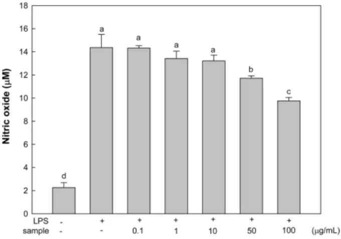
L-citru- lline으로 전환되는 과정에서 주로 생성되는 반응성이 강한 이원자 자유라디칼로 인체 내 생리적이나 병적인 반응에서 중요한 물질이다 [27,28]. 적절한 수준에서는 생리적으로 평 활근의 이완, 혈소판억제, 면역조절, 신경전달 매개, 혈관확 장에 관여하여 혈압을 조절하는 등의 역할을 하지만 지속적 인 염증반응에 의해 NO가 과도하게 증가할 시, 면역질환을 포함한 관절염, 기관지염, 다발성 경화증과 같은 병적반응을 일으킨다 [29,30]. 본 연구에서는 참치심장 물 추출물의 NO 생성 억제효과를 알아보기 위하여 세포의 생존율에 영향을 미치지 않는 농도 (0.1, 1, 10, 50, 100 μg/mL)에서 LPS로 염 증반응을 유발한 RAW 264.7 세포에 추출물을 처리하여 배 양한 후, 배양액에 griess 시약을 반응시켜 확인하였다. LPS 는 Gram 음성 세균의 세포벽 물질로서 산화적 스트레스를 유발하여 NO의 생성을 증가시킨다고 보고되어 있는데 [31], RAW 264.7 세포에 LPS만으로 염증을 유발한 결과, NO 분비 량이 14.37±1.13 μM로 아무것도 처리하지 않은 negative control 의 2.27±0.42 μM보다 약 6.3배 증가함을 보였다. 하지 만 THWE를 처리하였을 경우, positive control 군과 비교하 여 추출물 농도 의존적으로 NO 생성량이 억제된 것을 확인 하였으며, 특히, 최고 농도인 100 μg/mL에서 NO 생성량은 positive control 대비 32% 이상의 억제효과를 나타냈다 (Fig.
1). 이와 유사한 연구 결과로, 참치안구유 처리에 의해 10, 50 및 100 μg/mL 농도에서 50% 이상의 감소율을 보인다고 보 고되어 있다 [32]. 현재까지 참치의 항염증 효과는 참치에서 분리한 EPA 및 DHA에 대한 연구가 주를 이루고 있다 [33].
그러나 일반적으로 어류에는 많은 peptide가 함유되어 있는 데 Hwang 등 [34]과 Lee 등 [35]은 각각 참굴과 바지락의 해 양 생물 유래 peptide가 LPS로 유도된 RAW 264.7 세포의 염 증 반응에서 NO 생산을 억제하여 항염증 효과를 가진다고
Fig. 1. Effect of tuna heart water extract on nitric oxide production in RAW 264.7 cells. Cells were incubated in the presence of LPS (1
μ
g/mL) alone or in combination with THWE (0.1, 1, 10, 50, and 100μ
g/mL) for 24 h. The culture media of the treated cells were used to measure NO levels. a-dMeans with different superscripts are significantly different (p<0.05).보고한 바 있다. 따라서 LPS로 유도된 대식세포에서 증가된 NO에 대한 THWE의 분비 억제 효과는 참치 심장에 다량 함 유된 단백질 유래 생리활성 peptide 성분에서 기인될 것으로 사료된다.
3.3. Pro-inflammatory cytokines 생성 억제 효과
본 연구에서 이용된 LPS는 대식세포의 활성화에 관여하는 대표적인 유사분열물질 (mitogen)으로 마우스 대식세포주인 RAW 264.7 세포에 처리하면 대식세포의 표면 수용체인 toll- like receptor4가 이를 인식하여 결합하게 된다 [36]. 대식세포 에서 LPS에 의해 분비되는 pro-inflammatory cytokine에는 TNF-α, IL-6 및 IL-1β 등이 있다. 이러한 사이토카인들은 생 체의 염증반응에서 필연적으로 생성되어 다양한 면역 및 염 증반응을 조절하는 역할을 한다 [37]. 그 중 IL-6는 B cell이 형질세포로 분화하는 과정을 촉진하여 항체 생산을 증가시 키는데 체내에서 과잉 생산될 경우 악성 종양이나 자가 면역 질환 등을 비롯한 감염성 질환 등을 유발하는 것으로 알려져 있다 [38]. 특히 TNF-α는 대식세포의 활성화에 따라 mast cells, lymphoid cells, endothelial cells 등 생체에 존재하는 다 양한 세포에서 생산되는 사이토카인으로 LPS로 자극할 경 우 생산량이 더 많아진다 [39,40]. TNF-α의 분비가 증가함에 따라 열, 부종, 통증 및 발적 등의 염증의 기본적인 임상징후 를 일으키지만 지속적인 생성은 다른 염증세포를 활성화시 켜 NF-κB의 활성화를 유도함으로써 사이토카인 및 iNOS, COX-2의 생성을 증대시키고 만성염증을 유발하여 패혈성 쇼크, 염증, 염증성 장 질환과 같은 자가면역질환, 세포상해 성 등에 관여하는 것으로 알려져 있다 [41]. 또한 IL-1β는 염 증반응 시 생성되는 촉진성 사이토카인으로 세균감염에 대 한 염증성 응답의 개시 및 강화에 중요하다. 이는 낮은 농도
에서는 세포성장이나 항상성 유지에 필수적이지만 염증반 응 및 면역적 자극에 의해 대량생산될 경우 T cell을 활성화 시키고 B cell을 성숙시켜 증상을 악화시킨다 [42]. 본 연구 에서는 염증반응에서 전염증성 사이토카인들의 분비에 미 치는 THWE의 효과를 확인하기 위하여 LPS로 유도된 RAW 264.7 세포로부터 분비된 사이토카인의 분비량 억제 효과를 관찰하였다. THWE를 농도별 (0.1, 1, 10, 50, 100 μg/mL)로 처리하여 배양 상층액의 IL-6, TNF-α 및 IL-1β의 양을 ELISA kit를 이용하여 측정한 결과, THWE의 처리에 따라 각 cytokine의 분비량이 농도의존적인 감소를 보였다. IL-6의 경우 (Fig. 2), LPS 단독처리구가 596.71±2.64 pg/mL의 높은
Fig. 3. Effect of tuna heart water extract on production of IL-1β
in RAW 264.7 cells. Cells were incubated in the presence of LPS (1μ
g/mL) alone or with various concentrations of THWE (0.1, 1, 10, 50, and 100
μ
g/mL). a-dMeans with different superscripts are significantly different (p<0.05).Fig. 4. Effect of tuna heart water extract on production of TNF-
α
in RAW 264.7 cells. Cells were incubated in the presence of LPS (1μ
g/mL) alone or with various concentrations of THWE (0.1, 1, 10, 50, and 100
μ
g/mL). a-fMeans with different superscripts are significantly different (p<0.05).Fig. 2. Effect of tuna heart water extract on production of IL-6 in RAW 264.7 cells. Cells were incubated in the presence of LPS (1
μ
g/mL) alone or with various concentrations of THWE (0.1, 1, 10, 50, and 100
μ
g/mL). The levels of pro-inflammatory cytokine in the cell culture media were measured by ELISA. a-gMeans with different super- scripts are significantly different (p<0.05).분비량을 보인 것에 비하여 100 μg/mL의 농도로 처리 시 364.35±3.96 pg/mL로 약 39%의 억제율을 보인 것을 알 수 있 다. 또한, IL-1β 분비량은 LPS 단독 처리 시 33.59±0.87 pg/
mL의 높은 분비량를 보였으나, THWE를 100 μg/mL의 농도 로 처리 시 19.49±1.73 pg/mL로 약 42%의 억제율을 나타내 pro-inflammatory cytokine의 억제효과를 가짐을 확인하였다 (Fig. 3). TNF-α 분비량의 경우 또한 농도의존적인 분비량 감 소를 보였으며 50 μg/mL의 THWE 농도 이상의 처리시 20%
이상의 억제율을 나타내었다 (Fig. 4). 이와 유사한 결과로 Lin 등 [43]은 새우 유래 peptide가 NF-κB 활성을 억제하여 TNF-α 분비를 억제한다고 한다고 보고하였으며. Kang 등 [44]은 참치심장 에탄올 추출물이 100 μg/mL의 농도로 처리 시 pro-inflammatory cytokine에 대해 70% 이상 억제한 결과 를 확인하였다.
4. CONCLUSION
본 연구에서는 참치심장 물 추출물의 항염증 효과를 확인하 기 위해 LPS로 염증이 유도된 RAW 264.7 세포를 이용하여 염증 매개성 물질들의 분비량을 확인하였다. 그 결과, LPS만 처리한 대조군과 비교하였을 때, 참치심장 물 추출물이 농도 의존적으로 NO의 생성량을 감소시키는 것을 확인하였으며, 추출물의 최고 농도인 100 μg/mL에서 32% 이상의 억제효과 를 나타냈다. 또한, pro-inflammatory cytokine의 분비량을 확 인한 결과도 LPS만 처리한 대조군과 비교했을 때 참치심장 물 추출물이 농도 의존적으로 TNF-α, IL-6 및 IL-1β의 분비 량을 저해함을 보였으며, 특히 IL-1β 분비량은 100 μg/mL의 농도로 추출물 처리 시 42%의 비교적 높은 억제율을 나타내 었다. 이러한 결과가 세포 사멸에 의한 감소인지 알아보기 위 하여 MTT assay를 시행하였을 때, 세포 증식능은 참치심장 물 추출물 및 LPS를 처리하지 않은 무처리군과 유의적인 차이가 없음을 확인하였다. 이를 종합해 볼 때, 참치심장 물 추출물은 NO 및 pro-inflammatory cytokine과 같은 대식세포 유래 전염 증성 매개물질의 분비를 효과적으로 저해함으로써 추후 천 연물로서 염증치료제의 개발이 가능할 것으로 사료된다.
Acknowledgements
이 논문은 2015년도 해양수산부 재원으로 한국해양과학기술 진흥원의 지원을 받아 이루어진 것으로 이에 감사드립니다 (과제명: 수산가공부산물을 이용한 고부가가치 소재 및 식품 개발).
REFERENCES
1. Rocca, B. and G. A. FitzGerald (2002) Cyclooxygenases and pro-
staglandins: Shaping up the immune response. Int. Immunophara- col. 2: 603-630.
2. Lawrence, T., D. A. Willoughby, and D. W. Gilroy (2002) Anti- inflammatory lipid mediators and insights into the resolution of inflammation. Nat. Rev. Immunol. 2: 787-795.
3. Jeong, J. B., S. C. Hong, H. J. Jeong, and J. S. Koo (2012) Anti- inflammatory effects of ethyl acetate fraction from Cnidium offici- nale Makino on LPS-stimulated RAW 264.7 and THP-1 cells.
Korean J. Plant Res. 25: 299-307.
4. Jeong, D. H., K. B. W. R. Kim, M. J. Kim, B. K. Kang, and D. H.
Ahn (2014) Anti-inflammatory activity of methanol extract and n- hexane fraction mojabanchromanol b from Myagropsis myagroi- des. Life Sci. 114: 12-19.
5. Rankin, J. A. (2004) Biological mediators of acute inflammation.
AACN Clin. Issues 15: 3-17.
6. Guzik, T. J., R. Korbut, and T. Adamek-Guzik (2003) Nitric oxide and superoxide in inflammation and immune regulation. J. Phys- iol. Pharmacol. 54: 469-487.
7. Masaki, M., M. Matsushita, and K. Wakitani (1998) Inhibitory effect of JTE-522, a novel prostaglandin H synthase-2 inhibitor, on adjuvant-induced arthritis and bone changes in rats. Inflamm. Res.
47: 187-192.
8. Pedersen, M. H., C. Mφlgaard, L. I. Hellgren, and L. Lauritzen (2010) Effects of fish oil supplementation on markers of the meta- bolic syndrome. J. Pediatr. 157: 395-400.
9. Tenore, G. G., G. Calabrese, A. Ritieni, P. Campiglia, D. Gianetti, and E. Novellino (2014) functional food potentially safer than com- mercial fish oil based pharmaceutical formulations. Food Chem.
Toxicol. 71: 231-235.
10. McGahon, B. M., D. S. D. Martin, D. F. Horrobin, and M. A.
Lynch (1999) Age-related changes in synaptic function: analysis of the effect of dietary supplementation with ω-3 fatty acids. Neuro- sci. 94: 315-314.
11. Jang, J. R., K. K. Kim, S. B. Mun, and S. Y. Lim (2009) In vitro anticancer and antioxidant effect of solvent extracts from tuna dried at low temperature vacuum. J. Life Sci. 19: 633-638.
12. Hunter, E. (1987) PUFA and eicosanoid research. J. Anim. Oil Chem. Soc. 64: 1088-1092.
13. Hwang, W. I., N. G. Baik, Y. K. Hwang, and S. D. Lee (1992) Anti- tumor and immunological effects of tuna extract. J. Korean Soc.
Food Nutr. 21: 353-366.
14. Nestel, P. J. (1987) Polyunsaturated fatty acids (n-3, n-6). Am. J.
Clin. Nutr. 45: 1161-1167
15. Gnez-Guillnm, M. C., M. E. Lpez-Caballero, A. Alemn, A. Lpez de Lacey, B. Gimnez, and P. Montero (2010) Antioxidant and anti- microbial peptide fractions from squid and tuna skin gelatin. pp.
89-115. In: Le Bihan E. ed. Sea by-products as real material: New ways of application. Transworld Research Network, Trivandrum, India.
16. Kim, Y. M., I. H. Kim, J. W. Choi, M. K. Lee, and T. J. Nam (2015) The anti-obesity effects of a tuna peptide on 3T3-L1 adipo- cytes are mediated by the inhibition of the expression of lipogenic and adipogenic genes and by the activation of the Wnt/β-catenin signaling pathway. Int. J. Mol. Med. 36:327-334.
17. Ministry of Oceans and Fisheries (2003) Fisheries statistics year-
book. pp. 150-172. Korea.
18. Kang, C. H., H. Y. Jung, D. H. Lee, J. K, Park, J. H. Ha, S. C. Lee, and Y. I. Hwang (2000) Analysis of chemical compounds on tuna processing by-products. J. Korean Soc. Food Sci. Nutr. 29: 981- 986.
19. Park, S. E., H. W. Kim, S. R. Lee, and B. K. Kim (2000) Effects of nucleic acids complex of tuna testis on immunological activities. J.
Korean Assoc. Cancer Prev. 5: 15-23.
20. Shin, M. O., M. J. Ku, and S. J. Bae (2007) Cytotoxicity and qui- none reductase activity stimulating effects of fin of Thunnus thyn- nus extracts in various cancer cells. Korean J. Nutr. 40: 147-153.
21. Lee, H. S., H. J. Kim, J. I. Choi, J. H. Kim, J. G. Kim, B. S. Chun, D. H. Ahn, Y. J. Chung, Y. J. Kim, M. W. Byun, and J. W. Lee (2008) Antioxidant activity of the ethanol extract from cooking drips of Thunnus thynnus by gamma irradiation. J. Korean Soc.
Food Sci. Nutr. 37: 810-814.
22. Cheng, M. L., H. C. Wang, K. C. Hsu, and J. S. Hwang (2015) Anti-inflammatory peptides from enzymatic hydrolysates of tuna cooking juice. Food Agric. Immunol. 26: 770-781.
23. Park, Y. M., J. H. Won, K. J. Yun, J. H. Ryu, Y. N. Han, S. K. Choi, and K. T. Lee (2006) Preventive effect of Ginkgo biloba extract (GBB) on the lipopolysaccharide-induced expressions of induc- ible nitric oxide synthase and cyclooxygenase-2 via suppression of nuclear factor-κB in RAW 264.7 cells. Biol. Pharm. Bull. 29: 985- 990.
24. Lee, S. T., Y. R. Jeong, M. H. Ha, S. H. Kim, M. W. Byun, and S.
K. Jo (2000) Induction of nitric oxide and TNF-α by herbal plant extracts in mouse macrophages. J. Korean Soc. Food Sci. Nutr. 29:
342-348.
25. Kang, B. K., M. J. Kim, K. B. W. R. Kim, N. K. Ahn, Y. U. Choi, S. W. Bark, W. M. Pak, B. R. Kim, J. H. Park, N. Y. Bae, and D.
H. Ahn (2015) The anti-inflammatory effect of skipjack tuna (Kat- suwonus pelamis) oil in LPS-induced RAW 264.7 cells and mouse models. Microbiol. Biotechnol. Lett. 43: 45-55.
26. Kang, B. K., K. B. W. R. Kim, N. K. Ahn, Y. U. Choi, M. J. Kim, S. W. Bark, W. M. Pak, B. R. Kim, J. H. Park, N. Y. Bae, and D.
H. Ahn (2014) Immuno-stimulating activities of skipjack tuna Kat- suwounus pelamis cooking juice concentrates on mouse macroph- ages and spleen cells. Kor. J. Fish. Aquat. Sci. 47: 776-784.
27. Evans, C. H. (1995) Nitric oxide: what role does it play in inflam- mation and tissue destruction. Agents Actions Suppl. 47: 107-116.
28. Kubes, P. and D. M. McCafferty (2000) Nitric oxide and intestinal inflammation. Am. J. Med. 109: 150-158.
29. Kim, J. Y., K. S. Jung, and H. G. Jeong (2004) Suppressive effects of the kahweol and cafestol on cyclooxygenase-2 expression in macrophages. FEBS Lett. 569: 321-326.
30. Seo, J. S., T. H. Lee, S. M. Lee, S. E. Lee, N. S. Seong, and J. Y.
Kim (2009) Inhibitory effects of methanolic extracts of medicinal plants on nitric oxide production in activated macrophage RAW 264.7 cells. Korean J. Medicinal Crop Sci. 17: 173-178.
31. Weisz, A., I. Cicatiello, and H. Esumi (1996) Regulation of the
mouse inducible-type nitric oxide synthase gene promoter by inter- feron-gamma, bacterial lipopolysaccharide and NG-monomethyl- L-arginine. Biochem J. 316: 209-215.
32. Jeong, D. H. (2013) Anti-inflammatory activity of the tuna eyeball oil and Sargassum micracanthum extracts. Master Thesis. Puky- ong National University, Busan, Korea.
33. Khair-El-Din, T., S. C. Sicher, M. A. Vazquez, G. W. Chung, K. A.
Stallworth, K. Kitamura, R. T. Miller, and C. Y. Lu (1996) Tran- scription of the murine iNOS gene is inhibited by docosahexa- enoic acid, a major constituent of fetal and neonatal sera as well as fish oils. J. Exp. Med. 183: 1241-1246.
34. Hwang, J. W., S. J. Lee, Y. S. Kim, E. K. Kim, C. B. Ahn, Y. J.
Jeon, S. H. Moon, B. T. Jeon, and P. J. Park (2012) Purification and characterization of a novel peptide with inhibitory effects on colitis induced mice by dextran sulfate sodium from enzymatic hy- drolysates of Crussostrea gigas. Fish Shellfish Immunol. 33: 993- 999.
35. Lee, S. J., E. K. Kim, Y. S. Kim, J. W. Hwang, K. H. Lee, D. K.
Choi, H. Kang, S. H. Moon, B. T. Jeon, and P. J. Park (2012) Puri- fication and characterization of a nitric oxide inhibitory peptide from Ruditupes philippinarum. Food Chem. Toxicol. 50: 1660-1666.
36. Majdalawieh, A. and H. S. Ro (2010) Regulation of IκBα func- tion and NF-κB signaling: AEBP1 is a novel proinflammatory me- diator in macrophages. Mediators Inflamm. 2010: 1-27.
37. Tizard, I. R. (1988) Immunology: An introduction. 2nd ed., pp.
423-441. Saunders College Publishing, New York, NY, USA.
38. Delgado, A. V., A. T. McManus, and J. P. Chambers (2003) Pro- duction of tumor necrosis factor-alpha, interleukin 1-beta, interleu- kin 2 and interleukin 6 by rat leukocyte subpopulations after expo- sure to substance P. Neuropeptides. 37: 355-361.
39. Nathan, C. (1992) Nitric oxide as a secretory product of mamma- lian cells. FASEB J. 6: 3051-3064.
40. Walsh, L. J., G. Trinchieri, H. A. Waldorf, D. Whitaker, and G. F.
Murphy (1991) Human dermal mast cells contain and release tu- mor necrosis factor alpha, which induces endothelial leukocyte ad- hesion molecule 1. Proc. Natl. Acad. Sci. USA 88: 4220-4224.
41. Dinarello, C. A. (1999) Cytokines as endogenous pyrogens. J.
Infect. Dis. 179: 294-304.
42. Lebovic, D. I., F. Bentzien, V. A. Chao, E. N. Garrett, Y. G. Meng and R. N. Taylor (2000) Induction of an angiogenic phenotype in endometriotic stromal cell cultures by interleukin-1beta. Mol. Hum.
Reprod. 6: 269-275.
43. Lin, M. C., S. B. Lin, S. C. Lee, C. C. Lin, C. F. Hui, and J. Y.
Chen (2010) Antimicrobial peptide of an antilipopolysaccharide factor modulates of the inflammatory response in RAW264.7 cells.
Peptides 31: 1262-1272.
44. Kang, B. K. (2015) Anti-inflammatory activity of Sargassum fulv- ellum and skipjack tuna (Katsuwonus pelamis) heart ethanol ext- ract and purification of grasshopper ketone. Master Thesis. Puky- ong National University, Busan, Korea.