• 검색 결과가 없습니다.

Acute Stent Thrombosis and Heparin Induced Thrombocytopenia in a Patient With ST-Segment Elevation Myocardial Infarction

N/A
N/A
Protected

Academic year: 2021

Share "Acute Stent Thrombosis and Heparin Induced Thrombocytopenia in a Patient With ST-Segment Elevation Myocardial Infarction"

Copied!
4
0
0

로드 중.... (전체 텍스트 보기)

전체 글

(1)

646 Copyright © 2012 The Korean Society of Cardiology Korean Circulation Journal

Introduction

The incidence of stent thrombosis (ST) is relatively rare, with re- ported rates around 1-2%; however its consequences can be fatal, as most ST cases are associated with myocardial infarction or sud- den death.

1)

In accordance with the Academic Research Consortium definition, ST was subdivided into early ST (0 to 30 days), late ST (31 to 360 days), and very late ST (>360 days). Acute ST was defined as occurring during the 24 hours after the intervention.

2)3)

Early ST may be related to residual target lesion thrombus or dissection, stasis, st- ent underexpansion, or a combination of these.

4)

It is recommended for patients proceeding to primary percutane- ous coronary intervention (PCI) with ST-segment elevation myocar- dial infarction (STEMI) to be given supportive anticoagulant regi- mens such as unfractionated heparin (UFH).

5)

However ST, during

Case Report

http://dx.doi.org/10.4070/kcj.2012.42.9.646 Print ISSN 1738-5520 • On-line ISSN 1738-5555

Acute Stent Thrombosis and Heparin Induced Thrombocytopenia in a Patient With ST-Segment Elevation Myocardial Infarction

Hong Won Shin, MD, Hyuck Jun Yoon, MD, Sang Woong Choi, MD, Han Jun Bae, MD, Ji Hyun Sohn, MD, Ho Myung Lee, MD, Hyun Ok Cho, MD, Yun Kyeong Cho, MD, Hyoung Seob Park, MD, Hyungseop Kim, MD, Chang Wook Nam, MD, Seung Ho Hur, MD, Yoon Nyun Kim, MD, and Kwon Bae Kim, MD

Division of Cardiology, Department of Internal Medicine, Dongsan Medical Center, Keimyung University College of Medicine, Daegu, Korea

Heparin is an essential drug in the treatment of acute coronary syndrome and it is used during percutaneous coronary intervention (PCI).

Heparin-induced thrombocytopenia (HIT), albeit a serious complication of heparin therapy characterized by thrombocytopenia and high risk for venous and arterial thrombosis, has rarely been previously reported during PCI. We report a case of an acute stent thrombosis due to an unusual cause, HIT during primary PCI, in a patient with acute myocardial infarction. (Korean Circ J 2012;42:646-649)

KEY WORDS: Heparin; Thrombocytopenia; Thrombosis; Myocardial infarction.

Received: January 18, 2012 Revision Received: February 1, 2012 Accepted: February 6, 2012

Correspondence: Hyuck Jun Yoon, MD, Division of Cardiology, Depart- ment of Internal Medicine, Dongsan Medical Center, Keimyung University College of Medicine, 56 Dalseong-ro, Jung-gu, Daegu 700-712, Korea Tel: 82-53-250-7448, Fax: 82-53-250-7034

E-mail: [email protected]

• The authors have no financial conflicts of interest.

This is an Open Access article distributed under the terms of the Creative Commons Attribution Non-Commercial License (http://creativecommons.

org/licenses/by-nc/3.0) which permits unrestricted non-commercial use, distribution, and reproduction in any medium, provided the original work is properly cited.

coronary angioplasty in association with the abrupt onset of hepa- rin induced thrombocytopenia (HIT), has rarely been reported previ- ously.

6)

Further, we here report a case of the acute ST due to HIT dur- ing primary PCI.

Case

A 62-year-old woman was admitted with resting chest pain for 5 hours. As a cardiovascular risk factor, she had a history of type 2 di- abetes mellitus for 5 years. The initial electrocardiogram showed ST- segment elevation in II, III, and aVF. She was given aspirin 300 mg, clopidogrel 600 mg and UFH 3600 unit loading dose, and continu- ous UFH was injected for at a maintenance rate of 12 unit/kg/hour according to American College of Cardiology/American Heart As- sociation guideline.

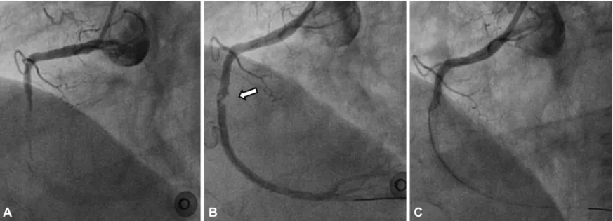
7)

With the diagnosis of STEMI, she was sent to cardiac catheterization laboratory. Right coronary angiogram re- vealed total occlusion of the mid portion of the right coronary artery (RCA) with thrombus (Fig. 1A). Left coronary angiogram also showed a tight stenotic lesion in the mid portion of the left anterior descend- ing artery (LAD, 65%) and left circumflex artery (LCX, 85%). We planned primary PCI at RCA as infarct-related artery and secondary PCI on LAD and LCX. We dilated the mid RCA lesion using a 2.0×15 mm balloon. Thereafter, abundant thrombus was shown in the mid- dle to distal RCA and thrombus aspiration was performed; a 2.75×

33 mm everdimus-eluting stent (Xience Prime

TM

, Abbott, Santa Clara,

CA, USA) was deployed at 10 atmospheres in the mid RCA. After

stent deployment, thrombus was shown at proximal end of the st-

(2)

647 Hong Won Shin, et al.

http://dx.doi.org/10.4070/kcj.2012.42.9.646 www.e-kcj.org

ent (Fig. 1B). We did thrombus aspiration and additional balloon di- latation; however, angiogram showed complete occlusion of the mid RCA stent site with progressed thrombus from the proximal portion of RCA (Fig. 1C). The activated clotting time (ACT) was 239 seconds at that time. Despite repeated thrombus aspiration and balloon dil- atation, coronary flow was not restored. We checked intravascular ultrasound (IVUS) on this site to exclude another reason to com- promise coronary flow, such as edge dissection or underexpansion.

In IVUS, there was total occlusion by extensive thrombus in RCA;

however, there was no evidence of edge dissection, stent malappo- sition or underexpasion (Fig. 2). A bolus dose of glycoprotein IIb/IIIa receptor antagonist abciximab (2.5 mg/kg) was given into the RCA followed by continuous infusion (0.125 mcg/kg/min). The RCA flow was not restored despite repeated efforts, and thrombus further pro- gressed proximal to the stented area. We scheduled delayed PCI on RCA and non-culprit lesion on LAD and LCX, after medical treatment.

As compared to the admission platelet count of 443000/mm

3

, a repeat platelet count 6 hours after PCI was 158000/mm

3

and even further decreased to 4000/mm

3

at 2 days after PCI. Aspirin, clopido- grel, abciximab and UFH were discontinued after a repeat count in an ethylenediaminetetraacetic acid-free citrate tube confirmed the finding. We used a clinical scoring system for identifying those with HIT.

8)

The patient had thrombocytopenia, proven new thrombosis, and timing of onset of platelet fall was under 1 day. Further, there was no other cause for the platelet count fall. Thus, we diagnosed heparin induced thrombocytopenia and administered the direct thrombin inhibitor, argatroban (2 µg/kg/min). Two units of platelets were transfused, due to bleeding concerns and profound thrombo- cytopenia. Post-transfusion platelet count was 7000/mm

3

and a few hours later dropped to 5000/mm

3

, but stabilized at 855000/mm

3

at

5 days after PCI, at which time, aspirin and clopidogrel were re- initiated and argatroban was discontinued.

Eleven days after admission, secondary PCI was performed using argatroban targeting ACT to 250-400 seconds. At that time, we used heparin free flushing solution to avoid recurrent heparin induc- ed thrombocytopenia. Right coronary angiogram revealed total oc- clusion from the RCA ostium (Fig. 3A). The RCA totally occluded le- sion was successfully crossed after several attempts and balloon angioplasty was performed using a 2.5 mm balloon. Despite repeat- ed balloon inflation and thrombus aspiration, RCA thrombolysis in myocardial infarction 3 flow could not be achieved (Fig. 3B). At the Fig. 2. In intravascular ultrasound on right coronary artery after stent de- ployment, there was total occlusion by extensive thrombus and there was no evidence of edge dissection, incomplete stent apposition or underex- pansion (arrow heads showed stent struts and arrow showed thrombus).

Fig. 1. Coronary angiogram demonstrated acute stent thrombosis during primary percutaneous coronary intervention. A: initial coronary angiogram re- vealed total occlusion of the mid portion of the right coronary artery (RCA) with thrombus. B: after stent deployment, thrombus (arrow) was shown at proxi- mal portion of the stent. C: a few minutes after, it showed complete occlusion of the mid RCA stent site with progressed thormbus from the proximal por- tion of RCA.

A   B   C  

(3)

648 Acute Stent Thrombosis and HIT in Patient With STEMI

http://dx.doi.org/10.4070/kcj.2012.42.9.646 www.e-kcj.org

LAD and LCX, 2.75×38 mm and 2.75×18 mm everolimus-eluting stents (XIENCE PRIME

TM

Abbott) were deployed, respectively. After left coronary PCI, a repeated right coronary angiogram was per- formed. It showed disappearance of the previously shallow coro- nary flow and we did not perform any further procedures in the RCA (Fig. 3C).

After the second PCI, platelet count was not decreased and the patient was subsequently discharged with a platelet count of 427000/mm

3

. There was no evidence of aspirin or clopidogrel resist- ance. Although there are concerns of increased bleeding risk from triple therapy, our patient was discharged on aspirin, clopidogrel and warfarin.

Discussion

Heparin induced thrombocytopenia carries a significant risk for venous and arterial thrombotic events. Very rarely, it has been de- scribed in the setting of coronary angioplasty for an acute myocar- dial infarction in association with the abrupt onset of HIT, such as in this case.

6)9)10)

Heparin induced thrombocytopenia is a prothrombotic disorder initiated by heparin administration and is related to antibody-me- diated platelet activation, causing thrombin generation and throm- botic complications. HIT can be regarded as a severe adverse drug reaction resulting from multicellular immune activation.

11)

The diag- nosis of HIT is based on its typical clinical feature. The ‘4Ts’ of HIT re- fers to the degree of Thrombocytopenia, the Timing of the platelet fall after heparin exposure, the presence of Thrombosis, and oTher causes for thrombocytopenia excluded.

8)

UFH has been the tradi- tional antithrombin agent during PCI. The American College of Car- diology recommends the use of UFH during PCI, with the dose ad-

justed for ACT (Class I, Level of Evidence: C), or enoxaparin (Class IIa, Level of Evidence: B).

12)

In patients with HIT, heparin should be avoided to prevent recur- rent HIT and PCI should be performed using alternative anticoagu- lants, despite the fact that their safety and efficacy have not been established.

13)

Direct thrombin inhibitors, such as argatroban, bi- valirudin, and hirudin, offer the theoretical advantages of inhibition of soluble and bound thrombin, ease of titration, and rapid reversal of anticoagulant effects upon cessation of therapy, without cross- reactivity with HIT antibodies.

14)

Argatroban is approved to treat both HIT complicated by thrombosis and isolated HIT. Lewis et al.

15)

reported that 91 HIT patients underwent 112 separate coronary in- terventions on 177 lesions using argatroban anticoagulation, and satisfactory outcome of the procedure was attained in 94.5%. They reported that argatroban enabled satisfactory outcomes of proce- dures and provided adequate anticoagulation for HIT patients un- dergoing PCI on single or multiple occasions. The anticoagulatory effects of argatroban and UFH were obtained with comparable safe- ty, since there were no significant differences in angiographic suc- cess rates, PCI-induced troponin level elevation, occurrence of the composite endpoint of death, MI, or revascularization, as well as incidence of bleeding events.

16)

Argatroban dose-dependently in- creases coagulation parameters and compared to UFH, demonst- rates a superior predictable anticoagulant effect in patients under- going elective PCI.

16)

This case report offers several unknowns. Several risk factors implicated in acute ST were present in our patient, such as multives- sel disease, PCI in the setting of acute coronary syndrome, and dia- betes mellitus. Moreover, acute ST is reported in up to 0.5-0.7% of the cases in the first 24 hours post PCI.

17)

Abciximab, which is a po- tent inhibitor of platelet aggregation, can also induce thrombocy- Fig. 3. Follow up coronary angiogram demonstrated persistent stent thrombosis. A: secondary stage coronary angiogram showed more progressed intra- coronary thrombosis from RCA ostium. B: despite repeated balloon and thrombus aspiration Thrombolysis in Myocardial Infarction 3 flow could not be ob- tained. C: a few minute after, coronary flow of RCA was disappeared. RCA: right coronary artery.

A   B   C  

(4)

649 Hong Won Shin, et al.

http://dx.doi.org/10.4070/kcj.2012.42.9.646 www.e-kcj.org

topenia within 24 hours of first exposure.

9)

The exposure to abcix- imab can result in a more rapid, severe thrombocytopenia in HIT. In our case, abciximab was given after ST. Therefore, we do not consid- er its cause. Although profound thrombocytopenia could have been an effect of abciximab, an acute ST with thrombus during PCI may account for HIT.

17)

Stent thrombosis was affected by various factors. This case shows that HIT could be one of the causes of acute ST. Prompt identifica- tion and management of acute ST with HIT is critically important to avoid serious complications.

References

1. Iakovou I, Schmidt T, Bonizzoni E, et al. Incidence, predictors, and out- come of thrombosis after successful implantation of drug-eluting st- ents. JAMA 2005;293:2126-30.

2. Mauri L, Hsieh WH, Massaro JM, Ho KK, D’Agostino R, Cutlip DE. Stent thrombosis in randomized clinical trials of drug-eluting stents. N Engl J Med 2007;356:1020-9.

3. Wenaweser P, Rey C, Eberli FR, et al. Stent thrombosis following bare- metal stent implantation: success of emergency percutaneous coro- nary intervention and predictors of adverse outcome. Eur Heart J 2005;

26:1180-7.

4. Fujii K, Carlier SG, Mintz GS, et al. Stent underexpansion and residual reference segment stenosis are related to stent thrombosis after si- rolimus-eluting stent implantation: an intravascular ultrasound study.

J Am Coll Cardiol 2005;45:995-8.

5. King SB 3rd. 2009 update of the ACC/AHA guidelines for the manage- ment of patients with ST-elevation myocardial infarction and guide- lines on percutaneous coronary intervention: what should we change in clinical practice? Pol Arch Med Wewn 2010;120:6-8.

6. Gupta BK, Savage MP, Brest AN. Acute myocardial infarction during coronary angioplasty associated with heparin-induced thrombocyto- penia. Cathet Cardiovasc Diagn 1995;35:42-6.

7. Antman EM, Anbe DT, Armstrong PW, et al. ACC/AHA guidelines for the management of patients with ST-elevation myocardial infarction--

executive summary. A report of the American College of Cardiology/

American Heart Association Task Force on Practice Guidelines (Writing Committee to revise the 1999 guidelines for the management of pa- tients with acute myocardial infarction). J Am Coll Cardiol 2004;44:

671-719.

8. Babuin L, Pengo V. Argatroban in the management of heparin-induced thrombocytopenia. Vasc Health Risk Manag 2010;6:813-9.

9. Dasari TW, Pappy R, Hennebry TA. Acute stent thrombosis associated with heparin-induced thrombocytopenia and abciximab-induced pro- found thrombocytopenia. J Invasive Cardiol 2011;23:E5-8.

10. Hussain F, Philipp R, Zieroth S. HITT and stent thrombosis: a “clinical”

diagnosis not to be missed. Int J Cardiol 2009;133:e11-3.

11. Warkentin TE. Heparin-induced thrombocytopenia: pathogenesis and management. Br J Haematol 2003;121:535-55.

12. Smith SC Jr, Feldman TE, Hirshfeld JW Jr, et al. ACC/AHA/SCAI 2005 guideline update for percutaneous coronary intervention--summary article: a report of the American College of Cardiology/American Heart Association Task Force on Practice Guidelines (ACC/AHA/SCAI Writing Committee to update the 2001 Guidelines for Percutaneous Coronary Intervention). Catheter Cardiovasc Interv 2006;67:87-112.

13. Matsue H, Masai T, Yoshikawa Y, Kawamura M. Serious acute coronary thrombosis associated with heparin-induced thrombocytopenia in off-pump coronary artery bypass grafting. Interact Cardiovasc Thorac Surg 2010;11:188-90.

14. Brieger DB, Mak KH, Kottke-Marchant K, Topol EJ. Heparin-induced thrombocytopenia. J Am Coll Cardiol 1998;31:1449-59.

15. Lewis BE, Matthai WH Jr, Cohen M, Moses JW, Hursting MJ, Leya F.

Argatroban anticoagulation during percutaneous coronary interven- tion in patients with heparin-induced thrombocytopenia. Catheter Cardiovasc Interv 2002;57:177-84.

16. Rössig L, Genth-Zotz S, Rau M, et al. Argatroban for elective percuta- neous coronary intervention: the ARG-E04 multi-center study. Int J Cardiol 2011;148:214-9.

17. Van Werkum JW, Heestermans AA, Zomer AC, et al. Predictors of coro-

nary stent thrombosis: the Dutch Stent Thrombosis Registry. J Am Coll

Cardiol 2009;53:1399-409.

수치

Fig. 1. Coronary angiogram demonstrated acute stent thrombosis during primary percutaneous coronary intervention

참조

관련 문서

Coronary angiography and intravascular ultrasound revealed multiple thrombotic occlusion of the mid left anterior descending artery, mid left circumflex artery and obtuse

Hypothesis: We analyzed data from the Korean Acute Myocardial Infarction Registry (KAMIR) to assess gender differences in in-hospital outcomes post ST-segment

Jong-Won Ha, Byoung-Wook Choi, Se-Joong Rim, Seok-Min Kang, Yangsoo Jang, Namsik With Persistent ST-Segment Elevation Simulating Acute Myocardial Infarction Extensive

OPCAB, off-pump coronary artery bypass grafting surgery; STEMI, ST elevation myocardial infarction; NSTEMI, non-ST elevation myocardial infarction; MACCE, major cardiovascular

It is uncertain that atorvastatin pretreatment can reduce myocardial damage in patients undergoing primary percutaneous coronary intervention (PCI) for ST-segment elevation

fixed stenosis, a coronary vasospasm was documented in nearly 50% of the patients tested with an intracoronary provocation test. Although coronary spasm has been implicated to cause

The follow up coronary angiography showed a fistulae from the septal branches of the distal LAD to the right ventricle (RV) and mild aneurysmal changes at the stent in the middle

Purpose: This study aimed to compare the incidence and clinical significance of transient versus persistent acute kidney injury (AKI) on acute ST elevation myocar- dial