Chung, Won-Heum Shim and Seung-Yun Cho
Jong-Won Ha, Byoung-Wook Choi, Se-Joong Rim, Seok-Min Kang, Yangsoo Jang, Namsik
With Persistent ST-Segment Elevation Simulating Acute Myocardial Infarction
Extensive Subepicardial Fibrosis in a Patient With Apical Hypertrophic Cardiomyopathy
Print ISSN: 0009-7322. Online ISSN: 1524-4539
Copyright © 2005 American Heart Association, Inc. All rights reserved.
is published by the American Heart Association, 7272 Greenville Avenue, Dallas, TX 75231 Circulation
doi: 10.1161/CIRCULATIONAHA.104.479386
2005;112:e49-e50
Circulation.
http://circ.ahajournals.org/content/112/3/e49
World Wide Web at:
The online version of this article, along with updated information and services, is located on the
http://circ.ahajournals.org/content/suppl/2005/07/18/112.3.e49.DC1.html
Data Supplement (unedited) at:
http://circ.ahajournals.org//subscriptions/
is online at: Circulation
Information about subscribing to Subscriptions:
http://www.lww.com/reprints
Information about reprints can be found online at: Reprints:
document.
Permissions and Rights Question and Answer
this process is available in the
click Request Permissions in the middle column of the Web page under Services. Further information about Office. Once the online version of the published article for which permission is being requested is located,
can be obtained via RightsLink, a service of the Copyright Clearance Center, not the Editorial Circulation
in
Requests for permissions to reproduce figures, tables, or portions of articles originally published Permissions:
at CONS KESLI on June 30, 2014 http://circ.ahajournals.org/
Downloaded from http://circ.ahajournals.org/ at CONS KESLI on June 30, 2014 Downloaded from
Extensive Subepicardial Fibrosis in a Patient With Apical
Hypertrophic Cardiomyopathy With Persistent ST-Segment
Elevation Simulating Acute Myocardial Infarction
Jong-Won Ha, MD, PhD; Byoung-Wook Choi, MD; Se-Joong Rim, MD, PhD;
Seok-Min Kang, MD, PhD; Yangsoo Jang, MD; Namsik Chung, MD, PhD;
Won-Heum Shim, MD, PhD; Seung-Yun Cho, MD, PhD
A
pical hypertrophic cardiomyopathy is a unique form ofhypertrophic cardiomyopathy (HCM), in which the hypertrophy of myocardium predominantly involves the apex of the left ventricle. The ECG in apical HCM typically shows repolarization changes in the anterolateral leads and some-times giant negative T waves. Previous reports have shown that apical HCM may mimic myocardial infarction, although its underlying mechanism is unclear. This case report dem-onstrates typical echocardiographic and MRI features of apical HCM, but with unusual electrocardiographic features characterized by chronic ST-segment elevation in the prec-ordial leads. Contrast-enhanced MRI showed unusual exten-sive subepicardial delayed hyperenhancement at the left ventricular apex, suggesting that subepicardial fibrosis may be a possible cause for this unusual ECG abnormality in patients with apical HCM. Because this condition can be misdiagnosed as acute infarction, resulting in unwarranted thrombolytic therapy or emergency angiography, we believe
this case is important as a reminder that acute infarction is not the only cause of ST-segment elevation.
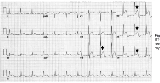
In March 2003, a 44-year-old man presented with exer-tional dyspnea. He was normotensive and had never experi-enced chest pain or other symptoms suggestive of heart disease. A routine 12-lead ECG (Figure 1) revealed an ST-segment elevation in V3 to V6 precordial leads, which made us suspect possible acute myocardial infarction. Phys-ical examination revealed no abnormal findings, and cardiac biomarkers were negative. Echocardiography and MRI (Fig-ure 2 and Movies) revealed a severe hypertrophy confined to the left ventricular apex, suggesting apical HCM. Coronary angiography by MRI showed no significant luminal narrow-ing (Figure 3). A contrast-enhanced image obtained by MRI showed prominent delayed hyperenhancement localized at the subepicardial area of the left ventricular apex (Figure 4). Follow-up ECG obtained 1 year later showed persistent ST elevation in precordial leads without interval change (Figure 5).
Figure 1. Initial ECG showing
ST-segment elevation in V3to V6 prec-ordial leads, indicative of possible acute myocardial infarction.
From Cardiology Division, Yonsei University College of Medicine, Yonsei Cardiovascular Center, Seoul, Korea.
The Data Supplement is available at http://www.circ.ahajournals.org/cgi/content/full/112/3/e49/DC1.
Correspondence to Jong-Won Ha, MD, PhD, Cardiology Division, Yonsei University College of Medicine, Yonsei Cardiovascular Center, CPO Box 8044, Seoul, Korea. E-mail [email protected]
(Circulation. 2005;112:e49-e50.)
© 2005 American Heart Association, Inc.
Circulation is available at http://www.circulationaha.org DOI: 10.1161/CIRCULATIONAHA.104.479386
e49
Images in Cardiovascular Medicine
at CONS KESLI on June 30, 2014 http://circ.ahajournals.org/
Figure 2. MRI revealed severe
hypertro-phy confined to left ventricular apex, suggesting apical HCM.
Figure 3. Coronary angiography by MRI
showed no significant luminal narrowing.
Figure 4. Contrast-enhanced image
obtained by MRI of 4-chamber view (left) and 2-chamber view (right) showed promi-nent delayed hyperenhancement localized at subepicardial area of left ventricular apex.
Figure 5. Follow-up ECG 1 y later showed
persistent ST elevation in precordial leads without interval change.
e50 Circulation July 19, 2005
at CONS KESLI on June 30, 2014 http://circ.ahajournals.org/