• 검색 결과가 없습니다.

A Case of Acute Myocardial Infarction With ST-Segment Elevation in a Lead Augmented Right Vector Caused by a Left Main Coronary Artery Vasospasm

N/A
N/A
Protected

Academic year: 2022

Share "A Case of Acute Myocardial Infarction With ST-Segment Elevation in a Lead Augmented Right Vector Caused by a Left Main Coronary Artery Vasospasm"

Copied!
4
0
0

로드 중.... (전체 텍스트 보기)

전체 글

(1)

50 Copyright © 2012 The Korean Society of Cardiology Korean Circulation Journal

Introduction

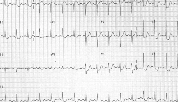
A ST-segment elevation in the aVR lead has been associated with fixed left main coronary artery (LMCA) lesions in patients with acute coronary syndrome (ACS).

1-4)

Although acute obstruction of the LMCA is infrequently encountered, predicting an LMCA obst- ruction is important for selecting the appropriate treatment strate- gy. Without a suitable management plan, acute LMCA obstruction usually causes severe hemodynamic deterioration resulting in a less favorable prognosis.

Coronary artery spasm is an infrequent but important cause of ACS. Its incidence among patients undergoing diagnostic coronary angiography for suspected ACSs is 3-4%.

1)

However, a spasm of

Case Report

http://dx.doi.org/10.4070/kcj.2012.42.1.50 Print ISSN 1738-5520 • On-line ISSN 1738-5555

A Case of Acute Myocardial Infarction With ST-Segment Elevation in a Lead Augmented Right Vector Caused

by a Left Main Coronary Artery Vasospasm

Kyong Yeun Jung, MD and Tae Soo Kang, MD

Division of Cardiovascular Medicine, Department of Internal Medicine, Dankook University College of Medicine, Cheonan, Korea

Diagnosing and selecting an appropriate treatment strategy for left main coronary artery (LMCA) obstruction is very important. Although this disease is not frequently encountered, it can cause severe hemodynamic deterioration resulting in a less favorable prognosis without a suitable management approach. Another aspect of LMCA that we must not overlook is coronary artery spasm, which can be an infrequent but important cause of acute coronary syndrome. Although it is rare, LMCA can cause critical complications. In this study, we report the case of a 35-year-old female who was admitted to the hospital with a diagnosis of acute myocardial infarction with ST-segment elevation in the aVR lead caused by a left main coronary spasm that was examined on intravascular ultrasound. (Korean Circ J 2012;42:50-53) KEY WORDS: Coronary artery vasospasm; Electrocardiography; Acute coronary syndrome.

Received: July 14, 2011

Revision Received: September 24, 2011 Accepted: October 28, 2011

Correspondence: Tae Soo Kang, MD, Division of Cardiovascular Medicine, Department of Internal Medicine, Dankook University College of Medicine, 16-5 Anseo-dong, Cheonan 330-715, Korea

Tel: 82-41-550-7690, Fax: 82-42-556-0524 E-mail: [email protected]

• The authors have no financial conflicts of interest.

This is an Open Access article distributed under the terms of the Creative Commons Attribution Non-Commercial License (http://creativecommons.

org/licenses/by-nc/3.0) which permits unrestricted non-commercial use, distribution, and reproduction in any medium, provided the original work is properly cited.

the LMCA is very rare. In this study, we report the case of a 35-year- old female who was admitted to hospital with a diagnosis of acute myocardial infarction with ST-segment elevation in the aVR lead caused by a left main coronary spasm demonstrated by intravas- cular ultrasound (IVUS).

Case

A 35-year-old female presented to our hospital with squeezing

chest pain for a duration of 15 minutes. She was a current smoker

(20 pack years) without any other risk factors of cardiovascular dis-

ease. On physical examination, her blood pressure was 145/80 mm Hg,

and pulse rate was 96 beats per minute. Chest radiography showed

mild cardiomegaly with mild pulmonary congestion. The initial elec-

trocardiogram (ECG) revealed 2-mm ST segment depression in leads

V3-6 and up to a 1-mm ST segment depression in leads I, aVL, II, aVF

with a 2-mm ST segment elevation in the aVR lead (Fig. 1). An initial

cardiac enzyme profile revealed elevation of creatine phosphoki-

nase (CK), 195 U/L (normal range, 26-174); CK-MB, 49 μg/L (normal

range, 0-3); and elevated troponin-I of 3.87 ng/mL (normal range,

0-0.05). Emergency coronary angiography showed a 90% narrowing

of the LMCA (Fig. 2A) and a normal right coronary artery without

significant narrowing (Fig. 3). During coronary angiography, the

patient complained of chest pain with a sudden decrease in systolic

blood pressure to 90 mm Hg. However, after administering 100 μg

(2)

51 Kyong Yeun Jung, et al.

http://dx.doi.org/10.4070/kcj.2012.42.1.50 www.e-kcj.org

of intracoronary nitrate, the chest pain was relieved and a re-angi- ography showed a completely resolved LMCA spasm without re- sidual luminal stenosis (Fig. 2B). IVUS showed only a minimal con- centric plaque in the LMCA without significant narrowing (Fig. 4).

Therefore, we stopped the procedure without intervention. After the procedure, an ST depression in leads I, aVL, II, aVF, and V3-6 and an ST elevation in aVR were completely resolved on the ECG (Fig. 5), and the cardiac enzymes had decreased to the normal range (CK, 41; CK- MB, 22 μg/L; and troponin-I, 1.31 ng/mL). The patient was disch- arged on the eighth hospital day receiving 40 mg of oral nitrate, 180

mg of diltiazem, 100 mg of aspirin, and 10 mg of rosuvastatin per day. A follow-up coronary angiography taken 9 months later show- ed no spasms or stenosis. The patient has exhibited no signs of an- gina for 2 years and has remained on medication in outpatient clinics.

Discussion

ST-segment elevation in the aVR lead of a patient with clinically suspected ACS suggests a strong possibility of an LMCA obstruc- Fig. 1. Initial electrocardiogram revealed a 2-mm ST segment depression in leads V3-6 and up to a 1-mm ST segment depression in leads I, aVL, II, aVF with a 2-mm ST-segment elevation in the aVR lead (arrows).

Fig. 2. Coronary angiography showing diffuse left main coronary artery vasospasm (A) and resolution of the spasm after administration of intracoronary nitrates (B).

A   B  

(3)

52 Left Main Coronary Artery Vasospasm

http://dx.doi.org/10.4070/kcj.2012.42.1.50 www.e-kcj.org

fixed stenosis, a coronary vasospasm was documented in nearly 50% of the patients tested with an intracoronary provocation test.

Although coronary spasm has been implicated to cause a progres- sion in atherosclerosis over years, few reports

9)

are available demon- strating a critical role for coronary artery spasm in rapid plaque pro- gression and the development of ACS. It was assumed that a focal spasm may have contributed to rupture an atheromatous plaque, leading eventually to a myocardial infarction. Previous experimental studies have shown that a coronary artery spasm can cause an in- traplaque hemorrhage and acute myocardial infarction in a swine model.

10)

Thus, coronary vasospasm may play an important role in the pathogenesis of not only variant angina but also ACS, as seen in our patient. Wakabayashi et al.

11)

defined coronary artery spasm as tion.

1)2)

In addition to the diagnosis of LMCA obstruction, the find-

ing of ST-segment elevation in the aVR lead also has prognostic sig- nificance.

6)

In a study by Kosuge et al.

7)

ST-segment elevation >0.5 mm on the aVR lead of an initial ECG and elevated troponin T levels were independent predictors of adverse clinical events at 90 days.

According to Barrabés et al.

8)

ST-segment elevation in the aVR lead at the time of admission is superior to ST-segment depression in other leads for predicting the risk of death in patients with a non- ST-segment elevation acute myocardial infarction.

Coronary artery spasm is an important cause of chest pain and myocardial ischemia. In a prospective study reported by Ong et al.,

5)

28% of 488 consecutive patients with ACS had no significant cul- prit lesions. Among the patients with ACS without any significant

Fig. 5. Follow up electrocardiogram (ECG) showing a normalized ST segment deviation after intracoronary nitrate.

Fig. 3. Coronary angiography showing normal right coronary artery. Fig. 4. Intravascular ultrasound showing a minimal concentric plaque in

the left main coronary artery.

(4)

53 Kyong Yeun Jung, et al.

http://dx.doi.org/10.4070/kcj.2012.42.1.50 www.e-kcj.org

transient total or subtotal occlusion with ECG changes and/or typ- ical chest symptoms. Ong et al.

5)

defined coronary artery spasm as when quantitative analysis confirms a 75% narrowing (compared with the relaxed state after nitroglycerin), and symptoms are re- produced. Although an ECG during a vasospasm is classically de- scribed with an ST elevation, ST depression can also be rarely docu- mented, as in our case. When coronary angiography is normal or shows only nonobstructive plaques, and if a transient ST segment deviation can be demonstrated at the time at which the patient has the discomfort, the presumptive diagnosis of variant angina can be made, and no further tests are necessary. Additionally, a cathe- ter-induced LMCA vasospasm should be meticulously considered.

12)

In our case, we distinguished a spontaneous spasm from a cathe- ter-induced spam for the following reasons. One, the initial present- ing ECG showed an aVR-lead ST segment elevation, which was re- solved by intracoronary nitroglycerin. The other reason was the an- giographic characteristics of the spasm. Usually, a catheter-induced spasm appears within 1 mm of the catheter tip in contrast to our case (spasm showed irregular and eccentric characteristics and was more than 5 mm from the ostium of the LMCA). IVUS at the time of angiography may help to identify minimal atherosclerotic disease suggesting a vasospasm.

In conclusion, coronary spasm is a frequent cause of ACS and sh- ould be regularly considered in the differential diagnosis. It is im- portant not to ignore the aVR lead because it could provide essen- tial diagnostic information in a range of clinical presentations.

REFERENCES

1. Kosuge M, Kimura K, Ishikawa T, et al. Predictors of left main or three-vessel disease in patients who have acute coronary syn- dromes with non-ST-segment elevation. Am J Cardiol 2005;

95:1366-9.

2. Duygu H, Yavuzgil O, Erturk U, Zoghi M, Ozerkan F. ST-segment elevation in lead augmented vector right may also be caused by diffuse left main coronary artery vasospasm without fixed ste- nosis. Clin Cardiol 2008;31:179-82.

3. Engelen DJ, Gorgels AP, Cheriex EC, et al. Value of the electrocar- diogram in localizing the occlusion site in the left anterior de- scending coronary artery in acute anterior myocardial infarction.

J Am Coll Cardiol 1999;34:389-95.

4. Yamaji H, Iwasaki K, Kusachi S, et al. Prediction of acute left main coronary artery obstruction by 12-lead electrocardiography: ST segment elevation in lead aVR with less ST segment elevation in lead V(1). J Am Coll Cardiol 2001;38:1348-54.

5. Ong P, Athanasiadis A, Hill S, Vogelsberg H, Voehringer M, Sech- tem U. Coronary artery spasm as a frequent cause of acute coro- nary syndrome: the CASPAR (Coronary Artery Spasm in Patients with Acute Coronary Syndrome) Study. J Am Coll Cardiol 2008;52:

523-7.

6. Nikus KC, Eskola MJ. Electrocardiogram patterns in acute left main coronary artery occlusion. J Electrocardiol 2008;41:626-9.

7. Kosuge M, Kmura K, Ishikawa T, et al. Combined prognostic utility of ST segment in lead aVR and troponin T on admission in non-ST- segment elevation acute coronary syndrome. Am J Cardiol 2006;

97:334-9.

8. Barrabés JA, Figureras J, Moure C, Cortadellas J, Soler-Soler J.

Prognostic value of lead VR in patients with a first non-ST-seg- ment elevation acute myocardial infarction. Circulation 2003;108:

814-9.

9. Nobuyoshi M, Tanaka M, Nosaka H, et al. Progression of coronary atherosclerosis: is coronary spasm related to progression? J Am Coll Cardiol 1991;18:904-10.

10. Kuga T, Tagawa H, Tomoike H, et al. Role of coronary artery spasm in progression of organic coronary stenosis and acute myocardi- al infarction in a swine model: importance of mode of onset and duration of coronary artery spasm. Circulation 1993;87:573-82.

11. Wakabayashi K, Suzuki H, Honda Y, et al. Provoked coronary sp- asm predicts adverse outcome in patients with acute myocardial infarction: a novel predictor of prognosis after acute myocardial infarction. J Am Coll Cardiol 2008;52:518-22.

12. Edris A, Patel PM, Kern MJ. Early recognition of catheter-induced

left main coronary artery vasospasm: implication of revasculariz-

ation. Catheter Cardiovasc Interv 2010;76:304-7.

수치

Fig. 2. Coronary angiography showing diffuse left main coronary artery vasospasm (A) and resolution of the spasm after administration of intracoronary  nitrates (B).
Fig. 5. Follow up electrocardiogram (ECG) showing a normalized ST segment deviation after intracoronary nitrate.

참조

관련 문서