Effects of High-dose Atorvastatin Pretreatment in Patients with ST-segment Elevation Myocardial Infarction Undergoing Primary Percutaneous Coronary Intervention: A Cardiac Magnetic
Resonance Study
It is uncertain that atorvastatin pretreatment can reduce myocardial damage in patients undergoing primary percutaneous coronary intervention (PCI) for ST-segment elevation myocardial infarction (STEMI). The aim of this study was to investigate the effects of atorvastatin pretreatment on infarct size measured by contrast-enhanced magnetic resonance imaging (CE-MRI) in STEMI patients. Patients undergoing primary PCI for STEMI within 12 hr after symptom onset were randomized to an atorvastatin group (n = 30, 80 mg before PCI and for 5 days after PCI) or a control group (n = 37, 10 mg daily after PCI).
The primary end point was infarct size evaluated as the volume of delayed
hyperenhancement by CE-MRI within 14 days after the index event. The median infarct size was 19% (IQR 11.1%-31.4%) in the atorvastatin group vs. 16.3% (7.2%-27.2%) in the control group (P = 0.27). The myocardial salvage index (37.1% [26.9%-58.7%] vs.
46.9% [39.9-52.4], P = 0.46) and area of microvascular obstruction (1.1% [0%-2.0%]
vs. 0.7% [0%-1.8%], P = 0.37) did not differ significantly between the groups. Frequency of the hemorrhagic and transmural infarctions was not significantly different in the 2 groups. Pretreatment with a high-dose atorvastatin followed by further treatment for 5 days in STEMI patients undergoing primary PCI failed to reduce the extent of myocardial damage or improve myocardial salvage.
Keywords: Atorvastatin; Myocardial Infarction; Percutaneous Coronary Intervention Eun Kyoung Kim,1 Joo-Yong Hahn,1
Young Bin Song,1 Sung-A Chang,1 Jin-Ho Choi,1 Seung-Hyuk Choi,1 Sang-Chol Lee,1 Yeon Hyeon Choe,2 Sang Hoon Lee,1 and Hyeon-Cheol Gwon1
1Division of Cardiology, Department of Medicine, Heart Vascular Stroke Institute, Cardiovascular Imaging Center, Samsung Medical Center, Sungkyunkwan University School of Medicine, Seoul; 2Department of Radiology and Center for Imaging Science, Cardiovascular Imaging Center, Samsung Medical Center, Sungkyunkwan University School of Medicine, Seoul, Korea
Received: 13 August 2014 Accepted: 6 November 2014 Address for Correspondence:
Joo-Yong Hahn, MD
Division of Cardiology, Department of Medicine, Samsung Medical Center, Sungkyunkwan University School of Medicine, 81 Irwon-ro, Gangnam-gu, Seoul 135-710, Korea Tel: +82.2-3410-6653, Fax: +82.2-3410-6278 E-mail: [email protected]
Funding: This study was supported by a grant from the IN- SUNG Foundation for Medical Research, Korea (CA88331).
http://dx.doi.org/10.3346/jkms.2015.30.4.435 • J Korean Med Sci 2015; 30: 435-441
INTRODUCTION
Since statins have a preventive role in primary and secondary prevention of coronary artery disease (1-3), several prospective trials have been conducted to identify the favorable effects of statin before percutaneous coronary intervention (PCI) (4-7).
In those trials, atorvastatin pretreatment in patients undergoing PCI for stable angina and non-ST-segment elevation myocardi- al infarction reduced myocardial damage and improved clini- cal outcomes. However, it is uncertain whether pretreatment of high dose of statins in patients with ST-segment elevation myo- cardial infarction (STEMI) can reduce infarct size. To answer this question, we conducted a prospective randomized trial to evaluate the effects of atorvastatin pretreatment on infarct size in patients with STEMI (AT-STEMI) (8). In this study, pretreat- ment with high-dose atorvastatin followed by further treatment for 5 days did not reduce infarct size measured by single-pho- ton emission computed tomography (SPECT). Although infarct
size measured by SPECT has been reported to be a surrogate for clinical outcomes (9), contrast enhanced magnetic resonance imaging (CE-MRI) is superior to myocardial SPECT in measur- ing infarct size, especially when the infarct burden is small and nontransmural (10-12). In addition, CE-MRI can assess the area at risk (AAR), microvascular obstruction (MVO), and myo- cardial hemorrhage in patients with myocardial infarction (13- 15). Therefore, we used CE-MRI to evaluate the efficacy of ator- vastatin pretreatment in patients undergoing primary PCI for STEMI. This study is a prespecified substudy of the AT-STEMI trial.
MATERIALS AND METHODS Study design and patients
The AT-STEMI trial was a prospective randomized, multicenter study designated to evaluate the impact of high-dose atorvas- tatin pretreatment on myocardial infarct size in patients under- Cardiovascular Disorders
going primary PCI for STEMI (8). This trial was registered with ClinicalTrials.gov, NCT00610870. Patients from 5 major coro- nary intervention centers in Korea were enrolled between Au- gust 2007 and February 2009. Patients were eligible if they had STEMI less than 12 hr after symptom onset. STEMI was defined as ST-segment elevation of at least 1 mm in two or more contin- uous leads on the electrocardiogram (ECG). Exclusion criteria were cardiogenic shock, previous MI, chronic liver disease, and current statin use. We planned several substudies including us- ing MRI before initiation of the AT-STEMI trial. All patients en- rolled at the Samsung Medical Center, one of the participating centers, were candidates for the MRI substudy.
Study protocol and procedure
The study protocol was described previously (8). Briefly, patients were randomly assigned to the atorvastatin group or the con- ventional primary PCI (control) group. Randomization was strat- ified by the hospitals and the location of infarction. In the atorv- astatin group, 80 mg of atorvastatin was administrated as early as possible after randomization. All patients received 300 mg aspirin and 600 mg clopidogrel before PCI if they had not previ- ously taken these medications. Intravenous heparin was admin- istered to maintain an activated clotting time of 300 sec. Glyco- protein IIb/IIIa receptor antagonist was used at the operator’s discretion. Coronary angiography and stent implantation were performed using standard interventional techniques. After the procedure, 80 mg atorvastatin was maintained for 5 days, and then the dose of atorvastatin was decreased to 10 mg/day for the atorvastatin group. In the control group, treatment of 10 mg atorvastatin was initiated the day after primary PCI. Thereafter, atorvastatin was titrated to reduce low-density lipoprotein cho- lesterol < 1.8 mM/L (70 mg/dL) in both groups. The myocardi- al band fraction of creatinine kinase (CK-MB) was measured before PCI, and every 8 hr for 2 days after the procedure. After 2 days, CK-MB was measured once daily until the level of enzyme was normalized. Twelve-lead ECGs were obtained before and at 60 min after the procedure (8).
Data analysis
Baseline characteristics, angiographic and procedural data, med- ication use, and outcome data were recorded and collected by dedicated research coordinators. ECG and angiographic data were analyzed independently at the angiographic core labora- tory of the Samsung Medical Center (Seoul, Korea). The total ST-segment elevation was measured manually from leads ex- ploring infarct area (16). Complete ST-segment resolution was defined as a decrease > 70% in ST-segment elevation by com- paring before and 60 min after PCI. Evaluation of myocardial blush grade (MBG) was performed using the angiograms made at the end of the index procedures as previously described: 0 = absence of contrast opacification in the myocardial infarct zone
or persistent staining without washout, 1 = minimal contrast opacification, 2 = reduced but clearly evident blush in the in- farct zone compared to the ipsilateral or contralateral nonin- volved epicardial vessels, and 3 = myocardial contrast filling equal to or greater than that seen in the noninvolved epicardial vessels (17).
CE-MRI protocol and analysis
Between days 5 and 14, CE-MRI consisting of cine, T2 weighted imaging, first pass perfusion and delayed enhancement imag- ing was performed. A 1.5 Tesla magnetic scanner (Achieva, Phil- ips Medical Systems, Best, the Netherlands) was used (19, 20).
Cine MRI images were obtained using a fast gradient echo se- quence after scout and localizer image acquisition. Slice thick- ness was set at 6 mm and short-axis images of the left ventricle (LV) were acquired from the apex to base. Temporal resolution was 25-30 frames per RR interval. T2-weighted MRI was per- formed in the cardiac short-axis using an inversion recovery fast-spin echo sequence. For the first pass images of contrast, a dose of 0.15 M/kg gadolinium-diethylenetriamine pentaacetic acid (Magnevist, Bayer Schering Pharma, Berlin, Germany) was injected intravenously followed by a saline flush. T1-weighted dynamic sequence was performed with 6 mm slice thickness, 40 × 40 cm field-of-view and 128 × 128 image matrix. Delayed enhancement and microvascular obstruction (MVO) were eval- uated 15 min after the administration of gadolinium in 10-12 continuous slices of 6 mm thickness with a 4 mm interslice gap using a multi-shot turbo field echo breath-hold sequence with nonselective inversion. The inversion delay time was varied in the range of 200-300 ms using a Look-Locker system.
Image analysis was performed using commercial analysis software, Cardiovascular Angiographic Analysis System (CAAS MRV version 1.0, Pie Medical Imaging B.V., the Netherlands) (19, 20). Left ventricular volume and ejection fraction were mea- sured by contouring the endocardial and epicardial border of myocardium in the short-axis images acquired at the end dias- tole and systole. Myocardial mass was calculated by multiply- ing the myocardial volume by myocardial density (1.05 g/mL).
The infarct volume was quantified by manual drawing of the delayed hyperenhanced lesion within each segment of the short- axis images, multiplied by the slice thickness to include the en- tire LV. The volume of perfusion defect and MVO were calculat- ed in same manner. The presence of myocardial hemorrhage, defined as a central core of hypointense signal within an area of increased T2 signal intensity, and myocardial AAR, defined as a myocardium with signal intensity greater than 2 SDs above the mean signal intensity of remote normal myocardium, were quan- tified in a T2-weighted image using a similar algorithm. Addi- tionally, the myocardial salvage index was computed as follows:
myocardial salvage index = (AAR-infarct size) × 100/AAR (18).
The infarct transmurality for each segment was calculated by
dividing hyperenhanced area by the total area of affected myo- cardium. The transmural infarction was defined as the segments with > 75% of infarct transmurality (19, 20).
Study end points
The primary end point was myocardial infarct size as assessed by CE-MRI. The secondary end points included myocardial sal- vage index and MVO area assessed by CE-MRI. MBG, complete ST resolution, and peak CK-MB level after the procedure were also compared between the atorvastatin group and control group.
Statistical analysis
The expected standard deviation of myocardial infarct size was estimated to be 10% based on the previous study (21), and a dif- ference > 7.5% would be regarded as significant. For a power of 0.80 and an α error of 0.05, the estimated sample size needed would be 29 patients in each group (8, 19, 20).
All analyses were performed with SPSS software version 17.0 (SPSS Inc, Chicago, IL, USA). Continuous variables were ana- lyzed using Student’s t-test or the Wilcoxon rank-sum test where applicable. Categorical data were tested using Fisher’s exact test or the chi-square test. The threshold for statistical significance was chosen as P = 0.05 in the 2-tailed tests.
Ethics statement
This study protocol was reviewed and approved by the institu- tional review board of Samsung Medical Center (IRB No.2007- 04-049), and performed according to the Declaration of Helsin- ki. All subjects provided their written informed consent before participation.
RESULTS
Baseline characteristics and procedural data
From August 2007 to February 2009, a total of 218 patients were enrolled in the AT-STEMI trial, and 87 patients enrolled in the Samsung Medical Center were candidates for the MRI substudy.
Of these, CE-MRI was not performed due to claustrophobia (n = 2) or metallic implant within the body (n = 1). Furthermore, 15 patients did not undergo CE-MRI because of hemodynamic in- stability after the enrollment (n = 6), or patient refusal (n = 9).
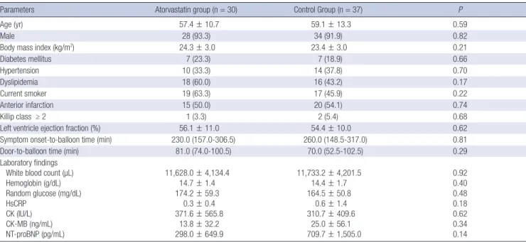
After excluding inadequate MRI images from analysis (n = 2), CE-MRI data were ultimately available for 30 patients in the ator- vastatin group and 37 patients in the control group. Overall sub- ject characteristics including clinical presentations and initial laboratory findings are presented in Table 1. There was no sig- nificant difference in baseline clinical and laboratory charac- teristics between the two groups.
Table 2 shows the angiographic and procedural findings of the two groups. The atorvastatin group tended to have a lower grade of TIMI flow than the control group, but this difference was not statistically significant. Most patients of the two groups underwent implantation of stents in the culprit coronary arter- ies. Drug-eluting stents were used predominantly, with no dif- ference between the study groups. The stent diameter and length were similar in the two groups. There were no significant differ- ences in medication during hospitalization and follow-up peri- od (Table 3).
Angiographic, ECG, biochemical, and clinical outcomes No angiographic reflow, final TIMI flow grade, and final MBG
Table 1. Baseline characteristics
Parameters Atorvastatin group (n = 30) Control Group (n = 37) P
Age (yr) 57.4 ± 10.7 59.1 ± 13.3 0.59
Male 28 (93.3) 34 (91.9) 0.82
Body mass index (kg/m2) 24.3 ± 3.0 23.4 ± 3.0 0.21
Diabetes mellitus 7 (23.3) 7 (18.9) 0.66
Hypertension 10 (33.3) 14 (37.8) 0.70
Dyslipidemia 18 (60.0) 16 (43.2) 0.17
Current smoker 19 (63.3) 17 (45.9) 0.22
Anterior infarction 15 (50.0) 20 (54.1) 0.74
Killip class ≥ 2 1 (3.3) 2 (5.4) 0.68
Left ventricle ejection fraction (%) 56.1 ± 11.0 54.4 ± 10.0 0.62
Symptom onset-to-balloon time (min) 230.0 (157.0-306.5) 260.0 (148.5-317.0) 0.81
Door-to-balloon time (min) 81.0 (74.0-100.5) 70.0 (52.5-102.5) 0.29
Laboratory findings White blood count (µL) Hemoglobin (g/dL) Random glucose (mg/dL) HsCRP
CK (IU/L) CK-MB (ng/mL) NT-proBNP (pg/mL)
11,628.0 ± 4,134.4 14.7 ± 1.4 174.2 ± 59.3
0.3 ± 0.4 371.6 ± 565.8
13.8 ± 32.2 298.0 ± 649.9
11,733.2 ± 4,201.5 14.4 ± 1.7 164.5 ± 50.8
0.6 ± 1.4 310.7 ± 409.6
25.0 ± 56.1 709.7 ± 1,505.0
0.92 0.40 0.48 0.18 0.62 0.34 0.14
Data are expressed as n (%), mean ± SD or median (interquartile range). HsCRP, Highly sensitive C-reactive protein; NT-proBNP, N-terminal pro-Brain natriuretic peptide.
Table 2. Angiographic and procedural findings
Findings Atorvastatin group
(n = 30) Control group
(n = 37) P
Culprit vessel
Left anterior descending artery, n (%) Left circumflex artery, n (%) Right coronary artery, n (%)
15 (50.0) 4 (13.3) 11 (36.7)
21 (56.8) 5 (13.5) 11 (29.7)
0.86
Number of diseased vessels 1
2 3
16 (53.3) 8 (26.7) 6 (20.0)
20 (54.1) 9 (24.3) 8 (21.6)
0.97
Lesion type B2/C 20 (66.7) 25 (67.6) 0.94
Baseline TIMI flow grade 0/1
2 3
24 (80.0) 5 (16.7) 1 (3.3)
22 (59.5) 6 (16.2) 9 (24.3)
0.06
PCI type Balloon only Stent
- 30 (100.0)
2 (5.4) 35 (94.6)
0.50
Type of stent Bare-metal stent Drug-eluting stent
4 (13.3) 26 (86.7)
7 (20.0) 28 (80.0)
0.48
Stent diameter (mm) 3.25 (3.0-3.5) 3.5 (3.0-3.5) 0.86 Stent length (mm) 24.0 (20.0-30.0) 24.0 (18.0-32.0) 0.72 Data are expressed as n (%) or median (interquartile range).
Table 3. Medications
Medications Atorvastatin group (n = 30)
Control group
(n = 37) P
During hospital stay Aspirin Clopidogrel β-blocker ACE inhibitor/ARBs Glycoprotein IIb/IIIa inhibitor
30 (100.0) 30 (100.0) 26 (86.7) 27 (90.0) 12 (40.0)
37 (100.0) 36 (97.3) 36 (97.3) 36 (97.3) 12 (32.4)
- 1.00 0.17 0.32 0.52 Follow up at 1 month
Aspirin Clopidogrel β-blocker ACE inhibitor/ARBs Statin
30 (100.0) 30 (100.0) 26 (86.7) 27 (90.0) 29 (96.7)
36 (97.3) 36 (97.3) 34 (91.9) 34 (91.9) 37 (100.0)
1.00 1.00 0.69 1.00 0.26 Follow-up at 6 months
Aspirin Clopidogrel β-blocker ACE inhibitor/ARBs Statin
30 (100.0) 29 (96.7) 26 (86.7) 28 (93.3) 28 (93.3)
36 (97.3) 35 (97.2) 31 (86.1) 32 (88.9) 34 (94.4)
1.00 1.00 1.00 0.68 1.00 Data are expressed as n (%). ACE, Angiotensin-converting enzyme; ARB, angiotensin- receptor blocker.
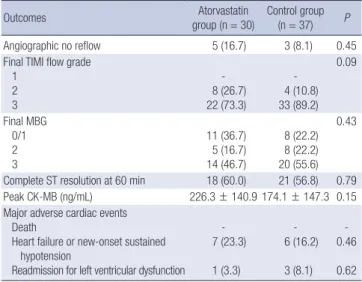
Table 4. Angiographic, ECG, biochemical and clinical outcomes
Outcomes Atorvastatin
group (n = 30) Control group (n = 37) P
Angiographic no reflow 5 (16.7) 3 (8.1) 0.45
Final TIMI flow grade 1
2 3
- 8 (26.7) 22 (73.3)
- 4 (10.8) 33 (89.2)
0.09
Final MBG 0/1 2 3
11 (36.7) 5 (16.7) 14 (46.7)
8 (22.2) 8 (22.2) 20 (55.6)
0.43
Complete ST resolution at 60 min 18 (60.0) 21 (56.8) 0.79 Peak CK-MB (ng/mL) 226.3 ± 140.9 174.1 ± 147.3 0.15 Major adverse cardiac events
Death
Heart failure or new-onset sustained hypotension
Readmission for left ventricular dysfunction - 7 (23.3) 1 (3.3)
- 6 (16.2) 3 (8.1)
- 0.46 0.62 Data are expressed as n (%) or mean ± SD.
Table 5. Results of CE-MRI analysis
Findings Atorvastatin group
(n = 30)
Control group
(n = 37) P
LVEDV (mL) 115.0 (98.2-130.4) 123.7 (94.9-148.8) 0.37 LVESV (mL) 49.8 (43.2-75.9) 66.0 (45.9-84.8) 0.36 LV Mass (g) 121.1 (96.8-135.6) 123.5 (102.9-137.7) 0.28 LV ejection fraction (%) 49.9 (39.8-60.2) 46.3 (39.9-52.4) 0.56 Infarct size (% of LV) 19.0 (11.1-31.4) 16.3 (7.2-27.2) 0.27 Area at risk (% of LV) 31.6 (24.7-51.3) 28.2 (20.2-41.4) 0.25 Myocardial salvage index 37.1 (26.9-58.7) 46.9 (31.1-60.4) 0.46 Hemorrhagic infarction, n(%) 17 (56.7) 18 (48.6) 0.51
MVO area (% of LV) 1.1 (0-1.97) 0.7 (0-1.8) 0.37
Transmurality 10 (33.3) 21 (56.8) 0.06
Data are expressed as n (%) or median (interquartile range). LV, left ventricle; LVEDV, left ventricle end diastolic volume; LVESV, left ventricle end systolic volume; MVO, mi- crovascular obstruction.
did not differ between the atorvastatin and the control group (P = 0.45 for no reflow, P = 0.09 for final TIMI flow grade, and P = 0.43 for final MBG) (Table 4). Similarly, both groups had comparable incidences of complete ST-segment resolution at 60 min (60% vs. 56.8%, P = 0.79). No significant difference was found in the level of peak CK-MB between the 2 groups (226.3 ± 140.9 vs. 174.1 ± 147.3, P = 0.15).
Complete clinical follow-up data were obtained for all patients.
The 6-month major cardiac event rate was not significantly dif- ferent between the atorvastatin group and control group.
CE-MRI findings
There was no difference in the intervals from procedure to MRI between the groups (9.5 [6-18.3] vs. 8 [5-12.5], P = 0.17). Table 5 shows data analyzed from the cine, T2-weighted and CE-MRI of the atorvastatin and control group. The LV end diastolic vol- ume, end systolic volume, and ejection fraction measured by MRI were similar in both groups. The primary end point, infarct size on CE-MRI after the atorvastatin pretreatment and proce- dure was not significantly different between the two groups (19%
[11.1-31.4] vs. 16.3% [7.2-27.2], P = 0.27). Additionally, the myo- cardial salvage index (37.1% [26.9-58.7] vs. 46.9% [39.9-52.4], P = 0.46) and the extent of MVO (1.1% [0-1.97] vs. 0.7% [0-1.8], P = 0.37) were not lower in the atorvastatin group compared to the control group. There were no significant differences of the incidence of hemorrhagic (56.7% vs. 48.6%, P = 0.51) or trans- mural infarctions in both groups (33.3% vs. 56.8%, P = 0.06).
DISCUSSION
In this randomized study, we compared CE-MRI recordings to evaluate the efficacy of high dose atorvastatin loading followed by further medication for 5 days after PCI on the extent of myo- cardial infarction in patients undergoing primary PCI for STE- MI. Our data indicate that infarction severity does not change after atorvastatin pretreatment in STEMI patients. There was no significant difference in the infarct size, AAR, MVO area, infarct transmurality, a frequency of hemorrhagic infarction, or the pro- portion of myocardial salvage between the atorvastatin group and controls. Moreover, post-procedural TIMI flow grade, MBG, complete ST-segment resolution at 60 min, and the peak CK- MB level was similar in both groups.
The AT-STEMI and STATIN STEMI trials report that high-dose atorvastatin pretreatment before PCI did not show significant improvement of clinical outcome (8, 22). However, these 2 trials have too few numbers of participants to draw conclusions re- garding the clinical outcome including major adverse cardiac events. Although infarct size measured by SPECT was primary end point in the AT-STEMI trial, SPECT has low sensitivity in cases of small myocardial infarctions or non-transmural infarc- tions. Furthermore, the STATIN STEMI study showed that sec- ondary end points such as MBG and complete ST segment res- olution were improved in the atorvastatin group. In the AT-STE- MI trial, the high-dose atorvastatin pretreatment group dem- onstrated absolutely 5.2% reduced incidence of major adverse cardiac events at 6 months (8). Considering these doubtful con- clusions, larger randomized trials or more obvious surrogate parameters as primary end points are required to confirm the effects of atorvastatin pretreatment. Therefore, our study may have strengths with respect to performing a planned substudy using CE-MRI-derived infarct size as the primary end point. In addition, CE-MRI is a reliable tool to detect morphologic and functional sequelae of myocardial infarction and provide a broad range of pathophysiological information such as the presence of scars, edema, and the viability of myocardium. CE-MRI might demonstrate more concrete impacts of atorvastatin pretreat- ment on injured myocardium that are not detected by other di- agnostic modalities.
Although statins are known to have anti-inflammatory, anti- thrombotic effects and also known to improve endothelial func- tion and myocardial circulation (24, 25), atorvastatin pretreat- ment did not reduce infarct size measured by CE-MRI in the pres- ent study. Moreover, the myocardial salvage index, which is an attractive CE-MRI parameter from a pathophysiological point of view, was not different between the atorvastatin and control group. MVO and hemorrhagic infarction, which are known to be predictors of adverse LV remodeling, as well as the risk of cardiovascular events did not show any differences between the 2 groups (26, 27). Our findings were supported by another
CE-MRI study. In the REPERATOR trial, Post et al. reported that pretreatment with atorvastatin in an acute myocardial infarc- tion does not result in an improved cardiac function, microvas- cular perfusion, or decreased myocardial infarct size (23). Al- though the comprehensive analysis of CE-CMR such as MVO and hemorrhagic infarction and a slightly larger number of en- rolled patients are advantages of our study over the REPERA- TOR trial, they elegantly performed serial CE-MRI studies with- in 1 day, at 1 week, and 3 months follow up. Based on the results of both studies, it seems unlikely that high dose atorvastatin pre- treatment in patients undergoing PCI for STEMI can reduce in- jury of myocardium.
This ineffectiveness of atorvastatin pretreatment in STEMI patients might be explained by severe ischemic burden or inju- ry which cannot be prevented or reduced by a single dose of atorvastatin before primary PCI. The interval between atorvas- tatin administration and reperfusion might be too short for ator- vastatin to initiate its action since the median door-to-balloon time was 81 min in the atorvastatin group (8, 20).
Our study has several limitations. First, the sample size is not large, although calculations were conducted by standard meth- ods based upon preliminary data. Second, only patients suit- able for MRI examination were included. Therefore, the num- ber of enrolled patients in each group was different. Unstable patients with hemodynamic compromise or severe arrhythmia were excluded from the study, which may have caused selec- tion bias. Third, the timing of CE-MRI was widely distributed.
However, CE-MRI was done within 14 days after primary PCI in all patients and there was no significant difference in interval from the index procedure to obtaining MRI between the 2 groups.
Moreover, we did not undergo follow up CE-MRI, which might be more closely related to final myocardial damage. Fourth, this was not a placebo-controlled study. However, the primary end point was assessed independently and blindly at the core labo- ratory, and therefore we believe that this study design has little effect on the results. Lastly, our study has broad inclusion crite- ria. Because of the small sample size, we could not perform the subgroup analysis on patients who have a short time from symp- tom onset to ballooning or who are present with anterior wall infarction, which might have a better outcome in the atorvas- tatin pretreatment group.
To conclude, in this CE-MRI substudy of AT-STEMI trial, a high dose atorvastatin pretreatment followed by 5 days of fur- ther treatment did not reduce the infarct size in in patients un- dergoing primary PCI for STEMI. The present study suggests that atorvastatin preloading does not improve clinical outcomes in patients with STEMI.
DISCLOSURE
All of the authors have no conflicts of interest to disclose
AUTHOR CONTRIBUTION
Conception and coordination of the study: Hahn JY; Design of ethical issues: Hahn JY, Kim EK, Song YB; Acquisition of data:
Kim EK, Hahn JY, Song YB, Chang SA, Choi JH, Choi SH, Lee SC; Data review: Kim EK, Hahn JY, Song YB, Chang SA, Choi JH, Choi SH, Lee SC, Choe YH; Statistical analysis: Kim EK, Song YB; Manuscript preparation: All authors; Manuscript approval:
all authors.
ORCID
Eun Kyoung Kim http://orcid.org/0000-0002-7653-3503 Joo-Yong Hahn http://orcid.org/0000-0002-4412-377X Young Bin Song http://orcid.org/0000-0002-2581-8891 Sung-A Chang http://orcid.org/0000-0001-8053-0068 Jin-Ho Choi http://orcid.org/0000-0002-5421-793X Seung-Hyuk Choi http://orcid.org/0000-0002-0304-6317 Sang-Chol Lee http://orcid.org/0000-0003-2176-0482 Yeon Hyeon Choe http://orcid.org/0000-0002-9983-048X Sang Hoon Lee http://orcid.org/0000-0003-1202-917X Hyeon-Cheol Gwon http://orcid.org/0000-0002-8967-4305 REFERENCES
1. Schwartz GG, Olsson AG, Ezekowitz MD, Ganz P, Oliver MF, Waters D, Zeiher A, Chaitman BR, Leslie S, Stern T, et al.; Myocardial Ischemia Reduction with Aggressive Cholesterol Lowering (MIRACL) Study In- vestigators. Effects of atorvastatin on early recurrent ischemic events in acute coronary syndromes: the MIRACL study: a randomized controlled trial. JAMA 2001; 285: 1711-8.
2. Shepherd J, Cobbe SM, Ford I, Isles CG, Lorimer AR, MacFarlane PW, McKillop JH, Packard CJ. Prevention of coronary heart disease with pravas- tatin in men with hypercholesterolemia. West of Scotland Coronary Pre- vention Study Group. N Engl J Med 1995; 333: 1301-7.
3. Patti G, Cannon CP, Murphy SA, Mega S, Pasceri V, Briguori C, Colom- bo A, Yun KH, Jeong MH, Kim JS, et al. Clinical benefit of statin pretreat- ment in patients undergoing percutaneous coronary intervention: a col- laborative patient-level meta-analysis of 13 randomized studies. Circu- lation 2011; 123: 1622-32.
4. Pasceri V, Patti G, Nusca A, Pristipino C, Richichi G, Di Sciascio G; AR- MYDA Investigators. Randomized trial of atorvastatin for reduction of myocardial damage during coronary intervention: results from the AR- MYDA (Atorvastatin for Reduction of MYocardial Damage during An- gioplasty) study. Circulation 2004; 110: 674-8.
5. Briguori C, Visconti G, Focaccio A, Golia B, Chieffo A, Castelli A, Mus- sardo M, Montorfano M, Ricciardelli B, Colombo A. Novel approaches for preventing or limiting events (Naples) II trial: impact of a single high loading dose of atorvastatin on periprocedural myocardial infarction. J Am Coll Cardiol 2009; 54: 2157-63.
6. Patti G, Pasceri V, Colonna G, Miglionico M, Fischetti D, Sardella G, Mon- tinaro A, Di Sciascio G. Atorvastatin pretreatment improves outcomes in patients with acute coronary syndromes undergoing early percutaneous
coronary intervention: results of the ARMYDA-ACS randomized trial. J Am Coll Cardiol 2007; 49: 1272-8.
7. Kim JW, Yun KH, Kim EK, Kim YC, Joe DY, Ko JS, Rhee SJ, Lee EM, Yoo NJ, Kim NH, et al. Effect of high dose rosuvastatin loading before primary percutaneous coronary intervention on infarct size in patients with ST- segment elevation myocardial infarction. Korean Circ J 2014; 44: 76-81.
8. Hahn JY, Kim HJ, Choi YJ, Jo SH, Kim HJ, Lee S, Ahn KJ, Song YB, Choi JH, Choi SH, et al. Effects of atorvastatin pretreatment on infarct size in patients with ST-segment elevation myocardial infarction undergoing primary percutaneous coronary intervention. Am Heart J 2011; 162: 1026- 33.
9. Miller TD, Christian TF, Hopfenspirger MR, Hodge DO, Gersh BJ, Gib- bons RJ. Infarct size after acute myocardial infarction measured by quan- titative tomographic 99mTc sestamibi imaging predicts subsequent mor- tality. Circulation 1995; 92: 334-41.
10. Kim RJ, Fieno DS, Parrish TB, Harris K, Chen EL, Simonetti O, Bundy J, Finn JP, Klocke FJ, Judd RM. Relationship of MRI delayed contrast en- hancement to irreversible injury, infarct age, and contractile function.
Circulation 1999; 100: 1992-2002.
11. Wagner A, Mahrholdt H, Holly TA, Elliott MD, Regenfus M, Parker M, Klocke FJ, Bonow RO, Kim RJ, Judd RM. Contrast-enhanced MRI and routine single photon emission computed tomography (SPECT) perfu- sion imaging for detection of subendocardial myocardial infarcts: an imaging study. Lancet 2003; 361: 374-9.
12. Chung KI, Chung TS, White RD, Weinmann HJ, Lim TH, Choi BI, Suh JH. Viable myocardium in reperfused acute myocardial infarction: rest and stress first-pass mr imaging. J Korean Med Sci 2001; 16: 294-302.
13. Perazzolo Marra M, Lima JA, Iliceto S. MRI in acute myocardial infarc- tion. Eur Heart J 2011; 32: 284-93.
14. Eitel I, Desch S, Fuernau G, Hildebrand L, Gutberlet M, Schuler G, Thiele H. Prognostic significance and determinants of myocardial salvage as- sessed by cardiovascular magnetic resonance in acute reperfused myo- cardial infarction. J Am Coll Cardiol 2010; 55: 2470-9.
15. de Waha S, Desch S, Eitel I, Fuernau G, Zachrau J, Leuschner A, Gut- berlet M, Schuler G, Thiele H. Impact of early vs. late microvascular ob- struction assessed by magnetic resonance imaging on long-term outcome after ST-elevation myocardial infarction: a comparison with traditional prognostic markers. Eur Heart J 2010; 31: 2660-8.
16. van’t Hof AW, Liem A, de Boer MJ, Zijlstra F. Clinical value of 12-lead electrocardiogram after successful reperfusion therapy for acute myocar- dial infarction. Zwolle Myocardial infarction Study Group. Lancet 1997;
350: 615-9.
17. van’t Hof AW, Liem A, Suryapranata H, Hoorntje JC, de Boer MJ, Zijlstra F. Angiographic assessment of myocardial reperfusion in patients treated with primary angioplasty for acute myocardial infarction: myocardial blush grade. Zwolle Myocardial Infarction Study Group. Circulation 1998;
97: 2302-6.
18. O’Regan DP, Ariff B, Neuwirth C, Tan Y, Durighel G, Cook SA. Assess- ment of severe reperfusion injury with T2* cardiac MRI in patients with acute myocardial infarction. Heart 2010; 96: 1885-91.
19. Xu J, Song YB, Hahn JY, Chang SA, Lee SC, Choe YH, Choi SH, Choi JH, Lee SH, Oh JK, et al. Comparison of magnetic resonance imaging find- ings in non-ST-segment elevation versus ST-segment elevation myocar- dial infarction patients undergoing early invasive intervention. Int J Car- diovasc Imaging 2012; 28: 1487-97.
20. Song YB, Hahn JY, Gwon HC, Chang SA, Lee SC, Choe YH, Choi SH, Choi JH, Lee SH, Oh JK. A high loading dose of clopidogrel reduces myo- cardial infarct size in patients undergoing primary percutaneous coro- nary intervention: a magnetic resonance imaging study. Am Heart J 2012;
163: 500-7.
21. Hahn JY, Gwon HC, Choe YH, Rhee I, Choi SH, Choi JH, Lee SH, Hong KP, Park JE. Effects of balloon-based distal protection during primary percutaneous coronary intervention on early and late infarct size and left ventricular remodeling: a pilot study using serial contrast-enhanced magnetic resonance imaging. Am Heart J 2007; 153: 665.e1-8.
22. Kim JS, Kim J, Choi D, Lee CJ, Lee SH, Ko YG, Hong MK, Kim BK, Oh SJ, Jeon DW, et al. Efficacy of high-dose atorvastatin loading before prima- ry percutaneous coronary intervention in ST-segment elevation myocar- dial infarction: the STATIN STEMI trial. JACC Cardiovasc Interv 2010; 3:
332-9.
23. Post S, Post MC, van den Branden BJ, Eefting FD, Goumans MJ, Stella PR, van Es HW, Wildbergh TX, Rensing BJ, Doevendans PA. Early statin treatment prior to primary PCI for acute myocardial infarction: REPER-
ATOR, a randomized placebo-controlled pilot trial. Catheter Cardiovasc Interv 2012; 80: 756-65.
24. Wassmann S, Faul A, Hennen B, Scheller B, Böhm M, Nickenig G. Rap- id effect of 3-hydroxy-3-methylglutaryl coenzyme a reductase inhibition on coronary endothelial function. Circ Res 2003; 93: e98-103.
25. Hinoi T, Matsuo S, Tadehara F, Tsujiyama S, Yamakido M. Acute effect of atorvastatin on coronary circulation measured by transthoracic Dop- pler echocardiography in patients without coronary artery disease by angiography. Am J Cardiol 2005; 96: 89-91.
26. Mather AN, Fairbairn TA, Ball SG, Greenwood JP, Plein S. Reperfusion haemorrhage as determined by cardiovascular MRI is a predictor of ad- verse left ventricular remodelling and markers of late arrhythmic risk.
Heart 2011; 97: 453-9.
27. Tarantini G, Razzolini R, Cacciavillani L, Bilato C, Sarais C, Corbetti F, Marra MP, Napodano M, Ramondo A, Iliceto S. Influence of transmu- rality, infarct size, and severe microvascular obstruction on left ventricu- lar remodeling and function after primary coronary angioplasty. Am J Cardiol 2006; 98: 1033-40.