582
Copyright © 2016 The Korean Society of Fisheries and Aquatic Science pISSN:0374-8111, eISSN:2287-8815
서 론
장염비브리오
(Vibrio parahaemolyticus)
는 그람음성,
간균,
무포자,
저도호염성균으로해수또는기수에서서식하며이균 에오염된어패류를생식하거나불충분하게가열처리된수산 물을섭취하면주로복통,
설사,
구토,
오한및미열등을동반하 는급성위장염증상을유발하는식중독원인세균이다(Honda and Iida, 1993; Zhang and Orth, 2013).
식품의약품안전처식 품안전정보포털의 식중독통계에의하면2006
년부터2015
년 까지10
년동안우리나라에서발생한장염비브리오에의한식 중독사고는주로6
월에서10
월사이의하절기에발생하였으며사고건수및환자수는전체세균성식중독사고의
12.5%
및5.4%
를차지하며이균에의한식중독사고는과거보다는감 소추세이나지속적으로발생하고있다(MFDS, 2016).
이균이 생산하는대표적인병원성독소는내열성용혈독(thermostable direct hemolysin, TDH),
내열성용혈독관련용혈독(TDH-re- lated hemolysin, TRH)
및type III secretion systems (TTSS 1
및2)
를통해분비되는각종effector
단백질등이보고되어있으 나정확한병원성메커니즘에관한설명은아직도부족한실정 이다(Honda and Iida, 1993; Park et al., 2004; Letchumanan et al., 2014; Wang et al., 2015).
해수또는어패류등자연계에서 분리한대부분의장염비브리오는병원성인자를보유하지않은곰소만 해역 해수에서 분리한 장염비브리오(Vibrio parahaemolyticus)의 항균제 내성 및 최소발육억제농도의 구명
김태옥·엄인선·김희대
1·박권삼*
군산대학교 식품생명공학과, 1충북도립대학 바이오생명의약과
Antimicrobial Resistance and Minimum Inhibitory Concentrations of Vibrio parahaemolyticus Strains Isolated from Gomso Bay, Korea
Tae-Ok Kim, In-Seon Um, Hee-Dai Kim
1
and Kwon-Sam Park*Department of Food Science and Biotechnology, Kunsan National University, Gunsan 54150, Korea
1
Department of Biotechnology and Biomedicine, Chungbuk Provincial College, Cheongju 28160, Korea
Seventy-nine Vibrio parahaemolyticus isolates from surface seawater from Gomso Bay, west coast of Korea, were analyzed for the presence of virulence genes and their susceptibility to 30 different antimicrobials. All 79 isolates were examined for the presence of two virulence genes ( tdh or trh ) using polymerase chain reaction (PCR); how- ever, no isolates possessed either the tdh or trh gene. According to a disk diffusion susceptibility test, all of the strains studied were resistant to oxacillin, penicillin, and vancomycin, followed by ticarcillin (97.5%), ampicillin (96.2%), clindamycin (86.1%), erythromycin (10.1%), streptomycin (7.6%), cefoxitin (6.3%), amikacin (2.5%), and cephalothin (2.5%). However, all of the strains were susceptible to 19 other antimicrobials including cefepime, ce- fotaxime, chloramphenicol, gentamycin, nalidixic acid, sulfamethoxazole/trimethoprim, and trimethoprim. All 79 isolates (100%) were resistant to four or more classes of antimicrobials, and two strains exhibited resistance to eight antimicrobial agents. The average minimum inhibitory concentrations (MICs) for V . parahaemolyticus for ampicil- lin, penicillin, ticarcillin, and vacomycin were 946.5, 1,305.9, 1,032.3, and 45.0 µg/mL, respectively.
Key words: Vibrio parahaemolyticus , Antimicrobial resistance, Virulence genes, Minimum inhibitory concentration, Gomso Bay
This is an Open Access article distributed under the terms of the Creative Commons Attribution Non-Commercial Licens (http://creativecommons.org/licenses/by-nc/3.0/) which permits unrestricted non-commercial use, distribution, and reproduction in any medium, provided the original work is properly cited.
http://dx.doi.org/10.5657/KFAS.2016.0582 Korean J Fish Aquat Sci 49(5) 582-588, October 2016
Received 8 August 2016; Revised 31 August 2016; Accepted 1 September 2016
*Corresponding author: Tel: +82. 63. 469. 1822 Fax: +82. 63. 469. 7448
E-mail address: [email protected]
비병원성균주인반면식중독사고의환자가검물에서분리한 장염비브리오는대부분병원성유전자를보유하고있는것으로 알려져있다
(Sakazaki et al., 1968; Honda and Iida, 1993).
따 라서장염비브리오의위해평가또는기준규격을설정하는경 우에는시료중의장염비브리오전체균수보다는병원성유전 자를보유하는장염비브리오의균수로고려해야할필요가있 다고판단된다.
페니실린발견이후다양한종류의항균제는사람과동물의 치료
,
질병예방및성장촉진등의목적으로사용되고있다.
지 속적인항균제의사용은양식어류및해수유래장염비브리오 에서다양한항균제내성균의증가를가중시키는결과로나타 나고있다(Son et al., 2005; Lee et al., 2007; Oh et al., 2008;
Lee et al., 2009; Ryu et al., 2010; Yu et al., 2010; Kim et al., 2014; Kang et al., 2016; Kim et al., 2016).
세균이항균제내성 을갖게되는이유는분해효소에의한항균제의불활성화,
표 적항균물질의변화,
세포막의항균제투과성변화및세포밖 으로항균제의유출등의다양한방법에의한것으로알려져있 으며이들메커니즘이단독또는복합적으로작용하여세균은 항균제에내성을갖게된다.
획득내성은세균염색체의유전자 변이, plasmid
또는transposon
에매개되는내성유전자의획득 에의해생기며,
내성유전자는염색체또는plasmid
에존재한 다(Kuhl et al., 1978).
그람음성세균에서항균제다제내성유 전자가삽입되어있는integron
은세균염색체에서이동성을가 진DNA
단편인transposon
을통하여유전자의한복제단위에 서다른복제단위로이동되는데일반적으로접합을통하여동 종및이종세균으로확산된다고보고되어있다(Rowe-Magnus and Mazel, 2002).
다양한유래의장염비브리오에대한항균제내성연구보고는 다수존재하나내성항균제에대한최소발육억제농도에관한 연구논문은거의없는실정이다
.
따라서본논문은장염비브리 오의각종항균제내성및내성항균제의최소발육억제농도에 대한기초자료를얻기위하여2014
년6
월부터2015
년10
월까 지전북곰소만해역의표층해수에서분리한장염비브리오79
균주를대상으로항균제내성양상및내성항균제에대한최소 발육억제농도를검토하였다.
재료 및 방법
사용 균주 및 시약
실험에사용한장염비브리오는
2014
년6
월부터2015
년10
월까지전북곰소만해역의표층해수에서분리한장염비브리 오79
균주와병원성유전자유무를확인하기위하여장염비브 리오RIMD2210633 (Makino et al., 2003)
및TH3996 (Park et al., 2000)
를표준균주로사용하였다.
항균제감수성정도관 리를위하여Escherichia coli ATCC 25922
와Staphylococcus aureus ATCC 25923
을사용하였다.
유전자증폭을위한각종효소는
Takara (Otsu, Japan)
사의제품,
항균제디스크는Bec- ton Dickinson (BBL Sensi-Disk, Sparks, MD, USA)
사의제 품및각종항균제는Sigma (St. Louis, MO, USA)
사의제품 을사용하였다.
장염비브리오의 분리 및 동정
표층해수의장염비브리오는식품공전에서제시한방법에준 하여분리하였다
(MFDS, 2016).
표층해수는선박을이용하여 채수기로멸균채수병에채수후얼음이채워진아이스박스에 넣어4℃
로유지하면서실험실로옮겨실험에사용하였다.
해 수10 mL
를double strength alkaline peptone water (Merck, Darmstadt, Germany) 10 mL
에접종하여35℃
에서18-24
시 간 배양후thiosulfate-citrate-bile-salts (TCBS) agar (Difco, Detroit, MI, USA)
에백금이로접종후35℃
에서18-24
시간 배양하였다. TCBS
배지에서장염비브리오로추정되는전형적 인3-5 mm
크기의청록색집락을triple sugar iron agar (Difco, Detroit, MI, USA)
에접종하고35℃
에서24
시간배양한후전 형적인반응을나타내는균주는API 20E system (BioMerieux, Marcy-I'Etoil, France)
으로생화학적시험을실시하여일차적 으로장염비브리오로동정하였다.
또한장염비브리오동정에 이용되고있는toxR (Kim et al., 1999)
및hns (No et al., 2011)
유전자의 존재유무는
PCR assay
로확인하였으며두유전자존재가확인된균주에한하여최종적으로장염비브리오로동 정하였다
.
동일균주의중복분리를배제하기위하여하나의해 수시료에서한균주의장염비브리오만을분리하였다.
동정이 완료된장염비브리오는Luria-Bertani (tryptone 1%, yeast-ex- tract 0.5%, NaCl 3%) broth
에배양후최종농도15%
가되도 록멸균된글리세린을첨가하여cryovial storage box (Simport, Canada)
에넣어-80℃
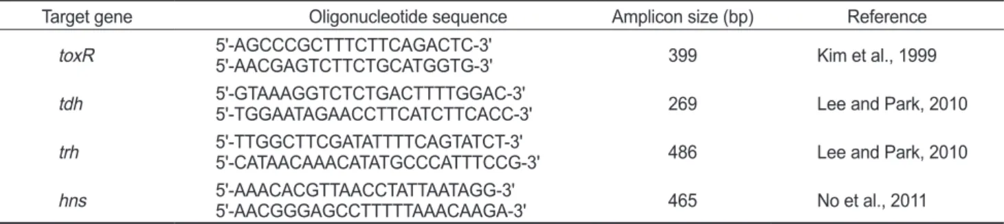
에보관하면서실험에사용하였다. DNA 증폭용 primer set
실험에사용한
DNA
증폭용primers
의염기서열및예상증폭DNA
크기등은Table 1
과같다. Primers
는Bioneer (Daejon, Korea)
에의뢰합성하였다. TDH, TRH, toxR
및H-NS
유전 자DNA
증폭을위한PCR
조건은95℃
에서3
분간1
회열변 성후95℃ 30
초, 55℃ 30
초, 72℃ 30
초를한단위로하여이 를30
회반복하여DNA
를 증폭하였다.
증폭된DNA
산물은1.5% agarose gel
에서전기영동후ethidium bromide
로염색하 여Vilber Lourmat (Bio-Paint ST4, France)
사Gel-Doc system
으로확인하였다.
항균제 감수성 시험
각종항균제에대한감수성은
Acar and Goldstein (1991)
의 디스크확산법과미국NCCLS (National Committee for Clini-
cal Laboratory Standards, 2002)
에준하여시험하였다.
식염3%
첨가된LB broth
에시험균주를접종하여35℃
에서하룻 밤배양한후멸균생리식염수로2
회세정하고농도를McFar-
land No. 0.5
로조정하여두께0.4 mm
의Muller Hinton agar (Merck, Germany)
평판에균을도말하였다.
여기에검사항 균제디스크를고착하여35℃
에서16-18
시간배양한후각항 균제에의해형성된생육저지환의크기를측정하고표준지표 에따라감수성여부를평가하였다.
시험항균제는amikacin (AK; 30 µg), ampicillin (AMP; 10 µg), cefepime (FEP; 30 µg), cefotaxime (CTX; 30 µg), cefotetan (CTT; 30 µg), cefox- itin (FOX; 30 µg), cefuroxime (CXM; 30 µg), ceftriaxone (CRO; 30 µg), cephalothin (KF; 30 µg), cephazolin (KZ; 30 µg), chloramphenicol (C; 30 µg), ciprofloxacin (CIP; 5 µg), clindamycin (CC; 2 µg), erythromycin (E; 15 µg), gentamicin (GN; 10 µg), imipenem (IPM; 10 µg), kanamycin (K; 30 µg), nalidixic acid (NA; 30 µg), nitrofurantoin (F; 100 µg), nor- floxacin (NOR; 10 µg), oxacillin (OX; 1 µg), penicillin (P; 10 µg), pipemidic acid (PIP; 20 µg), rifampin (RD; 5 µg), strep- tomycin (S; 10 µg), sulfamethoxazole/trimethoprim (SXT;
23.75/1.25 µg), tetracycline (TE; 30 µg), ticarcillin (TIC; 75 µg), trimethoprim (W; 5 µg), vancomycin (VA; 30 µg)
등30
종의항균제디스크를사용하였다.
최소발육억제농도(Minimum Inhibitory Concentration, MIC) 측정
최소발육억제농도는미국
NCCLS (National Committee for Clinical Laboratory Standards, 2002)
에기초하여변법으로측 정하였다.
멸균된Muller Hinton broth (Merck, Germany)
에2,048 µg/mL
에서1 µg/mL
까지절반씩농도를달리한항균제 를첨가한후멸균된소형시험관에각농도의항균제가 첨가 된배지를2 mL
씩분주하였다.
여기에식염이3%
첨가된LB broth
에서하룻밤전배양한시험균액3 µL
를접종하여35℃
에 서18
시간정치배양한후균증식여부는육안으로확인하여최 소발육억제농도를측정하였다.
결과 및 고찰
곰소만 표층해수에서 분리한 장염비브리오의 특성
해수유래장염비브리오의항균제내성양상및최소발육억제농도를검토하기위하여
2014
년6
월부터2015
년10
월까지전 북곰소만해역10
개지점에서10
회시료를채취하여총100
개 해수시료에서79
균주의장염비브리오를분리하였다.
해수채 취지점은Fig. 1
에나타내었다.
장염비브리오의분리는식품공 전에서제시하는방법에따라실시하였으며동정은생화학적 시험및유전학적방법즉, toxR (Kim et al., 1999)
및hns (No et al., 2011)
유전자의존재유무로확인하였으며두유전자를 보유하는 균주는최종적으로장염비브리오로동정하였다.
그 결과79
균주에서는toxR
및hns
유전자의예상DNA
증폭산물과동일한크기의
DNA
단편이전기영동결과확인되었다(
자료미제시
).
생화학적시험에서장염비브리오로동정되었으 나toxR
및hns
유전자모두증폭이확인되지않은6
균주는장 염비브리오가아닌것으로판명되었기에실험에사용하지않았 다.
과거대부분의연구에서장염비브리오동정을생화학적결 과만으로동정한사례가많았기때문에장염비브리오가아닌 비브리오속의균주도실험에포함됐을가능성이대두된다.
따 라서장염비브리오의분리및동정의정확성을높이기위해서 는생화학적결과뿐만아니라toxR
및hns
유전자의존재유무 까지확인하는것이반드시필요하다고판단된다.
병원성유전 자의보유성을PCR assay
로검토한결과,
표준균주에서는병 원성유전자의증폭이확인된반면,
분리된79
균주에서는TDH
Table 1. Primers used in this studyTarget gene Oligonucleotide sequence Amplicon size (bp) Reference
toxR 5'-AGCCCGCTTTCTTCAGACTC-3'
5'-AACGAGTCTTCTGCATGGTG-3' 399 Kim et al., 1999
tdh 5'-GTAAAGGTCTCTGACTTTTGGAC-3'
5'-TGGAATAGAACCTTCATCTTCACC-3' 269 Lee and Park, 2010
trh 5'-TTGGCTTCGATATTTTCAGTATCT-3'
5'-CATAACAAACATATGCCCATTTCCG-3' 486 Lee and Park, 2010
hns 5'-AAACACGTTAACCTATTAATAGG-3'
5'-AACGGGAGCCTTTTTAAACAAGA-3' 465 No et al., 2011
Fig. 1. Location of sampling stations in Gomso Bay, Korea, from June 2014 to October 2015.
또는
TRH
유전자가증폭된균주는없었다(
자료미제시).
환경 유래장염비브리오의병원성유전자보유율은매우낮은것으 로보고되어있었으나(Shirai et al., 1990),
최근에는새로운검 출방법의개발로인하여환경유래장염비브리오에서병원성 유전자를보유하고있는균주의검출율은높아지고있는추세 이다(Deepanjali et al., 2005; Jones et al., 2012; Ellingsen et al., 2013; Gutierrez et al., 2013; Kang et al., 2016; Kim et al.,
2016).
결과적으로곰소만해역의표층해수에서분리된79
균주는병원성유전자를보유하지않은비병원성장염비브리오 인것으로확인되었다
.
장염비브리오의 항균제 내성 양상
해수및어패류등다양한환경시료에서분리한장염비브리 오는
amikacin, ampicillin, cephalothin, cefoxitin, gentami- cin, kanamycin, rifampin, streptomycin
및vancomycin
등의 단독항균제에 내성을 나타낼뿐만아니라 다제내성균의검 출빈도도높은것으로보고되어있다(Son et al., 2005; Lee et al., 2009; Ryu et al., 2010; Han et al., 2012; Kim et al., 2014;
Kang et al., 2016; Kim et al., 2016).
곰소만해역해수에서분 리한장염비브리오79
균주를대상으로30
종의항균제에대한 감수성여부를디스크확산법으로측정한결과는Table 2
와같 다. 30
종의항균제중11
종의항균제는79
균주또는일부균 주에서내성을나타내었으며,
나머지19
종의항균제에대해서 는모든균주에서 감수성을나타내었다.
내성율이 높은항균 제는oxacillin, penicillin, vancomycin, ticarcillin, ampicillin, clindamycin, erythromycin, streptomycin, cefoxitin, amikacin
및cephalothin
순서였다.
내성율은oxacillin, penicillin
및van- comycin
은100%
의내성을나타내며, ticarcillin (97.5%), am- picillin (96.2%), clindamycin (86.1%), erythromycin (10.1%), streptomycin (7.6%), cefoxitin (6.3%), amikacin (2.5%)
및cephalothin (2.5%)
이였다. Cefotaxime
을포함한19
종의항균 제에대해서는모든균주는감수성을나타내었다.
이결과는과 거에보고된여러결과와크게다르지않았으며,
각종항균제에 대한장염비브리오의내성및감수성비율은분리원,
분리시기 및분리장소등의요인에따라차이가있다는기존의연구결과 와대체로일치하는결과였다(Son et al., 2005: Lee et al., 2007;
Lee and Park, 2010; Ryu et al., 2010; Han et al., 2012; Kim et al., 2014; Kang et al., 2106; Kim et al., 2016).
또한실험에사 용한79
균주에대한항균제내성양상에관한결과는Table 3
과 같다. 4
종의항균제에내성을나타내는균주는1
균주(1.3%)
였 으며, 5
종의항균제에내성을나타내는균주는9
균주(11.4%), 6
종의항균제에내성을나타내는균주는54
균주(68.4%), 7
종 의항균제에 내성을나타내는 균주는13
균주(16.5%)
및8
종 의항균제에내성균은2
균주(2.5%)
로파악되었다.
가장높은 빈도의항균제내성조합은AMP-CC-OX-P-TIC-VA
로50
균 주(63.3%)
이였으며,
다음으로AMP-OX-P-TIC-VA
은8
균주(10.1%)
및AMP-CC-OX-P-S-TIC-VA
은6
균주(7.6%)
로 파 악되었으며기타항균제내성조합빈도는4
균주이하로낮은 편이였다(Table 3).
결과적으로곰소만해역표층해수에서분리 한장염비브리오는최소4
종이상의항균제에내성을나타내고 있다는점에서심각성은매우크다고판단된다.
이렇게다제내 성을나타내는이유로는곰소만으로유입되는육상유입수에항 균제가포함되어있어해수중의장염비브리오가항균제내성 을획득하게되었거나,
항균제내성을갖고있는균주가보유하 Table 2. Antimicrobial susceptibility and resistance of Vibrio para- haemolyticus strains isolated from surface seawater in Gomso Bay Antimicrobials Disccontent (μg)
No. of isolates Resis-
tant Intermedi-
ate Suscep- tible
Amikacin (AK) 30 2 4 73
Ampicillin (AMP) 10 76 3 0
Cefepime (FEP) 30 0 2 77
Cefotaxime (CTX) 30 0 0 79
Cefotetan (CTT) 30 0 2 77
Cefoxitin (FOX) 30 5 16 58
Cefuroxime (CXM) 30 0 5 74
Ceftriaxone (CRO) 30 0 0 79
Cephalothin (KF) 30 2 7 70
Cephazolin (KZ) 30 0 11 68
Chloramphenicol (C) 30 0 1 78
Ciprofloxacin (CIP) 5 0 8 71
Clindamycin (CC) 2 68 11 0
Erythromycin (E) 15 8 6 65
Gentamicin (GN) 10 0 1 78
Imipenem (IPM) 10 0 0 79
Kanamycin (K) 30 0 1 78
Nalidixic acid (NA) 30 0 1 78
Nitrofurantoin (F) 100 0 22 57
Norfloxacin (NOR) 10 0 3 76
Oxacillin (OX) 1 79 0 0
Penicillin (P) 10 79 0 0
Pipemidic acid (PIP) 20 0 2 77
Rifampin (RD) 5 0 4 75
Streptomycin (S) 10 6 2 71
Sulfamethoxazole
/Trimethoprim (SXT) 25 0 0 79
Tetracycline (TE) 30 0 1 78
Ticarcillin (TIC) 75 77 2 0
Trimethoprim (W) 5 0 2 77
Vancomycin (VA) 30 79 0 0
고있는
plasmid
가장염비브리오에수평적전이가있었거나또 는해수에존재하는장염비브리오의특이적인bacteriophage
에 존재하는항균제내성유전자가장염비브리오에수평적전이를 통해내성유전자를획득하였을것으로추정된다.
장염비브리오의 최소발육억제농도 측정
내성을나타내는
11
종항균제의장염비브리오에대한최소발 육억제농도를측정한결과는Table 4
과같다. Amikacin
에내성 을나타내는2
균주의MIC
는32
및16 µg/mL
이였으며,
감수성 을나타내는나머지77
균주의MIC
는1 µg/mL
이하로측정되 었다. Ampicillin
에내성을나타내는균주의MIC
가2,048 µg/
mL
인균주는8
균주(10.1%), 1,024 µg/mL
의MIC
를나타내는 균주는41
균주(51.9%), 512 µg/mL
의MIC
를나타내는균주는26
균주(32.9%)
및256 µg/mL
의MIC
를나타내는균주는1
균 주(1.3%)
로파악되었으며감수성을나타내는3
균주(3.8%)
의MIC
는2.0 µg/mL
이였다.
Ampicillin
에내성을나타내는균주의MIC
의평균은946.5
µg/mL
로확인되었다.
이결과는자연계에서분리한장염비브리오는 일반적으로
ampicillin
에고도 내성을나타내고 있다 는기존의결과와대체로유사하였다(Tanil et al., 2005; Lee et al., 2009; Lee and Park, 2010; Kim et al., 2014). Clindamycin
에내성을나타내는균주의MIC
는대체로낮아평균16.0 µg/
Table 3. Antimicrobial resistance patterns of Vibrio parahaemolyti- cus strains isolated from surface seawater in Gomso Bay Resistance type No. of resistant strains
CC-OX-P-VA 1
AMP-OX-P-TIC-VA 8
CC-FOX-OX-P-VA 1
AK-CC-E-OX-TIC-VA 1
AMP-E-OX-P-TIC-VA 2
AMP-CC-OX-P-TIC-VA 50
AMP-KF-OX-P-TIC-VA 1
AMP-CC-OX-P-S-TIC-VA 6
AMP-CC-E-OX-P-TIC-VA 3
AMP-CC-FOX-OX-P-TIC-VA 3
AMP-CC-KF-OX-P-TIC-VA 1
AK-AMP-CC-E-OX-P-TIC-VA 1
AMP-CC-E-FOX-OX-P-TIC-VA 1
Total 79
AK, amikacin; AMP, ampicillin; CC, clindamycin; E, erythromy- cin; FOX, cefoxitin; KF, cephalothin; OX, oxacillin; P, penicillin;
S, streptomycin; TIC, ticarcillin; VA, vancomycin.
Table 4. Minimum inhibitory concentration of Vibrio parahaemolyticus strains isolated from surface seawater in Gomso Bay
Antimicrobials µg/mL
>1 1 2 4 8 16 32 64 128 256 512 1,024 2,048
Amikacin 77
(97.4%) 1
(1.3%) 1 (1.3%)
Ampicillin 3
(3.8%) 1
(1.3%) 26 (32.9%) 41
(51.9%) 8 (10.1%)
Cindamycin 11
(13.9%) 26
(32.9%) 29 (36.7%) 13
(16.5%) Erythromycin 71
(89.8%) 3
(3.8%) 4 (5.1%) 1
(1.3%) Cefoxitin 74
(93.7%) 3
(3.8%) 2 (2.5%)
Cephalothin 16
(20.3%) 61
(77.2%) 2
(2.5%)
Oxacillin 31
(39.2%) 42 (53.2%) 6
(7.6%)
Penicillin 4
(5.1%) 6
(7.6%) 41 (51.9%) 28
(35.4%) Streptomycin 73
(92.4%) 5
(6.3%) 1 (1.3%)
Ticarcillin 2
(2.5%) 1
(1.3%) 23
(29.1%) 40 (50.6%) 13
(16.5%)
Vancomycin 6
(7.6%) 43 (54.4%) 30
(38.0%)
mL
로확인되었으며감수성을나타내는11
균주의MIC
는2.0 µg/mL
이였다.
또한erythromycin, streptomycin
및cefoxitin
에대한MIC
는매우낮은농도로파악되었다. Cephalothin
에 대한MIC
는평균256 µg/mL
로확인되었으며, oxacillin
의평 균MIC
는225.2 µg/mL
로확인되었다. Penicillin
및ticarcil- lin
에대한MIC
는대체로높은농도로평균1,305.9 µg/mL
및1,032.3 µg/mL
로확인되었으며, vancomycin
에대한MIC
는45.0 µg/mL
로확인되었다.
결과적으로장염비브리오는ampi- cillin, penicillin, ticarcillin oxacillin
및cephalothin
등의항균 제에대해서는MIC
가대체로높은반면amikacin, clindamy- cin, erythromycin, cefoxitin, streptomycin, vancomycin
등의MIC
는상대적으로낮은것으로확인되었다(Table 4).
본실험에제공된곰소만해역에서분리한
79
균주모든장염 비브리오는4
종이상의항균제에내성을나타낼뿐만아니라ampicillin, penicillin,
및ticarcillin
에대한MIC
는다른항균 제에비해대체로높은것으로확인되었다.
또한다수의항균제 에대해서도내성보유현상이보편화되어있다는점에서장염 비브리오의항균제내성에관한꾸준한모니터링은필요하다 고판단된다.
더불어다양한항균제내성유전자의동정및염 색체DNA
상에서의존재양상등의파악은내성유전자의획득 및확산메커니즘을이해하는데큰도움이될것으로기대된다.
References
Acar JF and Goldstein FW. 1991. Disk susceptibility test. In:
Antibiotics in Laboratory Medicine, Lorian V, ed. Williams
& Wilkins, Baltimore, U.S.A., 17-52.
Deepanjali A, Kumar HS, Karunasagar I and Karunasagar I.
2005. Seasonal variation in abundance of total and patho- genic Vibrio parahaemolyticus bacteria in oysters along the southwest coast of India. Appl Environ Microbiol 71, 3575- 3580.
Ellingsen AB, Olsen JS, Granum PE, Rørvik LM and González- Escalona N. 2013. Genetic characterization of trh positive
Vibrio spp. isolated from Norway. Front Cell Infect Micro-
bial 3, 1-10. http://dx.doi.org/10.3389/fcimb.2013.00107.Gutierrez West CK, Klein SL and Lovell CR. 2013. High fre- quency of virulence factor genes tdh, trh, and tlh in Vibrio
parahaemolyticus strains isolated from a pristine estu-
ary. Appl Environ Microbiol 79, 2247-2252. http://dx.doi.org/10.1128/AEM.03792-12.
Han AR, Yoon YJ and Kim JW. 2012. Antibiotic resistance and plasmid profile of Vibrio parahaemolyticus strains isolated from Kyunggi-Incheon coastal area. Korean J Microbiol 48, 22-28.
Honda T and Iida T. 1993. The pathogenicity of Vibrio parahae-
molyticus and the role of the thermostable direct haemolysin
and related haemolysins. Rev Med Microbiol 4, 106-113.Jones JL, Ludeke CH, Bowers JC, Garrett N, Fischer M, Par- sons MB, Bopp CA and DePaola A. 2012. Biochemical,
serological, and virulence characterization of clinical and oyster Vibrio parahaemolyticus isolates. J Clin Microbiol 50, 2343-2352. http://dx.doi.org/10.1128/JCM.00196-12.
Kang CH, Shin Y, Kim W, Kim Y, Song K, Oh EG, Kim S, Yu H and So JS. 2016. Prevalence and antimicrobial suscepti- bility of Vibrio parahaemolyticus isolated from oysters in Korea. Environ Sci Pollut Res Int 23, 918-926. http://dx.doi.
org/10.1007/s11356-015-5650-9.
Kim SK, An SR, Park BM, Oh EG, Song KC, Kim JW and Yu HS. 2016. Virulence factors and antimicrobial suscep- tibility of Vibrio parahaemolyticus isolated from the oyster
Crassostrea gigas. Korean J Fish Aquat Sci 49, 116-123.
http://dx.doi.org/10.5657/KFAS.2016.0116.
Kim TO, Eum IS, Jo SM, Kim HD and Park KS. 2014. Anti- microbial-resistance profiles and virulence genes of Vibrio
para haemolyticus isolated from seawater in the Wando
area. Korean J Fish Aquat Sci 47, 220-226. http://dx.doi.org/10.5657/KFAS.2014.0220.
Kim YB, Okuda J, Matsumoto C, Takahashi N, Hashimoto S and Nishibuchi M. 1999. Identification of Vibrio parahae-
molyticus strains at the species level by PCR targeted to the toxR gene. J Clin Microbiol 37, 1173-1177.
Kuhl SA, Pattee PA and Baldwin NJ. 1978. Chromosomal map location of the methicillin resistance determinant in Staphy-
lococcus aureus. J Bacteriol 135, 460-465.
Lee H, Oh YH, Park SG and Choi SM. 2007. Antibiotic suscep- tibility and distribution of Vibrio parahaemolyticus isolated from the seafood. Kor J Env Hlth 33, 16-20.
Lee HW, Lim SK and Kim MN. 2009. Characteristics of ampi- cillin-resistant Vibrio spp. isolated from a west coastal area of Korea peninsula. J Kor Fish Soc 42, 20-25.
Lee KW and Park KS. 2010. Antibiotic-resistance profiles and the identification of the ampicillin-resistance gene of
Vibrio parahaemolyticus isolated from seawater. Korean
J Fish Aquat Sci 43, 637-641. http://dx.doi.org/10.5657/KFAS.2010.0637.
Letchumanan V, Chan KG and Lee LH. 2014. Vibrio parahae-
molyticus: a review on the pathogenesis, prevalence, and ad-
vance molecular identification techniques. Front Microbiol 5, 705. http://dx.doi.org/10.3389/fmicb.2014.00705.Makino K, Oshima K, Kurokawa K, Yokoyama K, Uda T, Tagomori K, Iijima Y, Najima M, Nakano M, Yamashita A, Kubota Y, Kimura S, Yasunaga T, Honda T, Shinagawa H, Hattori M and Iida T. 2003. Genome sequence of Vibrio
parahaemolyticus: a pathogenic mechanism distinct from
that of V. cholerae. Lancet 361, 743-749.Ministry of Food and Drug Safety (MFDS). 2016. Retrieved from http://www.foodsafetykorea.go.kr/portal/healthy- foodlife/foodPoisoningStat.do?menu_no=519&menu_
grp=MENU_GRP02 on August 1.
National Committee for Clinical Laboratory Standards (NC- CLS). 2002. Performance standards for antimicrobial sus-
ceptibility testing. Twelfth informational supplement M100- S12. Wayne, Pennsylvania, U.S.A., 19087-19098.
No AR, Okada K, Kogure K and Park KS. 2011. Rapid detec- tion of Vibrio parahaemolyticus by PCR targeted to the histone-like nucleoid structure (H-NS) gene and its genetic characterization. Lett Appl Microbiol 53, 127-133. http://
dx.doi.org/10.1111/j.1472-765X.2011.03072.x.
Oh EG, Yu HS, Shin SB, Son KT, Park KB, Kwon JY, Lee TS and Lee HJ. 2008. Trimethoprim resistance of Vibrio para-
haemolyticus isolated from the fish farm. J Kor Fish Soc 41,
324-329.Park KS, Iida T, Yamaichi Y, Oyagi T, Yamamoto K and Honda T. 2000. Genetic characterization of DNA region containing the trh and ure genes of Vibrio parahaemolyticus. Infect Im- mun 68, 5742-5748.
Park KS, Ono T, Rokuda M, Jang MH, Okada K, Iida T and Honda T. 2004. Functional characterization of two type III secretion systems of Vibrio parahaemolyticus. Infect Im- mun 72, 6659-6665.
Rowe-Magnus DA and Mazel D. 2002. The role of integrons in antibiotic resistance gene capture. Int J Med Microbiol 292, 115-125.
Ryu SH, Hwang YO, Park SG and Lee YK. 2010. Antibiotic susceptibility of Vibrio parahaemolyticus isolated from commercial marine products. Korean J Food Sci Technol 42, 508-513.
Sakazaki R, Tamura K, Kato T, Obara Y and Yamai S. 1968.
Studies on the enteropathogenic, facultatively halophilic bacterium, Vibrio parahaemolyticus. 3. Enteropathogenici- ty. Jpn J Med Sci Biol 21, 325-331.
Shirai H, Ito H, Hirayama T, Nakamoto Y, Nakabayashi N, Kumagai K, Takeda Y and Nishibuchi M. 1990. Molecular epidemiological evidence for association of thermostable direct hemolysin (TDH) and TDH-related hemolysin of Vib-
rio parahaemolyticus with gastroenteritis. Infect Immun 58,
3568-3573.Son KT, Oh EG, Lee TS, Lee HJ, Kim PH and Kim JH. 2005.
Antimicrobial susceptibility of Vibrio parahaemolyticus and
Vibrio alginolyticus from farms on the southern coast of Ko-
rea. J Kor Fish Soc 38, 365-371.Tanil GB, Radu S, Nishibuchi M, Rahim RA, Napis S, Mau- rice L and Gunsalam JW. 2005. Characterization of Vibrio
parahaemolyticus isolated from coastal seawater in penin-
sular Malaysia. Southeast Asian J Trop Public Health 36, 940-945.Wang R, Zhong Y, Gu X, Yuan J, Saeed AF and Wang S. 2015.
The pathogenesis, detection, and prevention of Vibrio
parahaemolyticus. Front Microbiol 6, 144. http://dx.doi.
org/10.3389/fmicb.2015.00144.
Yu HS, Park KB, Oh EG, Lee TS, Shin SB, Kwon JY, Kim JH and Son KT. 2010. Trimethoprim resistance by class I inte- gron in Vibrio parahaemolyticus from a fish farm. Korean J
Fish Aquat Sci 43, 125-130.
Zhang L and Orth K. 2013. Virulence determinants for Vibrio
parahaemolyticus infection. Curr Opin Microbiol 16, 70-77.
http://dx.doi.org/10.1016/j.mib.2013.02.002.