220
Copyright © 2014 The Korean Society of Fisheries and Aquatic Science pISSN:0374-8111, eISSN:2287-8815
서 론
장염비브리오(Vibrio parahaemolyticus)는비브리오과(Fam- ily Vibrionaceae)에속하는저도호염성해양세균으로이균에 오염된어패류를생식하거나불충분하게가열처리된수산물 을섭취하면주로설사, 복통, 구토, 오한및미열등을동반하는 급성위장염증상을유발하는식중독원인세균이다(Sakazaki et al., 1968; Honda and Iida, 1993). 식품의약품안전처의식중독 통계시스템자료에의하면 2002년부터 2013년까지 12년간우
리나라에서발생한장염비브리오에의한식중독사고는주로 여름철에집중적으로발생하며사고건수및환자수는전체세 균성식중독사고의 16.5% 및 8.2%를차지하며, 사고건수로 는병원성대장균, 살모넬라에이어 3위에환자수로는병원성 대장균, 황색포도상구균, 살모넬라에이어 4위를차지하고있 다(MFDS, 2014). 소득향상과건강에대한관심고조는수산물 에대한소비자의선호가증가하여수산물소비량은꾸준히증 가하는추세이다. 통계청자료에의하면국민 1인당연간수산 물소비량은 2005년 49.5 kg, 2007년 56.5 kg, 2009년 49.8 kg
완도해역 해수에서 분리한 장염비브리오(Vibrio parahaemolyticus)의 항균제 내성 및 병원성 유전자의 특징
김태옥·엄인선·조상만
1·김희대
2·박권삼*
군산대학교 식품생명공학과, 1군산대학교 해양생명과학과, 2충북도립대학 바이오생명의약과
Antimicrobial-resistance Profiles and Virulence Genes of Vibrio para- haemolyticus Isolated from Seawater in the Wando Area
Tae-Ok Kim, In-Seon Eum, Sang-Man Jo1, Hee-dai Kim2 and Kwon-Sam Park*
Department of Food Science and Biotechnology, Kunsan National University, Gunsan 573-701, Korea
1Department of Marine Life and Aquaculture, Kunsan National University, Gunsan 573-701, Korea
2Department of Biotechnology and Biomedicine, Chungbuk Provincial College, Okcheon 373-807, Korea
Sixty-seven
Vibrio parahaemolyticus
isolates from surface seawater from the Wando area, on the southern coast of Korea, were analyzed for their susceptibility to 15 different antimicrobials and the presence of virulence genes.According to the disk diffusion susceptibility test, all of the strains studied were resistant to ampicillin and oxacil- lin, while decreasing percentages were resistant to vancomycin (64.2%), streptomycin (56.7%), amikacin (31.3%), kanamycin (22.3%), cephalothin (20.9%), erythromycin (10.4%), ciprofloxacin (4.5%), and tetracycline (3.0%). All of the strains were susceptible to five antimicrobials: chloramphenicol, gentamycin, nalidixic acid, sulfamethoxazole/
trimethoprim, and trimethoprim. Fifty-nine isolates (88.1%) were resistant to three or more classes of antimicrobial and defined as multidrug resistant, and two strains were resistant to seven antimicrobial agents. The minimum inhibi- tory concentration (MIC) of the 67
V
.parahaemolyticus
isolates to ampicillin and oxacillin ranged from 512−2,048 and 64−512 µg/mL, respectively. All 67 isolates were also examined for the presence of thetdh
andtrh
virulence genes using the polymerase chain reaction (PCR). However, no isolates possessed eithertdh
ortrh
. TheVPA0477
(β-lactamase) gene, present in all of the tested strains, was validated as a new specific marker gene in PCR assays for the accurate detection and identification ofV
.parahaemolyticus
.Key words:
Vibrio parahaemolyticus
, Antimicrobial resistance, Virulence genes, Minimum inhibitory concentrationThis is an Open Access article distributed under the terms of the Creative Commons Attribution Non-Commercial Licens (http://creativecommons.org/licenses/by-nc/3.0/)which permits unrestricted non-commercial use, distribution, and reproduction in any medium, provided the original work is properly cited.
http://dx.doi.org/10.5657/KFAS.2014.0220 Kor J Fish Aquat Sci 47(3) 220-226, June 2014
Received 4 March 2014; Revised 29 April 2014; Accepted 27 May 2014
*Corresponding author: Tel: +82. 63. 469. 1822 Fax: +82. 63. 469. 7448 E-mail address: [email protected]
및 2011년 53.5 kg로매년다량의수산물을소비하고있기때문 에수산물의안전성확보는국민보건상절실히필요하다(Sta- tistics Korea, 2013).
장염비브리오는수온이 17℃ 이상으로상승하는하절기에는
자유유영의형태로해수에서쉽게검출되나수온이 10℃ 이
하로낮아지면해수에서의검출빈도는급격히떨어지며수온 이낮은동절기에는저질또는동물성플랑크톤의키틴질등 에부착하여월동한다고보고되어있다(Kaneko and Colwell,
1975). 해수및어패류등의자연계에서분리한대부분의장염
비브리오는병원성인자를보유하고있지않은비병원성균주 인반면식중독사고의환자가검물에서분리된대부분의장염 비브리오는병원성균주이다(Sakazaki et al., 1968; Honda and Iida, 1993). 이균이생산하는대표적인병원성독소는내열성 용혈독(thermostable direct hemolysin, TDH), 내열성용혈독 관련용혈독(TDH-related hemolysin, TRH), serine protease 및 type III secretion systems (TTSS 1 및 2)를통해분비되는각종
effector 단백질등이보고되어있으나정확한병원성메커니즘
에관한설명은아직도많이부족한실정이다(Honda and Iida, 1993; Lee et al., 2002; Makino et al., 2003; Park et al., 2004;
Kodama et al., 2007; Kodama et al., 2008).
페니실린 발견 이후 다양한 종류의 항생제는 사람과 동물 의치료, 질병예방및성장촉진등의목적으로사용되고있다. 2012년농림수산검역검사본부(NVRQS, 2009) 에의하면돼 지, 소및닭등축산용항생제판매량은 2005년이래서서히감 소하는추세인데비해수산용항생제판매량은 2009년 178톤, 2010년 203톤및 2011년 239톤으로오히려사용량이소폭증 가하고있는것으로파악되고있다. 수산용항생제판매실적은 oxytetracycline, erythromycin, amoxicillin, 설파계열, cipro- floxacin, ampicillin 순으로집계되고있다(NVRQS, 2009). 지 속적이고다량의항생제사용은양식어류에서다양한항생제에 내성을나타내는장염비브리오의증가를가중시키는결과로나 타나고있다(Son et al., 2005; Lee et al., 2007; Oh et al., 2008;
Lee et al., 2009; Ryu et al., 2010; Yu et al., 2010).
세균이항생제내성을갖게되는이유는분해효소에의한항 생제의불활성화, 표적항생물질의변화, 세포막의항생제투과 성변화및세포밖으로항생제의유출등의다양한방법에의 한것으로알려져있으며이들메커니즘이단독또는복합적으 로작용하여세균은항생제에내성을갖게된다. 획득내성은세 균염색체의유전자변이, 플라스미드(plasmid) 또는트랜스포 존(transposon)에매개되는내성유전자의획득에의해생기며, 내성유전자는 염색체또는 plasmid DNA에 존재한다(Kuhl et al., 1978). 그람음성세균에서항생제다제내성유전자를 encoding하고있는 integron은세균염색체에서이동성을가진 DNA 단편인 transposon을통하여유전자의한복제단위에서 다른복제단위로이동되는데가장일반적인방법은접합을통 하여동종및이종세균으로확산된다고보고되어있다(Rowe-
Magnus and Mazel, 2002).
본논문은해수유래장염비브리오의각종항균제내성양상 및병원성유전인자보유성에대한기초자료를수집하기위하 여완도해역해수에서분리한장염비브리오 67 균주를대상으 로검토하였다. 또한실험에사용된모든균주에서내성을나타 내는 ampicillin 및 oxacillin 항균제에대해서는최소발육억제 농도도검토하였다.
재료 및 방법
균주 및 시약
실험에사용한장염비브리오는 2011년 6월부터 2012년 10월 까지전남완도군군외면영흥리연안표층해수에서분리한 67 균주및장염비브리오 RIMD2210633 (Makino et al., 2003)를 표준균주로사용하였다. 항균제감수성결과의정도관리를위 하여 Escherichia coli ATCC 25922와 Staphylococcus aureus ATCC 25923을사용하였다. 해수중의장염비브리오는식품공 전이제시한방법에준하여분리하였다(MFDS, 2002). 해수 10 mL를 double strength alkaline peptone water (Merck, Darm- stadt, Germany) 10 mL에접종하여 35℃에서 18-24시간배양 후 Thiosulfate-citrate-bile salts (TCBS) agar (Difco, Detroit, MI, USA)에한백금이획선접종하여 35℃에서 18-24시간배
양하였다. TCBS 배지상에장염비브리오로추정되는전형적인
3-5 mm 청록색콜로니에대해 oxidase 측정및 API 20E Kit (BioMerieux, Marcy I'Etoil, France)를사용하여생화학적시 험을실시하여일차적으로장염비브리오로동정하였다. 또한 유전학적으로장염비브리오동정에사용되고있는 toxR (Kim et al., 1999) 및 hns (No et al., 2011) 유전자의존재유무는 PCR
assay로확인하였으며이들유전자존재가모두확인된균주
에한하여최종적으로장염비브리오로동정하였다. 동일균주 의중복분리를배제하기위하여하나의해수시료에서하나의
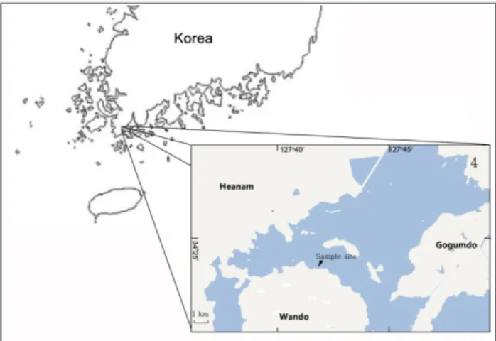
Fig. 1. Location of sample collection stations in Yeonghueng-ri, Wando coast, between June 2011 and October 2012.
장염비브리오를 분리하였다. 동정된 장염비브리오는 Luria- Bertani (tryptone 1%, yeast-extract 0.5%, NaCl 3%) broth에
배양한후 -70℃에보관하면서실험에사용하였다. 유전자증
폭을위한각종효소류는 Takara (Japan)사의제품, 항생제디스 크는 Becton Dickinson (BBL Sensi-Disk, Sparks, MD, USA) 사의제품, ampicillin과 oxacillin의항생제는 Sigma (St. Louis, MO, USA)사의제품을사용하였다.
DNA 증폭용 primer set
실험에사용한 DNA 증폭용 primers의염기서열및증폭예 상 DNA 크기등은 Table 1에제시하였다. Primers는 Bioneer (Daejon, Korea)에의뢰합성하였다. TDH, TRH, toxR 및 hns 유전자 DNA 증폭을위한 PCR 조건은 95℃에서 1회 3분열변 성후 95℃ 30초, 55℃ 30초, 72℃ 30초를한단위로하여이를 30회반복하여 DNA를증폭하였으며, VPA0477 (β-lactamase) 유전자는 95℃에서 1회 3분열변성후 95℃ 30초, 55℃ 30초, 72℃ 1분을한단위로하여이를 30회반복하여 DNA를증폭하 였다. 증폭된 DNA 산물은 1.5% agarose gel에서전기영동후 ethidium bromide로염색하여 Vilber Lourmat (Bio-Paint ST4, France)사의 Gel-Doc system으로관찰하였다.
항균제 감수성 시험
균주의항균제감수성은 Acar and Goldstein (1991)의디스크 확산법과미국 NCCLS (National Committee for Clinical Lab- oratory Standards, 2002)에준하여시험하였다. 식염 3% 첨가 된 LB broth에시험균주를접종하여 35℃에서하룻밤배양한 후멸균생리식염수로 2회세정하고농도를 McFarland 0.5로 조정하여 Muller Hinton Agar (Merck, Germany) 평판에도말 하였다. 여기에검사항균제디스크를고착하여 35℃에서 18시 간배양한후각항균제에의해형성된생육저지환의크기를측 정하고표준지표에따라감수성여부를평가하였다. 시험항균 제는 amikacin (AN; 30 µg), ampicillin (AM; 10 µg), cepha-
lothin (CF; 30 µg), chloramphenicol (C; 30 µg), ciprofloxacin (CIP; 5 µg), erythromycin (E; 15 µg), gentamicin (GM; 10 µg), kanamycin (K; 30 µg), nalidixic acid (NA; 30 µg), oxa- cillin (OX; 1 µg), streptomycin (S; 10 µg), sulfamethoxazole/
trimethoprim (SXT; 23.75/1.25 µg), tetracycline (TE; 30 µg), trimethoprim (T; 5 µg), vancomycin (VA; 30 µg) 등 15종의항 균제디스크를사용하였다.
최소발육억제 농도(Minimum Inhibitory Concen- tration, MIC) 측정
시험균주의최소발육억제농도는미국 NCCLS에기초하여 변법으로 측정하였다. 멸균된 Muller Hinton broth (Merck, Germany)에 1 µg/mL에서 4,096 µg/mL까지농도를달리한 ampicillin 및 1 µg/mL에서 1,024 µg/mL까지농도를달리한
oxacillin을첨가한후멸균된소형시험관에각농도의항생제
가첨가된배지를 2 mL씩분주하였다. 여기에식염이 3% 첨가 된 Luria-Bertani (LB) broth에서하룻밤전배양한시험균액 3 µL을접종하여 35℃에서 18시간정치배양한후균증식여부 는육안으로확인하였다.
결과 및 고찰
해수분리 장염비브리오의 특성
일반적으로임상분리장염비브리오의대부분은이균의대표
적인병원성인자인 TDH 또는 TRH 유전자를보유하고있는
데비해해수및어패류등의환경유래 장염비브리오에는이 들병원성유전자보유율은매우낮은것으로보고되어있다 (Shirai et al., 1990). 그러나새로운검출방법의개발로인하 여환경유래장염비브리오에서도병원성유전자를보유하고 있는균주검출율은높아지고있는추세이다(Deepanjali et al., 2005; Jones et al., 2012; Ellingsen et al., 2013; Gutierrez et al., Table 1. Primers used in this study
Target gene Oligonucleotide sequence Amplicon
size (bp) References
toxR 5'-AGCCCGCTTTCTTCAGACTC-3'
5'-AACGAGTCTTCTGCATGGTG-3' 399 Kim et al., 1999
TDH 5'-GTAAAGGTCTCTGACTTTTGGAC-3'
5'-TGGAATAGAACCTTCATCTTCACC-3' 269 Lee and Park, 2010
TRH 5'-TTGGCTTCGATATTTTCAGTATCT-3'
5'-CATAACAAACATATGCCCATTTCCG-3' 486 Lee and Park, 2010
hns 5'-AAACACGTTAACCTATTAATAGG-3'
5'-AACGGGAGCCTTTTTAAACAAGA-3' 465 No et al., 2011
VPA0477 5'-ATGAAAAAGTTATTCCTGTTG-3'
5'-TTAACTTTCTTTGTAGTGCTC-3' 852 Lee and Park, 2010
2013). 해수유래장염비브리오의항균성내성양상및병원성 유전자보유성을검토하기위하여 2011년 6월부터 2012년 10 월까지전남완도군군외면영흥리연안의표층해수에서 API 20E Kit등을이용하여 71균주의장염비브리오를분리하였다. 이들균주를대상으로장염비브리오동정을위한특이유전자 로알려져있는 toxR (Kim et al., 1999) 및 hns (No et al., 2011) 유전자의존재유무를 PCR assay로확인하였다. 그결과 67균
주에서는이들유전자의예상 DNA 증폭산물과유사한크기의
DNA 단편이확인되었으나나머지 4균주에서는 toxR 및 hns 유전자모두증폭이확인되지않았다(자료미제시). 이결과는 toxR 및 hns 유전자증폭이확인되지않은 4균주는장염비브리 오가아닌것으로판명되었기에실험에사용하지않았다. 과거 대부분의연구에서장염비브리오동정을 API 20E Kit를포함 한생화학적결과만으로동정한사례가많았기때문에장염비 브리오가아닌비브리오속의균주가실험에포함됐을가능성이 내포된다. 따라서장염비브리오동정의정확성을높이기위해 서는생화학적결과를포함한 toxR 및 hns 유전자의존재유무 까지확인하는것이반드시필요하다하겠다. 67균주를대상으 로병원성유전자(TDH 및 TRH)의존재유무는 Table 1에제시 한 primer를사용하여 PCR assay로검토한결과 67모든균주는
TDH 및 TRH 유전자를보유하지않은비병원성장염비브리오
로확인되었다(자료미제시).
장염비브리오의 항균제 내성
해수및어패류등다양한환경시료에서분리한장염비브리
오는 ampicillin, cephalothin, amikacin, cefoxitin, gentamicin, streptomycin, kanamycin, vancomycin 등의단독항균제에내 성을나타낼뿐만아니라다제내성균의검출빈도도높은것으 로보고되어있다(Son et al., 2005; Lee et al., 2009; Ryu et al., 2010; Han et al., 2012). 완도해역해수에서분리한장염비브 리오 67균주를대상으로 15종의항균제에대한감수성여부를 디스크확산법으로측정한결과는 Table 2에제시하였다. 15종 의항균제중 10종의항균제는일부또는모든균주에서내성 을나타내었으며, chloramphenicol, gentamicin, nalidixic acid, sulfamethoxazole/trimethoprim 및 trimethoprim 등 5종의항 균제는거의모든균주에서감수성을나타내었다. 특히, 실험 에사용된 67균주전부는 ampicillin과 oxacillin에내성을나 타내었다. 내성율이높은항생제순서는 ampicillin과 oxacillin 에이어 43균주에서내성을나타내는 vancomycin (64.2%), 38 균주에서내성을나타내는 streptomycin (56.7%), 21균주에서 내성을나타내는 amikacin (31.3%), 15균주에서내성을나타 내는 kanamycin (22.4%), cephalothin (20.9%), erythromycin (10.4%), ciprofloxacin (4.5%) 및 tetracycline (3.0%) 순서였 다. 본연구에사용된항균제중 vancomycin은그람양성균에 는항균효능을나타내나그람음성균에는항균효능이적기때 문에내성을나타내는장염비브리오가많은것으로사료된다. 실험에 사용한 67균주는 chloramphenicol와 trimethoprim 에는 100% 감수성을나타내었으며, sulfamethoxazole/trime- thoprim (97.0%), nalidixic acid (95.5%), tetracycline (91.0%), gentamicin (88.1%) 순으로감수성을나타내었다. 이결과는각 종항균제에대한장염비브리오의내성및감수성비율은분리 원, 분리시기및분리장소등의요인에따라차이가있다는기 존의연구결과와거의일치하는경향이다(Son et al., 2003; Son et al., 2005: Lee et al., 2007; Lee and Park, 2010; Ryu et al., 2010; Han et al., 2012).
장염비브리오의 다제 항균제 내성
실험에사용한 67균주에대한다제항균제내성에관한결과 는 Table 3에나타내었다. Ampicillin과 oxacillin의 2종의항균 제에대해서 67균주에서내성(100.0%)을보이며, 여기에 1종 의항균제즉전체 3종의항균제에내성을보이는균주는 8주 (11.9%), 4종의항균제에내성을보이는균주는 26주(38.8%), 5종의항균제에내성을보이는균주는 19주(28.4%), 6종의항 균제에내성을보이는균주는 4균주(6.0%) 및 7종의항생제에 내성을보이는균주는 2균주(3.0%) 였다. 3종또는그이상의 항균제에 대한 내성을나타내는 다제내성균은 전체 59균주 (88.1%)로상당히높은비율을차지하고있었다(Table 3). 가 장 높은빈도의항균제내성조합은 AM-OX-VA-S로 14균주 (20.9%) 이며, 다음으로 AM-OX-VA-S-CF의 5균주(7.5%) 및 AM-OX-VA-S-K의 4균주(6.0%)로 파악되었으며기타항균 제내성조합빈도는 3균주이하로매우낮은편이였다(Table 3).
Table 2. Antimicrobial susceptibility and resistance of Vibrio para- haemolyticus isolated from surface seawater in Wando area
Antimicrobials No. of isolates
Resistant Intermediate Susceptible
Amikacin (AN) 21 35 11
Ampicillin (AM) 67 0 0
Cephalothin (CF) 14 28 25
Chloramphenicol (C) 0 0 67
Ciprofloxacin (CIP) 3 49 15
Erythromycin (E) 7 46 14
Gentamicin (GM) 0 8 59
Kanamycin (K) 15 45 7
Nalidixic acid (NA) 0 3 64
Oxacillin (OX) 67 0 0
Streptomycin (S) 38 22 7
Sulfamethoxazole
/Trimethoprim (SXT) 0 2 65
Tetracycline (TE) 2 4 61
Trimethoprim (T) 0 0 67
Vancomycin (VA) 43 11 13
장염비브리오의 ampicillin 및 oxacillin의 최소발육 억제농도 측정
67균주에대한 ampicillin 및 oxacillin에대한최소발육억제 농도(MIC)를측정한결과는 Table 4 및 5에나타내었다. Table 4에나타낸바와같이 ampicillin의 경우, 2,048 µg/mL의 MIC 를나타내는균주는 20균주(29.8%), 1,024 µg/mL의 MIC를나
타내는균주는 28균주로전체의 41.8%를차지하였다. 512 µg/
mL의 MIC를나타내는균주는 19균주(28.4%)였다. 실험에사 용한장염비브리오 67균주의 ampicillin에대한최소발육억제 농도의평균치는 1,184 µg/mL로확인되었다(Table 4). 이결과 는자연계에서분리한장염비브리오는일반적으로 ampicillin 에고도내성을나타내고있다는기존의결과와대체로유사하 다(Tanil et al., 2005; Lee et al., 2007; Ferrini et al., 2008; Lee et al., 2009; Lee and Park, 2010; Lee et al., 2011).
또한 oxacilllin 에대한최소발육억제농도를측정한결과, 512 µg/mL의 MIC를나타내는균주는 3균주(4.5%), 256 µg/mL의 MIC를나타내는균주는 4균주(6.0%), 128 µg/mL의 MIC를 나타내는균주는 23균주(34.3%) 및 64 µg/mL의 MIC를나타
내는균주는 37균주(55.2%)로최소발육억제농도의평균치는
117.5 µg/mL로확인되었다(Table 5). 장염비브리오의 oxacil- lin에대한최소발육억제농도는 ampicillin에비해약 10배정도 낮은농도인것으로확인되었다.
장염비브리오의 ampicillin 내성유전자 VPA0477 (β-lactamase) 의존재유무를 PCR assay로확인한결과실험에사용한모든 균주에서양성으로확인되었으며, 일부균주에대한 PCR assay 결과는 Fig. 2에나타내었다. 이결과는장염비브리오에존재하
는 VPA0477 유전자는장염비브리오동정을위한표적유전자
로활용될가능성이제기된다.
본실험에제공된완도해역에서분리한 67균주장염비브리오 는모든균주에서 ampicillin 및 oxacillin에내성을나타낼뿐만 아니라다수의항균제에대해서도내성보유현상이보편화되어 있다는점에서장염비브리오의항균제내성에관한꾸준한모 니터링및감시가필요하다고판단된다. 또한다양한항균제내 성유전자의동정및염색체 DNA상에서의존재양상등의파악 은내성유전자의획득및확산메커니즘을이해하는데큰도움 Table 3. Antimicrobial resistance patterns of Vibrio parahaemolyti-
cus isolated from surface seawater in Wando area
Resistance type No. of resistant strains
AM-OX 67
AM-OX-AN 1
AM-OX-K 2
AM-OX-VA 2
AM-OX-CIP 1
AM-OX-CF 1
AM-OX-E 1
AM-OX-VA-S 14
AM-OX-VA-AN 2
AM-OX-VA-K 2
AM-OX-S-AN 2
AM-OX-S-K 1
AM-OX-S-CF 1
AM-OX-VA-CF 1
AM-OX-AN-K 2
AM-OX-AN-E 1
AM-OX-VA-S-CF 5
AM-OX-VA-S-K 4
AM-OX-VA-S-AN 3
AM-OX-VA-S-E 1
AM-OX-VA-AN-CF 1
AM-OX-VA-AN-E 2
AM-OX-VA-AN-K 1
AM-OX-S-AN-CF 1
AM-OX-S-K-CIP 1
AM-OX-VA-S-AN-CF 1
AM-OX-VA-S-CF-CIP 1
AM-OX-VA-AN-CF-E 1
AM-OX-S-AN-K-E 1
AM-OX-VA-S-AN-K-TE 1
AM-OX-VA-S-AN-CF-TE 1
Total 126
AM, ampicillin; OX, oxacillin; AN, amikacin; K, kanamycin; VA, vancomycin; CIP, Ciprofloxacin; CF, cephalothin; E, Erythromy- cin; S, streptomycin; TE, Tetracycline.
Table 5. Minimum inhibitory concentrations (MIC) to oxacillin of Vibrio parahaemolyticus isolated from surface seawater in Wando area
MIC to oxacillin (µg/mL) Ratio (%)
512 3/67 (4.5)
256 4/67 (6.0)
128 23/67 (34.3)
64 37/67 (55.2)
Table 4. Minimum inhibitory concentration (MIC) to ampicillin of Vibrio parahaemolyticus isolated from surface seawater in Wando area
MIC to ampicillin (µg/mL) Ratio (%)
2,048 20/67 (29.8)
1,024 28/67 (41.8)
512 19/67 (28.4)
이될것으로기대된다.
사 사
이연구는농림수산식품부수산실용화기술개발사업에의해 이루어진것이며, 현장조사에적극협조하여주신영흥어촌계 원여러분께깊이감사드립니다.
References
Acar JF and Goldstein FW. 1991. Disk susceptibility test. In:
Antibiotics in Laboratory Medicine, Lorian V, ed. Williams
& Wilkins, Baltimore, U.S.A., 17-52.
Deepanjali A, Kumar HS, Karunasagar I and Karunasagar I.
2005. Seasonal variation in abundance of total and patho- genic Vibrio parahaemolyticus bacteria in oysters along the southwest coast of India. Appl Environ Microbiol 71, 3575- 3580.
Ellingsen AB, Olsen JS, Granum PE, Rørvik LM and González- Escalona N. 2013. Genetic characterization of trh positive Vibrio spp. isolated from Norway. Front Cell Infect Micro- bial 3, 1-10.
Ferrini AM, Mannoni V, Suffredini E, Cozzi L and Croci L.
2008. Evaluation of antibacterial resistance in Vibrio strains isolated from imported seafood and Italian aquaculture set- tings. Food Anal Methods 1, 164-170.
Gutierrez West CK, Klein SL and Lovell CR. 2013. High fre- quency of virulence factor genes tdh, trh, and tlh in Vibrio parahaemolyticus strains isolated from a pristine estu- ary. Appl Environ Microbiol 79, 2247-2252. http://dx.doi.
org/10.1128/AEM.03792-12.
Han AR, Yoon YJ and Kim JW. 2012. Antibiotic resistance and plasmid profile of Vibrio parahaemolyticus strains isolated from Kyunggi-Incheon coastal area. Korean J Microbiol 48, 22-28.
Honda T and Iida T. 1993. The pathogenicity of Vibrio parahae- molyticus and the role of the thermostable direct haemolysin and related haemolysins. Rev Med Microbiol 4, 106-113.
Jones JL, Ludeke CH, Bowers JC, Garrett N, Fischer M, Par- sons MB, Bopp CA and DePaola A. 2012. Biochemical, serological, and virulence characterization of clinical and oyster Vibrio parahaemolyticus isolates. J Clin Microbiol 50, 2343-2352. http://dx.doi.org/10.1128/JCM.00196-12.
Kaneko T and Colwell RR. 1975. Adsorption of Vibrio para- haemolyticus onto chitin and copepods. Appl Microbiol 29, 269-274.
Kim YB, Okuda J, Matsumoto C, Takahashi N, Hashimoto S and Nishibuchi M. 1999. Identification of Vibrio parahae- molyticus strains at the species level by PCR targeted to the toxR gene. J Clin Microbiol 37, 1173-1177.
Kodama T, Hiyoshi H, Gotoh K, Akeda Y, Matsuda S, Park KS, Cantarelli VV, Iida T and Honda T. 2008. Identification of two translocon proteins of Vibrio parahaemolyticus type III secretion system 2. Infect Immun 76, 4282-4289.
Kodama T, Rokuda M, Park KS, Cantarelli VV, Matsuda S, Iida T and Honda T. 2007. Identification and characterization of VopT, a novel ADP-ribosyltransferase effector protein secreted via the Vibrio parahaemolyticus type III secretion system 2. Cell Microbiol 9, 2598-2609.
Kuhl SA, Pattee PA and Baldwin NJ. 1978. Chromosomal map location of the methicillin resistance determinant in Staphy- lococcus aureus. J Bacteriol 135, 460–465.
Lee CY, Cheng MF, Yu MS and Pan MJ. 2002. Purification and characterization of a putative virulence factor, serine prote- ase, from Vibrio parahaemolyticus. FEMS Microbiol Lett 19, 31-37.
Lee H, Oh YH, Park SG and Choi SM. 2007. Antibiotic suscep- tibility and distribution of Vibrio parahaemolyticus isolated from the seafood. Kor J Env Hlth 33, 16-20.
Lee HW, Lim SK and Kim MN. 2009. Characteristics of ampi- cillin-resistant Vibrio spp. isolated from a west coastal area of Korea peninsula. J Kor Fish Soc 42, 20-25.
Lee KW and Park KS. 2010. Antibiotic-resistance profiles and the identification of the ampicillin-resistance gene of Vibrio parahaemolyticus isolated from seawater. Kor J Fish Aquat Sci 43, 637-641.
Lee NH, Song HJ, Park CS, Kim HD and Park KS. 2011. Ge- netic characterization of β-lactamase (VPA0477) in Vibrio parahaemolyticus. Kor J Fish Aquat Sci 44, 597-604.
Makino K, Oshima K, Kurokawa K, Yokoyama K, Uda T, Tagomori K, Iijima Y, Najima M, Nakano M, Yamashita A, Kubota Y, Kimura S, Yasunaga T, Honda T, Shinagawa H, Hattori M and Iida T. 2003. Genome sequence of Vibrio parahaemolyticus: a pathogenic mechanism distinct from that of V. cholerae. Lancet 361, 743-749.
Ministry of Food and Drug Safety (MFDS). 2014. Korea food code. Retrieved from http://www.mfds.go.kr/e-stat/index.
M
852 bp
1 2 3 4 5 6
Fig. 2. Agarose gel electrophoresis of DNA products amplified in PCR using VPA0477 primers. M, 100 bp ladder marker; lane 1, RIMD2210633; lane 2, strain 3; lane 3, strain 10; lane 4, strain 41;
lane 5, strain 61; lane 6, strain 17.
do?nMenuCode=20 on February 6.
National Committee for Clinical Laboratory Standards (NC- CLS). 2002. Performance standards for antimicrobial sus- ceptibility testing. Twelfth informational supplement M100- S12. Wayne, Pennsylvania, U.S.A., 19087-19098.
National Veterinary Research and Quarantine Service (NVRQS). 2009. Monitoring of microbial resistance on the food animals and meats. National Veterinary Research and Quarantine Service, Korea, 1-92.
No AR, Okada K, Kogure K and Park KS. 2011. Rapid detec- tion of Vibrio parahaemolyticus by PCR targeted to the histone-like nucleoid structure (H-NS) gene and its genetic characterization. Lett Appl Microbiol 53, 127-133.
Oh EG, Yu HS, Shin SB, Son KT, Park KB, Kwon JY, Lee TS and Lee HJ. 2008. Trimethoprim resistance of Vibrio para- haemolyticus isolated from the fish farm. J Kor Fish Soc 41, 324-329.
Park KS, Ono T, Rokuda M, Jang MH, Okada K, Iida T and Honda T. 2004. Functional characterization of two type III secretion systems of Vibrio parahaemolyticus. Infect Im- mun 72, 6659-6665.
Rowe-Magnus DA and Mazel D. 2002. The role of integrons in antibiotic resistance gene capture. Int J Med Microbiol 292, 115-125.
Ryu SH, Hwang YO, Park SG and Lee YK. 2010. Antibiotic susceptibility of Vibrio parahaemolyticus isolated from commercial marine products. Korean J Food Sci Technol 42, 508-513.
Sakazaki R, Tamura K, Kato T, Obara Y and Yamai S. 1968.
Studies on the enteropathogenic, facultatively halophilic bacterium, Vibrio parahaemolyticus. 3. Enteropathogenici- ty. Jpn J Med Sci Biol 21, 325-331.
Shirai H, Ito H, Hirayama T, Nakamoto Y, Nakabayashi N, Kumagai K, Takeda Y and Nishibuchi M. 1990. Molecular epidemiological evidence for association of thermostable direct hemolysin (TDH) and TDH-related hemolysin of Vib- rio parahaemolyticus with gastroenteritis. Infect Immun 58, 3568-3573.
Son JC, Park SW and Min KJ. 2003. Environmental and an- timicrobial characteristics of Vibrio spp. isolated from fish, shellfish and brackish water samples in Gyeongbuk eastern coast. Kor J Env Hlth 29, 94-102.
Son KT, Oh EG, Lee TS, Lee HJ, Kim PH and Kim JH. 2005.
Antimicrobial susceptibility of Vibrio parahaemolyticus and Vibrio alginolyticus from farms on the southern coast of Ko- rea. J Kor Fish Soc 38, 365-371.
Statistics Korea. 2013. Retrieved from http://www.index.go.kr/
potal/main/EachDtlPageDetail.do?idx_cd=1317. on July 19.
Tanil GB, Radu S, Nishibuchi M, Rahim RA, Napis S, Mau- rice L and Gunsalam JW. 2005. Characterization of Vibrio parahaemolyticus isolated from coastal seawater in penin- sular Malaysia. Southeast Asian J Trop Public Health 36,
940-945.
Yu HS, Park KB, Oh EG, Lee TS, Shin SB, Kwon JY, Kim JH and Son KT. 2010. Trimethoprim resistance by class I inte- gron in Vibrio parahaemolyticus from a fish farm. Kor J Fish Aquat Sci 43, 125-130.