596
Copyright © 2019 The Korean Society of Fisheries and Aquatic Science pISSN:0374-8111, eISSN:2287-8815
서 론
V. parahaemolyticus는호염성균으로전세계해역에널리 분포하며 수산물섭취를통해 인체에식중독을 일으키는 것 으로알려져있다(Yeung and Boor, 2004). 1998년도에는집 계된식중독 119건가운데V. parahaemolyticus에의한발생 건수가전체의 26.8%였으나(KFDA, 1999), 2014년에는 7건 (2%), 2015년에는 330건 중 5건(1.5%)으로 지속적으로 감 소하다 2016년도에 399건 중 22건(5.5%), 2017년에는 9건 (2.6%)으로약간증가하는추세를보이고있다(MFDS, 2018).
V. parahaemolyticus의병원성과관련된인자로는내열성용혈 독(thermostable direct hemolysin, TDH) 및내열성용혈독관
련 용혈독(TDH-related hemolysin, TRH), type III secretion systems (TTSS) 등이있다(Makino et al., 2003; Yeung and Boor, 2004). 특히 TDH는 V. parahaemolyticus가장질환을 일으키는중요인자로서, Wagatsuma 혈액배지에서 β-용혈성 (Kanagawa phenomenon positive, KP+)을보이는것과관련 이 있다(Miyamoto et al., 1969). 임상검체로부터분리된 V.
parahaemolyticus는 대부분 KP+ 활성을보이는반면, 비임 상균주에서는 1-2%만이 KP+로알려져있다(Sakazaki et al., 1968; Miyamoto et al., 1969; Nishibuchi and Kaper, 1995). 또
한바늘형구조인 TTSS는그람음성세균의병원성단백질을
진핵숙주세포로주입하는데관여하는것으로 TDH 생산균주 에만나타나며 V. parahaemolyticus의인체병원성과관련이있
시판수산물에서 분리한 Vibrio parahaemolyticus의 병원성 인자와 항균제 내성 현황
이예지·김은희*
전남대학교 수산생명의학과
Virulence Factors and Antimicrobial Resistance of Vibrio parahaemo- lyticus Isolated from Commercial Fisheries Products
Ye Ji Lee and Eunheui Kim*
Department of Aqualife Medicine, Chonnam National University, Yeosu 59626, Korea
Vibrio parahaemolyticus
causes food poisoning, mainly via marine fisheries products. We investigated the virulence factors and drug resistance ofV. parahaemolyticus
isolated from fisheries products purchased from the Yeosu Fish- eries Market. The isolates were identified using a variety of biochemical tests and the detection oftoxR
andhns
gene. The presence of the virulence factor-encoding genestdh
andtrh
in the isolates was also investigated by PCR.The resistance of the isolates to 13 antibacterial agents was tested using the disc-diffusion method and carriage of β-lactamase genes and class 1 integrons by ampicillin-resistant isolates was investigated by PCR. Four of seventeen isolates identified as
V. parahaemolyticus
by biochemical tests produced a species-specific PCR band. Those isolates showed >98% 16S rRNA gene sequence homology withV. parahaemolyticus
and only one isolate harbored thetdh
gene. All of theV. parahaemolyticus
isolates were resistant to ampicillin and amoxicillin; moreover, VPA0477, a class A β-lactamase gene, and class 1 integrons were detected. Therefore,V. parahaemolyticus
from fisheries products represents a low risk to human health. Also,V. parahaemolyticus
is likely to develop multidrug resistance because it has class 1 integrons.Key words:
Vibrio parahaemolyticus
, Ampicillin resistance,tdh
, Integron*Corresponding author: Tel: +82. 61. 659. 7171 Fax: +82. 61. 659. 7179 E-mail address: [email protected]
This is an Open Access article distributed under the terms of the Creative Commons Attribution Non-Commercial Licens (http://creativecommons.org/licenses/by-nc/3.0/) which permits unrestricted non-commercial use, distribution, and reproduction in any medium, provided the original work is properly cited.
Received 14 October 2019; Revised 13 November 2019; Accepted 18 December 2019 저자 직위: 김예지(대학원생), 김은희(교수)
https://doi.org/10.5657/KFAS.2019.0596
Korean J Fish Aquat Sci 52(6), 596-604, December 2019
다고보고있다(Makino et al., 2003). 그러므로수산물에서분리 되는비임상 V. parahaemolyticus 균주들에서이러한병원성관 련인자를확인하는것은분리균주의장염유발가능성을알아 보는척도가될수있다.
더욱이최근항생제내성균의출현과그의빠른확산으로인 해세계각국은물론우리나라또한그위험성을감지하고체계 적인감시시스템구축을통해내성균발생및내성전이에대 응하고있다. 2015년세계보건기구(world health organization, WHO)는글로벌행동계획인항생제내성균감시체계(global antimicrobial resistance surveillance system, GLASS)을제시 하며국가별대책마련및국제공조를강력히촉구하고있다. 우리나라도 2016년 7월을시작으로임상분야뿐아니라비임 상분야인축·수산물에대한항생제내성균모니터링을지속적 으로실시하고있다(KCDC, 2016). 의료관련주요항균제에대 해내성을지닌비임상세균이인체유해세균과의접촉을통해 내성유전자를전이시킴으로써질병치료에어려움을가져올 가능성도제기되고있다(Barcelos et al., 2018). 실제 Liu et al.
(2013)은 V. parahaemolyticus를공여균으로하여내성전이실 험을한결과 qnrVC, blaPER-1 및 4개의 gene cassette (aacA3, catB2, dfrA1, aadA1)를가진 class 1 integron이 transconju- gant의 plasmid 상에서발견되었다고보고하였다.
따라서본연구에서는시판수산물에서분리되는 V. parahae-
molyticus의병독성인자유무와인체유용항균제에대한내
성현황을알아봄으로써분리균의인체건강위험도를평가하 고자하였다.
재료 및 방법
세균 분리
세균분리를위해사용된수산물은 2017년 4월부터 12월까지 총 16회에걸쳐여수인근수산시장에서무작위로구입하였으 며구입즉시아이스박스에담아실험실로운반하여 2시간이내 에실험시료를제작하였다. 시험균은식품공전의 V. parahae- molyticus (장염비브리오균) 미생물시험 법에 따라(MFDS, 2016) 분리하였다. 수산물검체 25 g에 alkaline peptone water (APW) 225 mL를첨가하고 stomacher (LEDEmbo, Hansol tech, Seoul, Korea)로마쇄한후 35°C에서 24시간증균배양 하였다. 증균시료는 thiosulfate citrate bile salts sucrose agar (TCBS, BD)에접종하여 35°C에서 24시간배양한후성장한 청록색집락을선택하였다.
분리 균의 생화학적 특성 분석
분리균들중그람음성간균이며 oxidase 양성이고, 호기적또 는혐기적조건에서모두 glucose 분해가가능하며 novobiocin 에감수성을보이는균주들을 Vibrio spp.로간이동정하였다. 이들을식품공전의시험법에따라 triple sugar iron (TSI, BD)
배지에접종하여 24시간배양후사면부가적색, 고층부가황 색(K/A)인결과를보이며, 가스를생성하지않는균을다시선 택하여 lysine indole motility (LIM, BD) 배지에접종하여 ly- sine decarboxylase 양성, indole 생성, 운동성양성을보이면 V.
parahaemolyticus로추정하였다(MFDS, 2016).
V. parahaemolyticus로추정된균들은 0, 3, 8, 10% NaCl을 첨가한 APW에 접종하여 내염성을 알아보았으며, API 20E kit (Biomerieux, France)를이용하여 VP (voges-proskauer), mannitol, arginine 및 ornithine, O-nitrophenyl-β-D-galacto pyranoside (ONPG) test 결과를확인하였다. 또한 V. parahae- molyticus로추정된균들을 tryptic soy agar (TSA) 배지에접종 하여 42°C에서 24시간배양하여균의성장여부를확인하였으 며식품공전의 V. parahaemolyticus 양성기준과비교하였다.
PCR에 의한 유전자 검출
분리균동정을위해, 시판되고있는 V. parahaemolyticus 검 출 kit (PowerChek, Kogenbiotech, Seoul, Korea)를이용하여 제조사의 protocol에 따라 polymerase chain reaction (PCR;
MyGenie96, Bioneer, Daejeon, Korea)을실시하였으며 375 bp의증폭산물을확인하였다. TSA 평판배지에서 24시간배 양한균의잘분리된 1-2개의집락으로 colony PCR을실시하 였다. 증폭산물은 1.5-2% agarose gel에서전기영동하여 0.5 ug/mL의 ethidium bromide (EtBr)로염색시킨후자외선조 사기(UVIvue, UVItec, UK)로 DNA band의크기를확인하였 다. 결과비교를위한표준균주는한국생명공학연구원의생물 자원센터에서분양받은 V. parahaemolyticus (KCTC 2471)를 사용하였다.
분리균주의 16S rRNA gene sequence를알아보기위하여 PCR premix (Bioneer)와 16S rRNA universal primer (fD1, AGA GTT TGA TCC TGG CTA G; rP2, ACG GCT ACC TTG TTA CGA CTT)를이용하여 PCR을실시하였다(Weis- burg et al., 1991). 95°C에서 10분동안열변성후 95°C에서 30 초, 45°C에서 30초, 72°C에서 30초의 cycle을 30회반복하고마
지막으로 72°C에서 10분간반응시킨후전기영동을실시하여
1400 bp의산물을확인하였다. PCR 산물은 PCR purification kit (AccuPrep, Bioneer)를이용하여정제한후 sequencing을 의뢰하였다(SolGent Co., Daejeon, Korea). 얻어진염기서열 은 NCBI (National center for biotechnology information)의 BLAST search를통해상동성을비교하였다.
V. parahaemolyticus 균동정에사용되는 toxR (Cholera toxin regulator)과 hns (Histone-like nucleoid structure) 유전자그리 고병원성관련유전자인 tdh와 trh의유무도특이 primer를이 용한 PCR로확인하였다(Table 1 and 2).
항균제 감수성 시험
분리 균주의 항균제 감수성 패턴은 디스크 확산법(NIFS,
2017)으로조사하였으며의료관련감염병관리에주요한항
균제 10종, ceftazidime (CAZ, Oxoid), cefotaxime (CTX, Ox- oid), imipenem (IPM, Oxoid), amoxicillin (AML, Oxoid), ampicillin (AMP, BD), amikacin (AK, Oxoid), gentamycin (CN, Oxoid), colistin (CT, Oxoid), ciprofloxacin (CIP, BD), nalidixic acid (NA, BD)와양식장에서사용빈도가높은약제 3종, erythromycin (E, BD), oxytetracycline (OT, Oxoid), tet- racycline (TE, BD)을사용하였다(Table 6). TSA배지에 24시 간배양한균주를 0.85% NaCl 용액으로 McFarland 0.5 탁도 (1.5×10⁸ CFU/mL)가되도록조정하였다. 탁도조정된균액을 0.5% NaCl이첨가된 MHA (muller hinton agar, BD)에멸균
면봉으로도말하고항균제디스크를평판위에고착시켜 35°C
에서 24시간배양한후생성된생장억제대의직경을측정하였 다. 항균제에대한감수성정도는 CLSI (clinical and laboratory standards institute)의 Enterobacteriaceae와 V. cholerae 의기 준에따라판정하였다(CLSI document, 2013).
β-lactamase 유전자와 Class 1 integron 검출
Ampicillin에대해내성을보이는균주는 β-lactamase 유전 자인 VPA0477과 blaCMY-2, blaPER-1, blaTEM 그리고blaVEB-2를 target으로하는 primer를이용하여 PCR을실시하여전기영동 한후특이 band 생성여부를확인하였다(Table 1 and 2). 또한 V. parahaemolyticus 분리균주에있는 β-lactamase 유전자 의 group을알아보기위해유전자 PCR 산물을정제하여 se- quencing을의뢰하여얻어진염기서열을분석하였다. Class 1 integron 구조의유무는 5'-conserved segment와 3'-conserved segment를 target으로하는 primer를사용하여 PCR로확인하 였다(Table 1 and 2).
결과 및 고찰
V. parahaemolyticus의 검출 특성
생화학적 특성에 근거한 검출균분리를위해채집한수산물은총 229개체이며, 이들로부터 138개의 sample을제작하여 TCBS배지에서청록색을띄는균 집락 97개를분리하였다(Table 3 and 4). 이들중 Vibrio spp.는 49 균주(50.51%)였으며, 식품공전의 V. parahaemolyticus로 추정하는기준을충족하는것은 17 균주(약 17.5%)였다(Table 5). 그러나표준균주와 V. parahaemolyticus로추정된 17 분리 균주는 8% 염분내성, mannitol 분해그리고 arginine 및 orni- thine 분해효소생성등식품공전의 V. parahaemolyticus를확 정하는추가시험결과에차이를보였다(Table 5). 그러므로생 화학적특성에근거하여분리균을동정하고출현비율을결정
Table 1. PCR primers used in this study and the size of products
Target gene Primer sequence (5'→3') Product (bp) Reference
toxR F1 AAC CCG CTT TCT TCA GAC TC
399 Kim et al., 1999
R AAC GAG TCT TCT GCA TGG TG
hns F AAA CAC GTT AAC CTA TTA ATA GG
465 No et al., 2011
R AAC GGG AGC CTT TTT AAA CAA GA
tdh F GTA AAG GTC TCT GAC TTT TGG AC
269 Lee and Park, 2010
Kim et al., 2016 R TGG AAT AGA ACC TTC ATC TTC ACC
trh F TTG GCT TCG ATA TTT TCA GTA TCT
R CAT AAC AAA CAT ATG CCC ATT TCC G 486
VPA0477 F CCT CAT CGA GAA ACA AAC AT
760 Park, 2014
R AGT GCT CTA AAA TCA GTT GG
blaCMY-2 F GCT GAG AGC TCA TGA TGA AAA AATCG
1,146 Li et al., 2015
R GGT ACG GAT CCT TAT TGC AGC
blaPER-1 F GCT CCG ATA ATG AAA GCG T
520 Dallenne et al., 2010
R TTC GGC TTG ACT CGG CTG A
blaTEM F TTG GGT GCA CGA GTG GGT TA
504 Arlet and Philippon, 1991
R TAA TTG TTG CCG GGA AGC TA
blaVEB-2 F CAT TTC CCG ATG CAA AGC GT
648 Dallenne et al., 2010
R CGA AGT TTC TTT GGA CTC TG
Int F GGC ATC CAA GCA GCA AG
various Levesque et al., 1995
R AAG CAG ACT TGA CCT GA
1F, forward; R, reverse. PCR, polymerase chain reaction.
하는것에대한보완필요성이제기된다. 한편 TCBS에서청록 색을형성하는세균들중에는 Vibrio spp.와매우유사한생화 학적특성을갖는 Morganella morganii가높은비율로나타나 므로 V. parahaemolyticus의검출에주의가요구되었다. 특이 유전자 검출
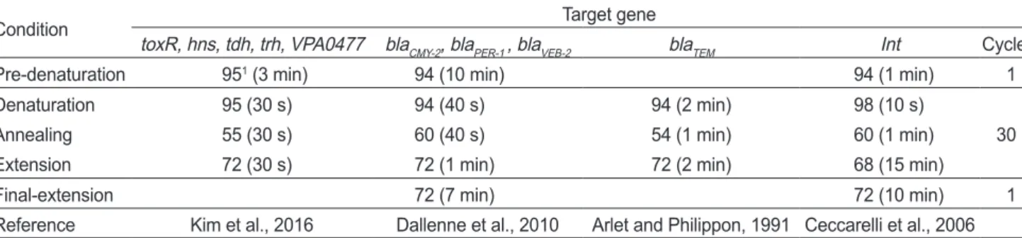
V. parahaemolyticus 검출 kit를이용한 PCR에서표준균주 와동일한 375 bp의산물이생성된균주는대하유래의 SS15- 5, 우렁쉥이유래의 SC19-5와 SC28-5, 그리고바지락유래의 MC23-1이였다(Table 5, Fig. 1). 이들의 16S rRNA 유전자염 기서열을분석한결과, 모두 V. parahaemolyticus와 98% 이상 의상동성을보였다(Data not shown). 한편 V. parahaemolyti- cus의분리동정에이용되는 toxR과 hns 유전자를 target으로 하는 primer로 PCR을실시하였을때에도 4 분리균주와표준 균주(KCTC 2471)는동일한결과를나타냈다(Fig. 2A, 2B). 따 라서생화학적시험과유전자분석결과에근거하여, V. para-
Table 3. Fisheries products collected from fish markets in Yeosu
Fisheries product No. of individual No. of sample1
Fish
Flatfish Paralichthys olivaceus 3 8
Fine spotted flounder Pleuronichthys cornutus 15 19
Rockfish Sebastes schlegeli 4 5
Black scraper Thamnaconus modestus 2 4
Spotty belly greenling Hexagrammos agrammus 1 1
Gizzard shad Konosirus punctatus 4 3
Shellfish
Manila clam Ruditapes philippinarum 86 24
Ark shell Scapharca subcrenata 10 2
Abalone Haliotis discus hannai 2 1
Scallop Patinopecten yessoensis 17 8
Spiny top shell Batillus cornutus 2 1
Crustacea Large shrimp Penaeus orientalis 52 31
Mollusk Sea squirt Halocynthia roretzi 31 31
Total 229 138
11 sample, pool of fisheries products (25 g).
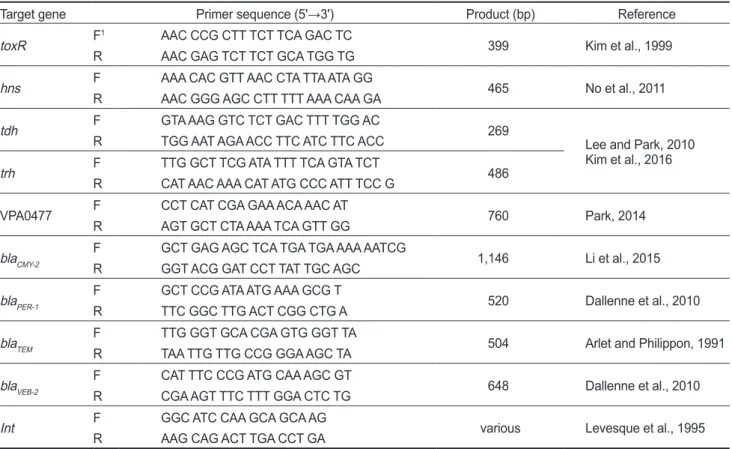
Fig. 1. Detection of Vibrio parahaemolyticus from 17 isolates by PCR using commercial detection kit. Lanes: M, 100 bp size mark- er; K, KCTC 2471 V. parahaemolyticus; 1, FSF 6-4; 2, SS 15-5;
3, SS 16-5; 4, RF 1-2; 5, SC 19-5; 6, MC 23-1; 7, FSF 9-4; 8, FSF 11-4; 9, SS 26-3; 10, SS 27-5; 11, SL 15-5; 12, SL 16-3; 13, SC 28-5; 14, SL 17-1; 15, SL 18-4; 16, SL 18-5; 17, SC 31-5. PCR, polymerase chain reaction; FSF, SS, RF, MC, SL, and SC refer to table 5.
A
D
B
E
C
F Table 2. PCR conditions for the detection of various genes
Condition Target gene
toxR, hns, tdh, trh, VPA0477 blaCMY-2, blaPER-1 , blaVEB-2 blaTEM Int Cycle
Pre-denaturation 951 (3 min) 94 (10 min) 94 (1 min) 1
Denaturation 95 (30 s) 94 (40 s) 94 (2 min) 98 (10 s)
30
Annealing 55 (30 s) 60 (40 s) 54 (1 min) 60 (1 min)
Extension 72 (30 s) 72 (1 min) 72 (2 min) 68 (15 min)
Final-extension 72 (7 min) 72 (10 min) 1
Reference Kim et al., 2016 Dallenne et al., 2010 Arlet and Philippon, 1991 Ceccarelli et al., 2006
1Temperature (°C). PCR, polymerase chain reaction.
haemolyticus는 4균주로확정되었다. TCBS배지에서 청록색
으로나타난집락에대해서는약 4.12%의분리비를나타내지
만시료에서검출되는총세균에대해서본다면더낮은분리비 로계산될것이다. V. parahaemolyticus 분리율을보고한다양 한연구들이있으나(Lee et al., 2007; Ryu et al., 2010; Park et al., 2016; Kang et al., 2017) 비율의단순한수치비교는어렵 다. 시료로사용된생물종류와각지역의해수특성및채집시 기와장소등의차이는물론, 전체시료중 V. parahaemolyticus 가분리된시료의비율, 또는시료중의총세균중 V. parahae- molyticus의출현수를계산하는등기준도다양하다. 본연구 에서식품공전의 V. parahaemolyticus 분류기준과일치하는분 리균은없었으나 8% 염분내성을제외한다른모든특성이식
품공전에서제시한결과와동일한분리균은 5균주였다(1, 4, 5, 12 and 13 in Table 5). 그러나이들중유전자구조를검출하 는 kit에서양성반응을보인균은 2균주뿐이었다(5 and 13 in
Table 5). 그러므로식품공전의기준에따른분류는분리비율
을실제보다높게나타나도록하는경향이있다고보아지며, 수 산물에서분리한균주들중 V. parahaemolyticus를확인할때, 식품공전시험법에더하여특이유전자를검출하는 PCR을실 시하는단계가추가로필요할것으로사료된다.
병독성 인자
병독성인자인 tdh 유전자는 V. parahaemolyticus 표준균주 와바지락에서분리된 MC 23-1 균주에서약 500 bp 크기의밴 드로나타났다(Fig. 2C). 이는 Nishibuchi and Kaper (1995)의 연구에서제시한 269 bp와달랐다. 이 PCR product의염기서 열을분석한결과 V. parahaemolyticus strain ATCC 17802와 FORC_006의 chromosome 2에있는유전자구조(Accession No. CP014047.2 and CP009766.1)와 99% 이상의상동성을 보였다. V. parahaemolyticus의 2번염색체는작은염색체라고 도불리며, 이염색체에는 tdh를포함한여러병원성유전자를 지니고있는 pathogenicity island가존재하는것으로알려져있 다(Makino et al., 2003). 따라서바지락유래의 MC23-1은인 체에대한유해성이있을것으로판단되었다. 그러나 trh 유전자 는표준균주를제외한분리균주에서는모두검출되지않았다 Table 4. Bacteria isolated from various fisheries products on TCBS
medium
Identification No. of isolate Detection rate (%)
Vibrio spp. 49 50.51
Morganella morganii 24 24.74
Shewanella spp. 13 13.4
Other strain 11 11.34
Total 97 100
TCBS, thiosulfate citrate bile salts.
Fig. 2. PCR assay for the detection of various gene structures in Vibrio parahaemolyticus. toxR (A) and hns genes (B) for identification; tdh (C) and trh (D) of virulence gene ; VPA0477 (E) of β-lactamase gene; the class 1 integron structure (F). Lanes: M, 100 bp size marker; 1, KCTC 2471; 2-5, isolates: 2, SS15-5; 3, SC19-5; 4, MC23-1; 5, SC28-5; 6-7, Morganella morganii isolates. SS, SC, and MC refer table 5.
A
D
B
E
C
F
(Fig. 2D). Lee and Park (2010)은해수에서분리한 V. parahae- molyticus 28 균주로부터 tdh와trh 유전자가모두검출되지않 았다고보고하였으며, Oh et al. (2011)은양식어류에서분리한 V. parahaemolyticus에서 tdh와trh 유전자는각각전체의 5.5%
와 0.5% 비율로검출되었다고보고한바가있다. 또한 Park et al. (2016)은패류양식장에서분리한 V. parahaemolyticus의병 원성유전자를확인한결과총 121 균주중 tdh 유전자는 2 균 주, trh 유전자는 8 균주에서검출되었다고보고하였고, Kang
et al. (2017)은시판되는패류에서분리한 V. parahaemolyticus 중 tdh 유전자를가지고있는균주는전체 9.1%이고 trh 유전자 는검출되지않았다고보고하였다. 이와같은결과는해수나각 종수산물에서 V. parahaemolyticus의검출율이높게나왔다할 지라도실제사람에게식중독을일으킬수있는 V. parahaemo- lyticus는훨씬적다는것을의미한다할수있으나, 계절과시 료의종류및채집장소에따른변동이심하므로지속적인모니 터링이필요하다.
Table 6. Drug susceptibility of Vibrio parahaemolyticus isolated from fisheries products
Drug (μg/disc) Isolate
SS15-5 SC19-5 MC23-1 SC28-5
β-lactams
Ceftazidime (30) S1 S S S
Cefotaxime (30) S S S S
Imipenem (10) S S S S
Amoxicillin (10) R R R R
Ampicillin (10) R R R R
Aminoglycosides Amikacin (30) S S S S
Gentamycin (10) S I S S
Polypeptides Colistin (10) R R S R
Quinolones Ciprofloxacin (5) S S S S
Nalidixic acid (30) S S S S
Macrolides Erythromycin (15) I I I I
Tetracyclines Oxytetracycline (30) S S S S
Tetracycline (30) S S S S
1S, Susceptibility; I, Intermediate; R, Resistance.
Table 5. Characterization to confirm Vibrio parahaemolyticus among 17 isolates Standard1 KCTC
2471 Isolate
12 2 3 4 5 6 7 8 9 10 11 12 13 14 15 16 17
Salinity tolerance (%)
0 - - - - - - - - - - - - - - - - - - -
3 + + + + + + + + + + + + + + + + + + +
8 + - - - - - - - - - - - - - - - - - -
10 - - - - - - - - - - - - - - - - - - -
VP - - - - - - - - - - - - - - - - - - -
Mannitol + + + - + + + - - - - - - + + - - - +
Arginine - - - + - - - + + + + + + - - + + + -
Ornithine + + + - - + + - - - - - - + + - - - -
ONPG - - - - - - - - - - - - - - - - - - -
Growth at 42°C + - + + + + + + + + + + + + + - + - -
1V. parahaemolyticus standard of Korean Food Standards Codex. 21-17, Isolates presumed as V. parahaemolyticus by biochemical tests: 1, fine spotted flounder (FSF)6-4; 2, large shrimp (SS)15-5; 3, SS16-5; 4, rockfish (RF)1-2; 5, sea squirt (SC)19-5; 6, manila clam (MC)23-1;
7, FSF9-4; 8, FSF11-4; 9, SS26-3; 10, SS27-5; 11, scallop (SL)15-5; 12, SL16-3; 13, SC28-5; 14, SL17-1; 15, SL18-4; 16, SL18-5; 17, SC31-5. The isolates 2, 5, 6, and 13 were identified as V. parahaemolyticus by PCR using commercial detection kit. ONPG, ornithine, O-nitrophenyl-β-D-galacto pyranoside; VP, Voges-Proskauer.
항균제 내성과 β-lactamase 유전자
V. parahaemolyticus 4 분리균주들은의료관련감염병관리 에주요한항균제들중 AMP, AML과 CT를제외한모든항균 제에대해감수성을보였다(Table 6). Penicillin 계통의항균제 에대한 V. parahaemolyticus의내성에관해서는많은연구보 고가있다. Ryu et al. (2017)은패류에서분리한 V. parahae- molyticus의 AMP 내성율을 41.8%로보고하였지만, Lee et al.
(2007)과 Ryu et al. (2010)은어패류에서분리한 V. parahae- molyticus 균주의 AMP 내성을각각 100%, 95.2%로보고한바 있다. 한편본연구의분리균에서 β-lactamase 유전자를확인 한결과 VPA0477은모든균주에서 760 bp 크기의특이밴드로 나타났으나, blaPER-1과 blaTEM은각각 2개의비특이밴드로나타 났고 blaCMY-2,과 blaVEB-2는검출되지않았다(Fig. 2E, Table 7).
VPA0477 증폭산물인 760 bp의 염기서열을분석한결과, blaCARB 유전자와 99% 이상의 상동성을 보였다. blaCARB는 carbenicillin계약물을가수분해하는효소(Labia et al., 1981) 인 class A β-lactamase에 속하는 CARB β-lactamase유전자 이다. 따라서본 연구에서 분리된V. parahaemolyticus는모 두 class A β-lactamase에의하여저항성을나타내는것으로판 단되었으며이는 Lee and Park (2010)이 AMP 분해유전자인 β-lactamase와상동성이있는유전자 VPA0477이 V. parahae- molyticus의 AMP 내성에관여한다고보고한것과일치하였다.
한편 blaPER-1 유전자 PCR 결과형성된비특이밴드들은염기서
열분석에서모두 V. parahaemolyticus 2번염색체에있는유 전자구조와 99% 이상의상동성이있고, blaTEM의비특이밴드
들은 1번염색체의유전자구조와 99% 이상의상동성을보였
다(Data not shown). 따라서이들이본분리균의항균제내성 과어떤관련이있는지에대해서는추가검토가필요하다. 또한 class 1 integron 구조가 V. parahaemolyticus 표준균주(KCTC 2471)와 4개의분리균주에서모두 750 bp의 DNA 밴드로검출 되었는데(Fig. 2F) 이들의구조확인과 V. parahaemolyticus의 내성전달과의관련성에대해서도추가연구가필요하다.
아미노산 서열의 유사도에 근거한 β-lactamase 분류에서
(Ambler, 1980; Bush and Jacoby. 2010), Class A β-lactamase 는 β-lactam계항균제의 target 부위인 DD-peptidase와구조 적유사성을가지며(Ghuysen, 1994), 활성메커니즘에서 serin 이중심활성부위로사용된다. 따라서본연구에서분리한 V.
parahaemolyticus의 β-lactamase는활성을위해 2가양이온은 필요로하지않는것으로생각된다.
한편 Pazhani et al. (2014)은임상분리균인 V. parahaemo- lyticus를대상으로한실험에서 AMP 내성인균주뿐아니라 감수성인균주에서도 VPA0477 유전자가검출되어, VPA0477 과 AMP 내성과의연관성에의문을제기한바있다. V. parahae- molyticus의 AMP 내성은고도내성으로서빈번히발생하고있 는실정이지만아직까지명확한발생기전은밝혀지지않고여 러주장이나오고있는상황으로앞으로추가적인연구가필요 할것으로사료된다.
이상의결과들로부터 수산물에서 분리되는 V. parahaemo-
lyticus에의해식중독을일으킬가능성은분리비율보다낮다
고볼수있으며, AMP, AML, CT 이외의의료용약물에대해서 는감수성이었으므로감염시항균제에의한치료가비교적용 이할것으로생각되나 class 1 integron 구조가확인됨으로써다 약제내성으로발전할가능성이높다고판단된다.
References
Ambler RP. 1980. The structure of β-lactamases. Phil Trans R Soc Lond B289, 321-331. https://doi.org/10.1098/
rstb.1980.0049.
Arlet G and Philippon A. 1991. Construction by polymerase chain reaction and intragenic DNA probes for three main types of transferable β-lactamases (TEM, SHV, CARB). FEMS Microbiol Lett 82, 19-26. https://doi.
org/10.1111/j.1574-6968.1991.tb04833.x.
Barcelos DHF, Knidel C and Fernandes CGL. 2018. Emer- gence and dispersion of resistance genes by the aquatic en- vironment: a review. Pollution 4, 305-315. https://doi.org/
10.22059/poll.2017.242030.320.
Bush K and Jacoby GA. 2010. Updated functional classification of β-lactamases. Antimicrob Agents Chemother 54, 969- 976. https://doi.org/10.1128/AAC.01009-09.
Ceccarelli D, Salvia AM, Sami J and Cappuccinelli P. 2006.
New cluster pf plasmid-located class I integrons in Vibrio cholera O1 and a dfrA15 cassette containing integron in V.
parahaemolyticus isolated in Angola. Antimicrob Agents Chemother 50, 2493-2499.
CLSI (Clinical and Laboratory Standards Institute). 2013. Per- formance standards for antimicrobial susceptibility testing.
CLSI document M100-S23 (M02-A11). CLSI, Wayne, PA, U.S.A.
Dallenne C, Costa AD, Decre D, Favier C and Arlet G. 2010.
Development of a set of multiplex PCR assays for the de- Table 7. PCR results for the detection of various β-lactamase genes
in V. parahaemolyticus isolated from commercial fisheries prod- ucts
β-lactamase Strain
KCTC2471 SS15-5 SC19-5 MC23-1 SC28-5 VPA0477
(760 bp) + + + + +
blaCMY-2 - - - - -
blaPER-1 NS1 NS NS NS NS
blaTEM NS NS NS NS NS
blaVEB-2 - - - - -
1non-specific bands.
tection of genes encoding important β-lactamases in Entero- bacteriaceae. J Antimicrob Chemother 65, 490-495. https://
doi.org/10.1093/jac/dkp498.
Ghuysen JM. 1994. Molecular structures of penicillin-binding proteins and β-lactamases. Trends Microbiol 2, 372-380.
Kang CH, Shin YJ, Jang SC, Yu HS, Kim SK, An S, Park K and So JS. 2017. Characterization of Vibrio parahaemolyticus isolated from oysters in Korea: Resistance to various antibi- otics and prevalence of virulence genes. Mar Pollut Bull 118, 261-266. https://doi.org/10.1016/j.marpolbul.2017.02.070.
Kim YB, Okuda J, Matsumoto C, Takahashi N, Hashimoto S and Nishibuchi M. 1999. Identification of Vibrio parahae- molyticus strains at the speices level by PCR targeted to the toxR gene. J Clin Microbiol 37, 1173-1177.
Kim TO, Um IS, Kim HD and Park KS. 2016. Antimicrobial resistance and minimum inhibitory concentrations of Vib- rio parahaemolyticus strains isolated from Gomso bay, Korea. Korean J Fish Aquat Sci 49, 582-588. http://dx.doi.
org/10.5657/KFAS.2016.0582.
KCDC (Korea Centers for Disease Control and prevention).
2016. One health collaboration project proposal. Retrieved from http://www.prism.go.kr/homepage/entire/retrieveEn- tireDetail.do?research_id=1351000-201700044 on Jun 5, 2018.
KFDA (Korea Food and Drug Administration). 1999. Status of food poisoning and measures. Food industry 72-84. Re- trieved from http://www.ndsl.kr/ndsl/search/detail/article/
articleSearchResultDetail.do?cn=JAKO199968317949050 on Mar 27, 2019.
Labia R, Guionie M, Barthelemy M and Pilippon A. 1981.
Properties of three carbenicillin-hydrolysing β-lactamases (CARB) from Pseudomonas aeruginosa: identification of a new enzyme. J Antimicrob Chemother 7, 49-56. https://doi.
org/10.1093/jac/7.1.49.
Lee H, Oh YH, Park SG and Choi SM. 2007. Antibiotic sus- ceptibility and distribution of Vibrio parahaemolyticus iso- lated from the seafood. Kor J Env Hlth 33, 16-20. https://doi.
org/10.5668/JEHS.2007.33.1.016.
Lee KW and Park KS. 2010. Antibiotic-resistance profiles and the identification of the ampicillin-resistance gene of Vibrio parahaemolyticus isolated from seawater. Ko- rean J Fish Aquat Sci 43, 637-641. https://doi.org/10.5657/
kfas.2010.43.6.637.
Levesque C, Piche L, Larose C and Roy PH. 1995. PCR map- ping of integrons reveals several novel combinations of re- sistance genes. Antimicrob Agents Chemother 39, 185-191.
https://doi.org/10.1128/AAC.39.1.185.
Li R, Lin D, Chen K, Wong MHY and Chen S. 2015. First detec- tion of AmpC β-lactamase blaCMY-2 on a conjugative IncA/C plasmid in a Vibrio parahaemolyticus isolates of food origin.
Antimicrob Agents Chemother 59, 4106-4111. http://dx.doi.
org/10.1128 /AAC.05008-14.
Liu M, Wong MHY and Chen S. 2013. Molecular characterisa- tion of a multidrug resistance conjugative plasmid from Vib- rio parahaemolyticus. Int J Microbiol Agents 42, 575-579.
https://doi.org/10.1016/j.ijantimicag.2013.08.014.
Makino K, Oshima K, Kurokawa K, Yokoyama K, Uda T, Tagomori K, Iijima Y, Najima M, Nakano M, Yamashita A, Kubota Y, Kimura S, Yasunaga T, Honda T, Shinagawa H, Hattori M and Iida T. 2003. Genome sequence of Vib- rio parahaemolyticus: a pathogenic mechanism distinct from that of V. cholerae. Lancet 361, 743-749. https://doi.
org/10.1016/S0140-6736(03)12659-1.
MFDS (Ministry of Food and Drug Safety). 2016. Notice on full revision of food standards and regulations, Chapter 7, Gen- eral test methods (No. 2016-154), 167-212. Retrieved from http://www.mfds.go.kr/brd/m_207/view.do?seq=11502&s on Dec 31, 2018.
MFDS (Ministry of Food and Drug Safety). 2018. Occurrence of food poisoning by causes and patients - year and city: 2008- 2017. Retrieved from http://www.mfds.go.kr/wpge/m_312/
de010603l0001.do on Oct 6, 2019.
Miyamoto Y, Kato T, Obara Y, Akiyama S, Takizawa K and Yamai S. 1969. In vitro hemolytic characteristic of Vibrio parahaemolyticus: its close correlation with human patho- genicity. J Bacterial 100, 1147-1149.
NIFS (National Institute of Fisheries Science). 2017. Antibiotic resistant bacteria inspection manual (11-1192266-000195- 01). GMK communication, Busan, Korea, 34-38.
Nishibuchi M and Kaper JB. 1995. Thermostable direct hemo- lysin gene of Vibrio parahaemolyticus: a virulence gene ac- quired by a marine bacterium. Infect Immun 63, 2093-2099.
No AR, Okada K, Kogure K and Park KS. 2011. Rapid detec- tion of Vibrio parahaemolyticus by PCR targeted to the histone-like nucleoid structure (H-NS) gene and its genetic characterization. Lett Appl Microbiol 53, 127-133. https://
doi.org/10.1111/j.1472-765X.2011.03072.x.
Oh EG, Son KT, Yu H, Lee TS, Lee HJ, Shin S, Kwon JY, Park K and Kim J. 2011. Antimicrobial resistance of Vibrio para- haemolyticus and Vibrio alginolyticus strains isolated from farmed fish in Korea from 2005 through 2007. J Food Prot 74, 380-386. https://doi.org/10.4315/0362-028X.JFP-10- 307.
Park KS. 2014. Application of the β-lactamase (VPA0477) gene for the detection of Vibrio parahaemolyticus by polymerase chain reaction. Korean J Fish Aquat Sci 47, 740-744. http://
dx.doi.org/10.5657/KFAS.2014.0740.
Park YS, Park K, Kwon JY, Yu HS, Lee HJ, Kim JH, Lee TS and Kim PH. 2016. Antimicrobial resistance and distribu- tion of virulence factors of Vibrio parahaemolyticus isolated from shellfish farms on the southern coast of Korea. Ko- rean J Fish Aquat Sci 49, 460-466. https://doi.org/10.5657/
KFAS.2016.0460.
Pazhani GP, Bhowmik SK, Ghosh S, Guin S, Dutta S, Rajendran
K, Saha DR, Nandy RK, Bhattacharya MK, Mukhopadhyay AK and Ramamurthy T. 2014. Trend in the epidemiology of pandemic and nonpandemic strain of Vibrio parahaemolyti- cus isolated from diarrheal patients in Kolkata, India. PLoS Negl Trop Dis 8, e2815. https://doi.org/10.1371/journal.
pntd.0002815.
Ryu AR, Park K, Kim SH, Ham IT, Kwon JY, Kim JH, Yu HS, Lee HJ and Mok JS. 2017. Antimicrobial resistance patterns of Escherichia coli and Vibrio parahaemolyticus isolated from shellfish from the west coast of Korea. Ko- rean J Fish Aquat Sci 50, 662-668. https://doi.org/ 10.5657/
KFAS.2017.0662.
Ryu SH, Hwang YO, Park SG and Lee YK. 2010. Antibiotic susceptibility of Vibrio parahaemolyticus isolated from commercial marine products. Korean J Food Sci Technol 42, 508-513.
Sakazaki R, Tamura K, Kato T, Yamai S and Hobo K. 1968.
Studies of the enteropathogenic, facultatively halophilic bacteria, Vibrio parahaemolyticus. III. Enteropathogenicity.
Jpn J Med Sci Biol 21, 325-331.
Weisburg WG, Barns SM, Pelletier DA and Lane DJ. 1991. 16S ribosomal DNA amplification for phylogenetic study. J Bac- teriol 173, 679-703. https://doi.org/10.1128/jb.173.2.697- 703.1991.
Yeung PSM and Boor KJ. 2004. Epidemiology, pathogenesis, and prevention of foodborne Vibrio parahaemolyticus.
Foodborne Pathog Dis 1, 74-88.