116
Copyright © 2016 The Korean Society of Fisheries and Aquatic Science pISSN:0374-8111, eISSN:2287-8815
서 론
장염비브리오균
(Vibrio parahaemolyticus)
은그람음성,
무포 자,
호염성간균으로기수와해양환경에널리분포하는자연상 재세균으로부적절하게취급된수산물을생식하거나덜익혀 섭취하여발생하는급성장염의주요원인균이다(Su and Liu, 2007; Gode-Potratz et al., 2011; Ceccarelli et al., 2013; Zarei et al., 2012; Zhang and Orth, 2013; KFDA, 2016).
과거에는 표현형과생화학적특성을바탕으로장염비브리오균을동정하 였으며수산물과해양환경에서분리된장염비브리오균총균 수를근거로잠재적위해를추정하였다(Malcolm et al., 2015).
그러나
PCR
과같은분자생물학적기법을발달로장염비브리 오균에서병원성과관련된독소유전자가확인이가능하게되 었는데,
이독소유전자는thermostable direct hemolysin (tdh) gene
과TDH related hemolysin (trh) gene
이며숙주세포에대 한 용혈과세포독성을유발하는 것으로밝혀졌다(Broberg et al., 2011; Zhang and Orth, 2013; Letchumanan et al., 2015).
그래서수산물에서장염비브리오균위해평가를실시하거나기 준규격을설정하는경우특정수산물에서독소유전자보유장 염비브리오균출현빈도를고려하고있다
.
일반적으로비브리오균은대부분의임상용항균제에감수성 이큰것으로알려져있다
(Shaw et al., 2014; Yu et al., 2014).
양식 굴(Crassostrea gigas)에서 분리된 장염비브리오균의 독소 유전자 보유 및 항균제 감수성
김수경·안세라·박보미·오은경
1·송기철·김정완
2·유홍식*
국립수산과학원 서해수산연구소, 1국립수산과학원 남해수산연구소, 2인천대학교 생명과학과
Virulence Factors and Antimicrobial Susceptibility of
Vibrio parahaemolyticus Isolated from the Oyster Crassostrea gigas
Sukyung Kim, Sera An, Bomi Park, Eun-Gyoung Oh
1
, Ki Cheol Song, Kim Jung-Wan2
and Hongsik Yu*West Sea Fisheries Research Institute, National Institute of Fisheries Science, Incheon 22383, Korea
1
South Sea Fisheries Research Institute, National Institute of Fisheries Science, Yeosu 59780, Korea
2
Division of Bioenginnering, University of Incheon, Incheon 22012, Korea
This study investigated the prevalence of Vibrio parahaemolyticus in the oyster Crassostrea gigas , which is com- monly consumed raw. The presence of virulence factors and the antimicrobial susceptibility of isolates were also investigated. The overall prevalence rate of V . parahaemolyticus in oysters was 37.5% (36/96) and the range of con- centrations was 30–11,000 MPN/100 g. PCR-based assays indicated that 9.6% (11/115) of the isolates were positive for the thermostable direct hemolysin-related hemolysin gene ( trh ), while none of the isolates were positive for the thermostable direct hemolysin gene ( tdh ). The Multiple Antibiotics Resistance (MAR) index was measured for 16 common antimicrobial agents and 46.1% (53/115) of the isolates had a MAR index > 0.2. The MAR index ranged from 0.07 to 0.73. The highest MAR index was observed with strain s150608, isolated in June 2015, which exhibited resistance to 11 antimicrobial agents. Our results demonstrate that oysters are high-risk sources of V . parahaemolyti- cus , although no antimicrobial agent was being used to promote growth or to treat bacterial infections in the sampled oyster-growing areas.
Key words: Vibrio parahaemolyticus , Oyster, Virulence factors, Antimicrobial susceptibility, MAR index
This is an Open Access article distributed under the terms of the Creative Commons Attribution Non-Commercial Licens (http://creativecommons.org/licenses/by-nc/3.0/) which permits unrestricted non-commercial use, distribution, and reproduction in any medium, provided the original work is properly cited.
http://dx.doi.org/10.5657/KFAS.2016.0116 Korean J Fish Aquat Sci 49(2) 116-123, April 2016
Received 29 February 2016; Revised 30 March 2016; Accepted 30 March 2016
*Corresponding author: Tel: +82. 32. -745. 0751 Fax: +82. 32. 745. 0619
E-mail address: email; [email protected]
그러나오랜기간임상
,
농업및수산양식분야에서항균제와화 학약품을과도하게사용함에따라항균제내성균주가환경에 유입되고있다는주장이제기되고있다(Cabello et al., 2013).
이러한주장은아시아지역수산양식분야에서는
oxytetracy- cline, tetracycline, quinolone, sulphonamides
와trimethoprim
등이생산성향상을위해지속적인사용되어왔고,
항균제내성 장염비브리오균이우리나라를비롯한태국,
말레이지아와중 국등에서분리되어있다는보고를근거로한것이다(Oh et al., 2011; Rico et al., 2012; Sani et al., 2013; Xu et al., 2014; Yano et al., 2014; Yu et al., 2014).
또한환경에서분리된장염비브리 오균을비롯한잠재적병원성세균이임상용항균제에대해서도내성을가질것이라는우려가최근확산되고있다
(Okamoto
et al., 2009; Letchumanan et al., 2015).
이는국가에따라차 이가있으나비브리오균감염증치료를위해임상에서사용되 는항균제에cephalothin, cefuroxime, cefotaxime ceftazidime, tetracycline, doxycycline
과fluoroquinolone
등이포함되어있 으며일부는수산양식분야에사용되는항균제와동일하거나 유사한계열이기때문이다(Tang et al., 2002).
이연구에서는주거지역과자연하천등육상오염원과인접한 개펄에서양식되고생식을위주로소비되고있는서해안양식 산굴의위생안전확보와항생제내성관리에필요한자료를확 보하고자장염비브리오균분포와병인인자보유여부및항생 제내성특성에대하여검토하였다
.
재료 및 방법
패류 시료
장염비브리오균
(Vibrio parahaemolyticus)
분리를 위하여2014
년1
월부터2015
년12
월까지서해안패류생산해역에설 정된4
개지점(
인천시옹진군덕적면및자월면연안)
에서양식 굴(Crassostrea gigas)
을매월채취하여시료로하였다.
패류시료는멸균된용기에채취한후
10℃
이하로유지하면서실험실로운반하여사용하였다
.
시험에사용된굴시료수는총96
개였 다.
시료채취시해수의수온과염분농도는YSI 556 multi- probe system (Yellow Springs, YSI Life Science, OH, USA)
을사용하여현장에서측정하였다.
장염비브리오균의 분리·동정 및 정량
장염비브리오균은
Bacteriological Analytical Manual
의방법 에따라3 tube MPN
법으로정량분석을실시하였다(Elliot et al., 2005).
패각을제거한후패육과패액200 g
에phosphate buffered saline (PBS: 140 mM NaCl, 5 mM anhydrous Na
2H- PO
4, and 1.5 mM KH
2PO
4pH 7.4)
을2
배비율로첨가하여균 질화하였다.
그리고80 mL
의PBS
에균질액20 g
을첨가하여10
배희석액을만들어시료로하였다.
희석액을이용하여100
배, 1,000
배등단계별추가희석용액을만들어사용하였다.
각희석단계별로
alkaline peptone water (pH 8.5±0.2, 2% NaCl
함유)
가들어있는3
개의시험관에각각접종하고35±0.5℃
에 서18-24
시간증균배양하였다.
배양한후Thiosulfate Citrate Bile Salt Agar (TCBS agar, Difco)
에획선도말하여, 35℃
에서
18-24
시간배양하였다.
전형적인청록색집락을도말하여Triple Sugar Iron Agar (Difco)
에접종하고35℃
에서24
시간 배양한후,
전형적인반응을나타내는균주를대상으로동정시 험을실시하였다.
동정시험에는내염성시험, 42℃
발육시험, ONPG (O-nitrophenyl-β-D-galacto-pyranoside)
시험및API 20E system (BioMerieux, Marcy-l'Etoile, France)
을이용하였 으며, 16S rDNA
부분염기서열을분석하여GeneBank
에등 록되어있는장염비브리오균의16S rDNA
염기서열과상동성 을비교하여 확정하였다.
동정결과장염비브리오균으로확정 된경우해당분리주가유래한희석단계별증균시험관을장염 비브리오균양성시험관으로하여그수를헤아려최확수표에 서정량치를얻고희석배율을곱하여100 g
당MPN
으로환산 한후정량치로하였다.
16S rDNA 및 독소 유전자 확인
16S rDNA
상동성비교및독소유전자확인을위하여분리균주를
2%
식염이함유된Triple Soy Broth (TSB, Oxoid, U.K.) 10 mL
에접종하여37℃
에서18-24
시간배양한후QIAamp DNA Mini Kit (Qiagen, Hilden, Germany)
를이용하여DNA
를추출하였다.
분리된DNA
는사용할때까지-18℃
에보관 하였다.
분리된DNA
로부터16S rDNA
증폭에는universal primer (Macrogen, Korea)
을사용하였으며독소유전자인tdh
및trh gene
의증폭에는각각VPD-1/VPD-2
와VPR-1/VPR-2 (Takara, Japan)
를사용하였다. PCR
은주형DNA 5 μL, deoxy- nucleoside triphosphate mixture (1 mM dATP, 1 mM dCTP, 1 mM dTTP, 1 mM dGTP) 2 μL, 10×PCR
용액5 μL, primer (100 pmol/μL) 1 μL, Taq polymerase 1.25 U
를혼합한후,
총 부피를50 μL
로조정하고Thermal Cycle (Takara, Japan)
로94℃
에서60
초, 60℃
에서60
초, 72℃
에서60
초의 조건에서35
회반복하여실시하였다.
증폭된PCR
산물은DE/QIAxcel advanced system (Qiagen, Germany)
를이용하여amplicon
크 기를확인하였다(16S rDNA, 663 bp; tdh, 251 bp; trh, 250 bp).
항균제 감수성 시험
분리동정된장염비브리오균의항균제감수성평가에는
Acar and Goldstein (1991)
의디스크확산법을이용하였다.
각균주 를Muller Hinton Broth (Merck, Germany)
에접종한후35℃
에서
18-24
시간전배양한다음균배양액의농도를McFarland
No. 0.5
로희석조정하였다.
희석된균배양액을미리1%
농 도가되도록식염을첨가한두께4 mm
의Muller Hinton Agar
(Merck, Germany)
평판에도말한후실온에5
분간방치하여 균액을흡수시키고항균제디스크(φ 8 mm)
를균접종후15
분이내에고착시켰다.
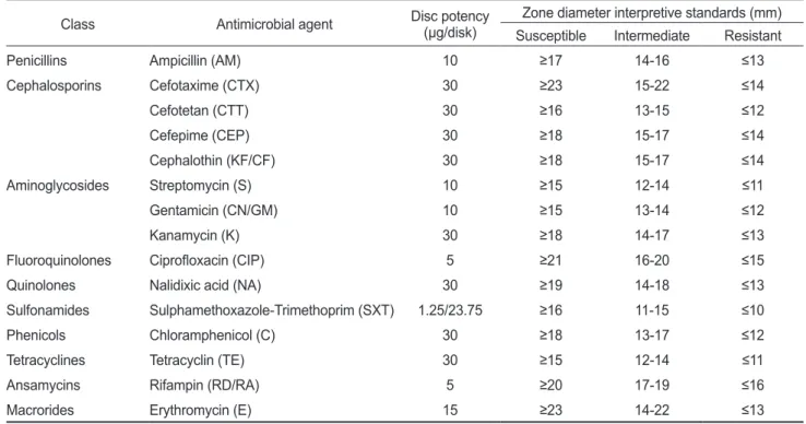
시험항균제는ampicillin
을비롯한15
종
10
계열을사용하였다(Table 1).
항균제디스크를고착시킨Muller Hinton Agar
평판은35℃, 16-18
시간배양한다음균 의증식저해대(inhibition zone)
의크기를caliper
로측정하여Table 1
에제시된기준(CLSI, 2015)
에따라감수성을판정하였 다.
감수성결과의정도관리를위하여Escherichia coli ATCC
25922
를대조균으로사용하여각항균제디스크에대한역가를확인하였다
.
MAR (Multiple Antibiotic Resistance) Index
분리균주의항균제에대한동시내성정도를비교하기위하여 분리균주에대한MAR index
를다음계산식에따라산출하였 다(Krumperman, 1983).
MAR
index= r/t
여기서
“r”
은분리균주가내성을나타낸항균제의개수, “t”
는 시험에사용된항균제의총개수이다.
결과 및 고찰
장염비브리오균 검출 현황 및 독소 유전자 보유율
장염비브리오균은전세계적으로분포하는식중독세균의일 종으로환경과수산물에서분포및농도는계절,
지역,
시료의유형과적용분석법에 따라다르게보고되고있다
(Martinez-
Urtaza et al., 2008; Zarei et al., 2012).
이연구에서는 서해 안산양식굴
(Crassostrea gigas) (n=96)
을대상으로alkaline
peptone water
에증균하여선택배지를사용하여분리한후표 현형과유전형분석을통하여동정하고최확수법으로정량하여 조사를실시하였다.
장염비브리오균으로추정되는총390
개의 균주가분리되었으며115
개균주가최종적으로확정되었다.
장 염비브리오균으로확정된모든분리주의16S rDNA
부분염기 서열은GeneBAnk
에등록된Vibrio parahaemolyticus RIMD 2210633
의16S rDNA
염기서열과99%
이상의상동성을보였 다(Makino et al., 2003).
2014
년도에는7
월부터10
월까지채집된양식굴에서검출되 었고 동절기에는 장염비브리오균이 검출되지 않았으며(<30 MPN/100 g), 2015
년도에는6
월부터10
월에까지채집된양식굴에서검출되었다
(Fig. 1).
장염비브리오균이검출된시기의 수온은20℃
이상이었다.
시료중연중평균검출율은2014
년에33.3% (16/48)
그리고2015
년에41.6% (20/48)
에달했 다.
그리고채취지점별검출율에는차이가없었다.
양식굴에 서장염비브리오균의농도는30-11,000 MPN/100 g
의범위를 나타내었으며2015
년(30-4,600 MPN/100 g)
에 비하여2014
년(62-11,000 MPN/100 g)
에다소큰검출범위를나타내었다(Fig. 1).
이연구에서장염비브리오검출율은인도의다양한수산물에 서장염비브리오검출율이
48-55%
였다는보고와중국의어패 류에서검출율이47.2%
였다는보고보다다소낮은것이며,
이 전에수행한연구에서남해안양식굴에서장염비브리오균검 출율을72.5%
로보고한것과많은차이가있었다(Chakraborty et al., 2008; Chao et al., 2009; Yu et al., 2014; Letchumanen
Table 1. Tested antimicrobial agents and breakpoints for the interpretation of the disk diffusion method
Class Antimicrobial agent Disc potency
(μg/disk) Zone diameter interpretive standards (mm) Susceptible Intermediate Resistant Penicillins
Cephalosporins
Aminoglycosides
Fluoroquinolones Quinolones Sulfonamides Phenicols Tetracyclines Ansamycins Macrorides
Ampicillin (AM) Cefotaxime (CTX) Cefotetan (CTT) Cefepime (CEP) Cephalothin (KF/CF) Streptomycin (S) Gentamicin (CN/GM) Kanamycin (K) Ciprofloxacin (CIP) Nalidixic acid (NA)
Sulphamethoxazole-Trimethoprim (SXT) Chloramphenicol (C)
Tetracyclin (TE) Rifampin (RD/RA) Erythromycin (E)
10 30 30 30 30 10 10 30 5 30 1.25/23.75
30 30 5 15
≥17
≥23
≥16
≥18
≥18
≥15
≥15
≥18
≥21
≥19
≥16
≥18
≥15
≥20
≥23
14-16 15-22 13-15 15-17 15-17 12-14 13-14 14-17 16-20 14-18 11-15 13-17 12-14 17-19 14-22
≤13
≤14
≤12
≤14
≤14
≤11
≤12
≤13
≤15
≤13
≤10
≤12
≤11
≤16
≤13
et al., 2015).
또한미국의Chesapeake Bay
에서채취한굴시 료에서 장염비브리오균 검출율이100%
였다는 보고도 있다(Parveen et al., 2008).
이는수산물에축적되는장염비브리오 균의농도가다양한연안해역의환경에영향을받으며통계적 유의성이확보될만큼충분한시료를확보하지못하는데기인 하는여겨진다(WHO/FAO, 2002; Kirs et al., 2011).
한편
,
분리된장염비브리오균115
균주중독소유전자(tdh/
trh gene)
보유율을조사한결과,
내열성용혈독소유전자인tdh gene
을보유한균주는없었고유사용혈독소유전자인trh gene
을보유한분리균은11
균주(9.6%)
로나타났다(Table 2).
유럽과아시아지역에서이루어진조사에의하면
tdh
및trh
gene
보유장염비브리오균의비율이약0-6%
정도라고 보고되고있다
(DePaola et al., 2000; Vuddhakul et al., 2000; Alam et al., 2002; Hervio-Heathetal., 2002; Haley et al., 2014; Yu et
al., 2014).
일반적으로장염비브리오균이수산물에서흔히검출되나모든균주가병원성을가지는것은아니라고알려져있 는데이는환경분리주중
tdh
및trh gene
등병인인자보유율이 다른병원성세균에비하여높지않기때문이다(Panicker et al., 2004; Deepanjali et al., 2005; Canizalez-Roman et al., 2011;
Gutierrez West et al., 2013; Johnson et al., 2008; Yamamoto et al., 2008; Malcolm et al., 2015).
그러나우리나라는수산물을생식하는식습관을가지고있어하절기를중심으로장염비브리 오균에의한식중독이지속적으로발생하고있기때문에
,
지역 별수산물품종별총장염비브리오균및독소유전자보유균주 출현율에대한지속적인모니터링과위해분석이필요할것으로 사료된다(KFDA, 2016).
항균제 감수성
이 연구에서는 미국
CDC
가 비브리오 감염증치료에사용 을 권고한cefotaxime, gentamicin, sulfamethoxazole-trime- thoprim
등16
종의항균제(Table 1)
를이용하여 분리된장염 비브리오균의항균제에대한감수성을조사하였다(Daniels et al., 2000; Shaw et al., 2014).
분리균주의94.8%
가ampicillin
에 내성을나타내었으며, cephalothin (53.9%), streptomycin
Fig. 1. Monthly prevalence of Vibrio parahaemolyticus in oystersCrassostrea gigas collected at the west coast of Korea. Limit of the detection = 30 MPN/100 g.
V. parahaemolyticus (MPN/100 g)
30 1000 2000 3000 4000 5000 6000 7000 8000 9000 10000 11000 12000
Water Temp. ( ℃ )
0 5 10 15 20 25 30 V. parahaemolyticus Water temperature
Jan Mar May Jul Sep Nov Jan Mar May Jul Sep Nov
Median Average
2014 2015
Table 3. Antimicrobial resistance of V. parahaemolyticus isolates from oysters Crassostrea gigas
Antimicrobial agent No. of isolates (%)1 Susceptible Intermediate Resistant Ampicillin (AM)
Cefotaxime (CTX) Cefotetan (CTT) Cefepime (CEP) Cephalothin (KF/CF) Streptomycin (S) Gentamicin (CN/GM) Kanamycin (K) Ciprofloxacin (CIP) Nalidixic acid (NA) Sulphamethoxazole- Trimethoprim (SXT) Chloramphenicol (C) Tetracyclin (TE) Rifampin (RD/RA) Erythromycin (E)
2 (1.7) 87 (75.7) 98 (85.2) 101 (87.8) 20 (17.4) 46 (40.0) 78 (67.8) 30 (26.1) 71 (61.7) 111 (96.6) 109 (94.8)
113 (98.3) 100 (87.0) 18 (15.7) 1 (0.9)
4 (3.5) 28 (24.3) 17 (14.8) 10 (8.7) 33 (28.7) 0 (0) 29 (25.2) 73 (63.5) 39 (33.9) 2 (1.7) 6 (5.2)
2 (1.7) 14 (12.1) 42 (36.5) 98 (85.2)
109 (94.8) 0 (0) 0 (0) 4 (3.5) 62 (53.9) 69 (60.0) 8 (7.0) 12 (0.4) 5 (4.4) 2 (1.7) 0 (0)
0 (0) 1 (0.9) 55 (47.8) 16 (13.9)
1the ratio of the numbers of strains susceptible/intermediate resis- tant/resistant to the tested antimicrobial agents to total numbers of tested isolates expressed as a fraction of 100.
Table 2. Presence of virulence factors in V. parahaemolyticus isolates from oysters Crassostrea gigas No. of isolates
2014 2015 Total
Jul Aug Sep Oct Jun Jul Aug Sep Oct (%)
trh 0 5 2 4 0 0 0 0 0 11 (9.6%)
tdh 0 0 0 0 0 0 0 0 0 0 (0.0%)
None 16 14 21 9 3 11 16 8 6 104 (90.4%)
(60.0)
과rifampin (47.8)
에높은내성율을보였다(Table 3).
그 리고cefotaxime, cefotetan, sulfamethoxazole-trimethoprim
과tetracyclin
에는감수성또는중간내성을나타내었다.
수산물에서분리된장염비브리오균의높은
ampicillin
내성에 관한보고는쉽게찾아볼수있으며,
그원인을1
세대항생제로서과다노출에따른내성획득으로보는의견도있다
(Okuda
et al., 1997; Han et al., 2007; Oh et al., 2011; Al-Othrubi et al., 2014; Sudha et al., 2014).
또한sulfamethoxazole-trime- thoprim
과tetracyclin
에 높은 감수성을 보인 결과는수산물 을대상으로한다른조사결과와유사하다(Han et al., 2007;
Sahilah et al., 2014; Sudha et al., 2014; Letchumanen et al., 2015).
그리고분리균주의MAR index
범위는0.07-0.73
로나 타났다(Table 4).
가장높은MAR index
를보인균주는2015
년6
월덕적면연안에서분리된장염비브리오균(s150608)
으로11
개의항균제에내성을나타내었다.
그리고분리균주의46.1%
(53/115)
가MAR index 0.2
이상을나타내었다.
Krumperman (1983)
은MAR index 0.2
를해당시료가고위 험도오염원에영향을받았는지여부를결정하는기준점으로 제시한바있다.
제시된이값은객관성이있는절대적기준은 아니라고하더라도,
항균제를사용하여양식된새우류에서분 리된장염비브리오균의50%
이상이MAR index 0.2
이상을나 타내었다는보고를고려한다면,
양식굴에서MAR index 0.2
이상을나타낸장염비브리오균의비율이거의유사하게나타 내고있어조사해역굴양식장이위험도가높은육해상오염원 에영향을받고있다는것으로추측된다(Furtula et al., 2013;
Letchumanen et al., 2015).
비브리오균중일부는내성인자들을수계침전물이나주변 환경으로부터획득할수있다는보고도있으며
,
주거지가밀집 하여있는지역에서발생한오수가유입되는지역에서분리된 장염비브리오균은상대적으로높은내성균검출경향을나타내 었다는보고도있다(Neela et al., 2007; Oh et al., 2009).
서식지 가연안에위치하고있으며여과섭식의생리적특성을가진패 류의경우는각종식중독원인균의전달매개체로서나아가해 양세균과육상세균간내성인자전달매개체로서도위험군에 속하는수산물이기때문에생산단계와수확후단계에서적절 한위생관리가이루어져야할것으로사료된다.
사 사
이 논문은
2016
년도 국립수산과학원 수산과학연구사업(R2016059)
의지원으로수행된연구이며연구비지원에감사드립니다
.
References
Acar JF and Goldstein FW. 1991. Disk susceptibility test. In:
Antibiotics in laboratory medicine. Lorian V, ed. Williams Table 4. Antibiograms and MAR indices of the V. parahaemolyti-
cus isolates from oysters Crassostrea gigas Antibiograms
No. of isolates MAR
index Antimicrobial agents No. of
antimicrobials AM
1
17
20 0.07
CF 2
S 1
AM/CF
2
15
42 0.13
AM/E 2
AM/GM 1
AM/K 2
AM/RA 11
AM/S 9
CF/S 1
RA/CF 1
AM/CF/RA
3
11
24 0.20
AM/CF/S 4
AM/GM/CF 2
AM/RA/E 3
AM/RA/S 4
AM/CF/E/S
4
2
20 0.27
AM/CF/K/S 1
AM/GM/CF/S 1
AM/RA/CF/GM 1
AM/RA/CF/K 1
AM/RA/CF/S 7
AM/RA/CIP/CF 1
AM/RA/E/CF 5
AM/RA/K/S 1
AM/RA/CIP/CF/K
5
1
5 0.33
AM/RA/S/CF/CTT 1
AM/RA/S/CF/K 2
AM/RA/S/E/GM 1
AM/RA/CIP/CF/K/S 6 1 1 0.40
AM/CF/GM/E/RA/NA/K/S
8 1
2 0.53
CF/GM/E/RA/CIP/CTT/K/S 1
AM/TE/CF/GM/E/RA/CIP/
CTT/NA/K/S 11 1 1 0.73
AM, Ampicillin; C, Chloramphenicol; CEP, Cefepime; CIP, Cip- rofloxacin; GM, Gentamicin; CTT, Cefotetan; CTX, Cefotaxime;
E, Erythromycin; K, Kanamycin; CF, Cephalothin; NA, Nalidixic acid; RA, Rifampin; S, Streptomycin; SXT, Sulphamethoxazole- Trimethoprim; TE, Tetracyclin.
& Wilkins, Baltimore, U.S.A., 17-52.
Alam MJ, Tomochika KI, Miyoshi SI and Shinoda S. 2002.
Environ- mental investigation of potentially pathogenic Vib-
rio parahaemolyticus in the Seto-Inland Sea,Japan. FEMS
Microbiol Lett 208, 83-87. http://dx.doi.org/10.1111/j.1574- 6968.2002.tb11064.x.Broberg CA, Calder TJ and Orth K. 2011. Vibrio parahaemo-
lyticus cell biology and pathogenicity determinants. Mi-
crobes Infect 13, 992-1001. http://dx.doi.org/10.1016/j.micinf.2011.06.013.
Cabello FC, Godfrey HP, Tomova A, Ivanova L, Dölz H, Milla- nao A and Buschmann AH. 2013. Antimicrobial use in aqua- culture re-examined: its relevance to antimicrobial resistance and to animal and human health. Appl Eviron Microbiol 15, 1917-1942. http://dx.doi.org/10.1111/1462-2920.12134.
Canizalez RA, Flores VH, Zazueta BJ, Muro AS and Leon SN.
2011.Comparative evaluation of achromogenic agar medi- um-PCR protocol with a conventional method for isolation of Vibrio parahaemolyticus strains from environmental and clinical samples. Can J Microbiol 57, 136-142. http://dx.doi.
org/10.1139/W10-108.
Ceccarelli D, Hasan NA, Hug A and Colwell RR. 2013. Dis- tribution and dynamics of epidemic and pandemic Vibrio
parahaemolyticus virulence factors. Front Cell Infect Mi-
crobio 3, 1-9. http://dx.doi.org/10.3389/fcimb.2013. 00097.Chakraborty RD, Surendran PK and Joseph TC. 2008. Isola- tion and characterization of Vibrio parahaemolyticus from seafoods along the southwest coast of India. World J Micro- biol Biotechnol 24, 2045-2054. http://dx.doi.org/10.1007/
s11274-00809708-4.
Chao G, Jiao X, Zhou X, Yang Z, Huan J, Pan Z, Zhou L and Qian X. 2009. Serodiversity, pandemic O3:K6 clone, mo- lecular typing, and antibiotic susceptibility of foodborne and clinical Vibrio parahaemolyticus isolates in Jiangsu, China. Foodborne Pathog Dis 6, 1021-1028. http://dx.doi.
org/10.1089/fpd.2009.0295.
CLSI (Clinical and Laboratory Standard Institute). 2015. Sus- ceptibility testing process. In: Performance standards for an- timicrobial disk susceptibility tests. Approved standard 12th edition. CLSI document M2-A10. Jean BP, ed. Clinical and Laboratory Standard Institute, Wayne Pa, U.S.A., 15-39.
Daniels NA, MacKinnon L, Bishop R, Altekruse S, Ray B, Hammond RM, Thompson S, Wilson S, Bean NH, Griffin PM and Slutsker L. 2000. Vibrio parahaemolyticus infec- tion in the United States, 1973-1998. J Infect Dis 181, 1661- 1666. http://dx.doi.org/10.1086/315459.
Deepanjali A, Kumar HS, Karunasagar I and Karunasagar I.
2005. Seasonal variation in abundance of total and patho- genic Vibrio parahaemolyticus bacteria in oysters along the south west coast of India. Appl Environ Microbiol 71, 3575- 3580. http://dx.doi.org/10.1128/AEM.71.7.3575-3580.2005.
DePaola A, Kaysner CA, Bowers J and Cook DW. 2000. En-
vironmental investigations of Vibrio parahaemolyticus in oysters after outbreaks in Washington, Texas, and New York (1997 and 1998). Appl Environ Microbiol 66, 4649-4654.
http://dx.doi.org/10.1128/AEM.66.11.4649-4654.2000.
Elliot EL, Kaysner CA, Jackson L and Tamplin ML. 1995. Vib-
rio cholerae, V. parahaemolyticus, V. vulnificus and other Vibrio spp. In: Bacteriological Analytical Manual. Asso-
ciation of Official Analytical Chemists ed. FDA, Arlington, U.S.A., 9.01-9.27.Furtula V, Charlene RJ, Erin GF, John BB, Lari MH and Patricia AC. 2013. Antimicrobial Resistance in Enterococcus spp.
Isolated from Environmental Samples in an Area of Inten- sive Poultry Production Int J Environ Res Public Health 10, 1020-1036. http://dx.doi.org/10.3390/ ijerph10031020.
Gode-Potratz CJ, Kustusch RJ, Breheny PJ, Weiss DS and Mc- Carter LL. 2011. Surface sensing in Vibrio parahaemolyti-
cus triggers a programme of gene expression that promotes
colonization and virulence. Mol Microbiol 79, 240-263.http://dx.doi.org/10.1111/j.1365-2958.2010.07445.x.
Gutierrez WCK, Klein SL and Lovell CR. 2013. High frequency of virulence factor genes tdh, trh, and tlh in Vibrio parahae-
molyticus strains isolated from a pristine estuary. Appl En-
viron Microbiol 79, 2247-2252. http://dx.doi.org/10.1128/AEM.03792-12.
Haley BJ, Kokashvili T, Tskshvediani A, Janelidze N, Mitaish- vili N, Grim CJ, Constantin de MG, Chen AJ, Taviani E, Eliashvili T, Tediashvili M, Whitehouse CA, Colwell RR, Huq A. 2014. Molecular diversity and predictability of Vib-
rio parahaemolyticus along the Georgian coastal zone of the
BlackSea. Front Microbiol 5, 1-9. http://dx.doi.org/10.3389/fmicb.2014.00045.
Han F, Walker RD, Janes ME, Prinyawinwatkul W and Ge B.
2007. Antimicrobial susceptibilities of Vibrio parahaemo-
lyticus and Vibrio vulnificus isolates from Louisiana Gulf
and retail raw oysters. Appl Environ Microbiol 73, 7096- 7098. http://dx.doi.org/10.1128/AEM.01116-07.Hervio-Heath D, Colwell RR, Derrien JM, Robert-Pillot A, Fournier JM and Pommepuy M. 2002. Occurrence of pathogenic vibrios in coastal areas of France. J Appl Mi- crobiol 92, 1123-1135. http://dx.doi.org/10.1046/j.1365- 2672.2002.01663.x.
Johnson CN, Flowers AR, Young VC, Gonzalez-Escalona N, DePaola A, Norie NFIII and Grimes DJ. 2008. Genetic re- latedness among tdh+ and trh+ Vibrio parahamolyticus cul- tured from Gulf of Mexico oysters (Crassostrea virginica) and surrounding water and sediment. Microb Eco 57, 437- 443. http://dx.doi.org/10.1007/s00248- 008-9418-3.
KFDA. 2016. Food poisoning statistics by pathogen and month. Retrieved from https://www.foodsafetykorea.
go.kr/portal/healthyfoodlife/foodPoisoningStat.do?menu_
no=519&menu_grp=MENU_GRP02 on February 22.
Kirs M, DePaola A, Fyfe R, Jones JL, Krantz J, Van Laanen
A, Cotton D and Castle M. 2011. A survey of oysters (Crassostrea gigas) in New Zealand for Vibrio parahaemo-
lyticus and Vibrio vulnificus. Int J Food Microbiol 147, 149-
153. http://dx.doi.org/10.1016/j.ijfoodmicro.2011. 03.012.Krumperman PH. 1983. Multiple antibiotic resistance indexing of Escherichia coli to identify high-risk sources of fecal con- tamination of foods. Appl Environ Microbiol 46, 165-170.
Letchumanan V, Yin WF, Lee LH and Chan KG. 2015. Preva- lence and antimicrobial susceptibility of Vibrio parahaemo-
lyticus isolated from retail shrimps in Malaysia. Front Micro-
biol 6, 1-11. http://dx.doi.org/10.3389/fmicb.2016.00277.Makino K, Oshima K, Kurokawa K, Yokoyama K, Uda T, Tagomori K, Iijima Y, Najima M, Nakano M, Yamashita A, Kubota Y, Kimura S, Yasunaga T, Honda T, Shinagawa H, Hattori M and Iida T. 2003. Genome sequence of Vib-
rio parahaemolyticus: a pathogenic mechanism distinct
from that of V. cholerae. Lancet 361, 743-749. http://dx.doi.org/10.1016/S0140-6736(03)12659-1.
Malcolm TTH, Cheah YK, Radzi CWJWM, Kasim FA, Kan- tilal HK, John TYH, Martinez-Urtazaf J, Nakaguchig Y, Nishibuchig M and Sona R. 2015. Detection and quantifi- cation of pathogenic Vibrio parahaemolyticus in shellfish by using multiplex PCR and loop-mediated isothermal am- plification assay. Food Control 47, 664-671. http://dx.doi.
org/10.1016/j.foodcont.2014.08. 010.
Martinez-Urtaza J, Huapaya B, Gavilan RG, Blanc-Abad V, An- sede-Bermejo J, Cadarso-Suarez C, Figueiras A, Trinanes J.
2008. Emergence of Asiatic vibrio disease in south America in phase with ElNino. Epidemiol 19, 829–837. http://dx.doi.
org/10.1097/EDE.0b013e3181883d43.
Neela FA, Nonaka L and Suzuki S. 2007. The diversity of multi- drug resistance profiles in tetracycline-resistant Vibrio spe- cies isolated from coastal sediments and seawater. J Micro- biol 45, 64-68.
Oh EG, Son KT, Ha KS, Yoo HD, Yu HS, Shin SB, Lee HJ and Kim JH. 2009. Antimicrobial resistance of Vibrio strains from brackish water on the coast of gyeongsangnamdo. J Kor Fish Soc 42, 335-343. http://dx.doi.org/10.5657/kfas.
2009.42.4.335.
Oh EG, Son KT, Yu HS, Lee TS, Lee HJ, Shin SB, Kwon JY, Park KBW and KIM JH. 2011. Antimicrobial resistance of
Vibrio parahaemolyticus and Vibrio alginolyticus strains
isolated from Farmed Fish in Korea during 2005-2007.J Food Protec 74, 380-386. http://dx.doi.org/10.5657/
KFAS.2014.0508.
Okamoto AS, AndreattiFilho RL, Rocha TS, Menconi A and Marietto-Goncalves GA. 2009. Detection and transfer of an- timicrobial resistance gene integron in Salmonella enteritidis derived from avian material. Rev Brasil Ciên Avíc 11, 195- 201. http://dx.doi.org/10.1590/S1516-635X200900030009.
Panicker G, Myers ML and Bej AK. 2004. Rapid detection of
Vibrio vulnificus in shellfish and Gulf of Mexico water by
real-time PCR. Appl Environ Microbiol 70, 498-507. http://
dx.doi.org 10.1128/AEM.70.1.498-507.2004.
Parveen S, Hettiarachchi KA, Bowers JC, Jones JL, Tamplin ML, McKay R, Beatty W, Brohawn K, Dasilva LV, Depaola A. 2008. Seasonal distribution of total and pathogenic Vibrio
parahaemolyticus in Chesapeake Bay oysters and waters. Int
J Food Microbiol 128, 354-361. http://dx.doi.org/10.1016/j.ijfoodmicro.2008.09.019.
Rico A, Satapornvanit K, Haque MM, Min J, Nguyen PT, Telfer T and van den Brink PJ. 2012. Use of chemicals and biologi- cal products in Asian aquaculture and their potential envi- ronmental risks: acritical review. Rev Aquac 4, 75-93. http://
dx.doi.org/10.1111/j.1753-5131.2012.01062.x.
Sahilah AM, Laila RA, Sallehuddin HM, Osman H, Aminah A and Ahmah AA. 2014. Antibiotic resistance and molecular typing among cockle (Anadaragranosa) strains of Vibrio
parahaemolyticus by polymerase chain reaction (PCR)-
based analysis. World J Microbiol Biotech 30, 649–659.http://dx.doi.org/10.1007/s11274-013-1494-y.
Sani NA, Ariyawansa S, Babji AS and Hashim JK. 2013. The risk assessment of Vibrio parahaemolyticus in cooked black tiger shrimps (Penaeusmonodon) in Malaysia. Food Control 31, 546-552. http://dx.doi.org/10.1016/j.food- cont.2012.10.018.
Shaw KS, RosenbergGoldstein RE, He X, Jacobs JM, Crump BC and Sapkota AR. 2014. Antimicrobial susceptibility of
Vibrio vulnificus and Vibrio parahaemolyticus recovered
from recreational and commercial areas of Cheaspeake Bay and Maryland coastal bay. PLoS One 9:e89616. http://dx.doi.org/10.1371/journal.pone.0089616.
Su CY and Liu C. 2007. Vibrio parahaemolyticus: a concern of seafood safety. Food Microbiol 24, 549-558. http://dx.doi.
org/10.1016/j.fm.2007.01.005.
Sudha S, Mridula C, Silvester R and Hatha AAM. 2014. Preva- lence and antibiotic resistance of pathogenic vibrios in shell- fishes from Cochin market. Indian J Mar Sci 43, 815-824.
Tang HJ, Chang MC, Ko WC, Huang KY, Lee CL and Chuang YC. 2002. In vitro and in vivo activities of newer fluoro- quinolones against Vibrio vulnificus. Antimicrob Agents Chemother 46, 3580-3584. http://dx.doi.org/10.1128/
AAC.46.11.3580-3584.2002.
Vuddhakul V, Chowdhury A, Laohaprertthisan V, Pungrasamee P, Patararun-grong N and Thianmontri P. 2000. Isolation of a pandemic O3:K6 clone of a Vibrio parahaemolyticus strain from environmental and clinical sources in Thai- land. Appl Environ Microbiol 66, 2685-2689. http://dx.doi.
org/10.1128/AEM.66.6.2685- 2689.2000.
WHO/FAO, 2002. Hazard identification, exposure assessment and hazard characterization of Vibrio spp. in seafood In:
Joint FAO/WHO Expert Consultation on Risk Assessment of Microbiological Hazards in Foods. Geneva, Switzerland, 3-27.
Xu X, Wu Q, Zhang J, Cheng J, Zhang S and Wu K. 2014.
Prevalence, pathogenicity, and serotypes of Vibrio parahae-
molyticus in shrimp from Chinese retail markets. Food Con-
trol 46, 81-85. http://dx.doi.org/10.1016/j.foodcont.2014.04.042.
Yamamoto A, Iwahori J, Vuddhakul V, Charernjiratrag W, Vose D, Osaka K, Shigematsu M, Toyofuku H, Yamamoto S, Nishibuchi M and Kasuga F. 2008. Quantitative modeling for risk assessment of Vibrio parahaemolyticus in bloody clams in southern Thailand. Int J Food Microbiol 124, 70- 78. http://dx.doi.org/10.1016/j.ijfoodmicro.2008.02.021.
Yano Y, Hamano K, Satomi M, Tsutsui I, Ban M and Aue-um- neoy D. 2014. Prevalence and antimicrobial susceptibility of Vibrio species related to food safety isolated from shrimp cultured at inland ponds in Thailand. Food Control 38, 30- 45. http://dx.doi.org/0.1016/j.foodcont.2013. 09.019.
Yu HS, Oh EG, Shin SB, Park YS, Lee HJ, Kim JH and Song KC. 2014. Distribution and Antimicrobial Resistance of Vib-
rio parahaemolyticus Isolated from Korean Shellfish. Kore-
an J Fish Aquat Sci 47, 508-515. http://dx.doi.org/10.5657/KFAS.2014.0508.
Zarei M, Borujeni MP, Jamnejad A and Khezrzadeh M. 2012.
Seasonal prevalence of Vibrio species in retail shrimps with an emphasis on Vibrio parahaemolyticus. Food Control 25, 107-109. http://dx.doi.org/10.1016/j.foodcont.2011. 10.024.
Zhang L and Orth K. 2013. Virulence determinants for Vibrio
parahaemolyticus infection. Curr Opin Microbiol 16, 70-77.
http://dx.doi.org/10.1016/j.mib.2013.02.002.