elSSN 2287-1683 plSSN 1738-8767
Journal of Trauma and Injury Vol. 26, No. 3, September, 2013
� Original Article � [ J Trauma Inj 2013;26:151-156 ]
� Address for Correspondence : Chun Kee Chung, M.D., Ph.D.
Department of Neurosurgery, Seoul National University College of Medicine, 28 Yeongun-Dong, Jongno-Gu, Seoul 110-744, Korea
Tel : 82-2-2072-2352, Fax : 82-2-744-8459, E-mail : [email protected]
Submitted : May 14, 2013 Revised : August 15, 2013 Accepted : September 2, 2013
경추 손상 후 뇌척수액 유출에 대한 관리
서울대학교 병원 신경외과학교실 이수언, 정천기, 장태안, 김치헌
- Abstract -
Management of Cerebrospinal Fluid Leak after Traumatic Cervical Spinal Cord Injury
Soo Eon Lee, M.D., Chun Kee Chung, M.D., Ph.D., Tae-Ahn Jahng, M.D., Ph.D., Chi Heon Kim, M.D., Ph.D.
Department of Neurosurgery, Seoul National University Hospital, Seoul National University College of Medicine, Seoul, Korea
Purpose: Traumatic cervical SCI is frequently accompanied by dural tear and the resulting cerebrospinal fluid
(CSF) leak after surgery can be troublesome and delay rehabilitation with increasing morbidity. This study evalu- ated the incidence of intraoperative CSF leaks in patients with traumatic cervical spinal cord injury (SCI) who underwent anterior cervical surgery and described the reliable management of CSF leaks during the periopera- tive period.Methods: A retrospective study of medical records and radiological images was done on patients with CSF
leaks after cervical spine trauma.Results: Seven patients(13.2%) were identified with CSF leaks during the intraoperative period. All patients
were severely injured and showed structural abnormalities on the initial magnetic resonance image (MRI) of the cervical spine. Intraoperatively, no primary repair of dural tear was attempted because of a wide, rough defect size. Therefore, fibrin glue was applied to the operated site in all cases. Although a wound drainage was insert- ed, it was stopped within the first 24 hours after the operation. No lumbar drainage was performed.Postoperatively, the patients should kept their heads in an elevated position and early ambulation and rehabili- tation were encouraged. None of the patients developed complications related to CSF leaks during admission.
Conclusion: The incidence of CSF leaks after surgery for cervical spinal trauma is relatively higher than that of
cervical spinal stenosis. Therefore, one should expect the possibility of a dural tear and have a simple and effec- tive management protocol for CSF leaks in trauma cases established.Key Words: Spine trauma, Cerebrospinal leakage, Dura tear, Cervical spine
I.
Introduction
Traumatic spinal cord injury (SCI) leads to varying degrees of motor and/or sensory deficits and reduces the quality of life, as well as increases the morbidity and mortality.(1-3) According to extensive literature on descriptive epidemiology of traumatic SCI, the proportion of cervical spinal injuries is increasing, and the average age at injury is also increasing according to an aging general population which is at risk.(3) Traumatic cervical SCI develops from cervi- cal fractures and dislocations, acute intervertebral disc ruptures, vertebral body fractures, and some- times from spinal stenosis without spinal instability after injury.(4-7) Traumatic central cord syndrome is the most common clinical feature and accounts for up to 70% of all incomplete cervical cord injuries.(8,9) Although there are controversies on the optimal timing of the surgery, surgical tech- niques and approaches for traumatic cervical SCI have been facilitated by the development of the Subaxial Injury Classification (SLIC) scoring sys- tem.(6,10) Traumatic cervical SCI is frequently accompanied by a dura tear; consequentially, a cerebrospinal fluid (CSF) leak after anterior cervical spine surgery can be troublesome and delay rehabil- itation with increasing morbidity. Although various techniques have been described to manage CSF leak related complications after surgery for degenerative spinal disease,(11-17) there are no reports describing
the management of CSF leaks after traumatic cervi- cal SCI. Therefore, we reviewed the incidence of CSF leaks after traumatic cervical SCI and described an effective method to manage CSF leaks during the perioperative period.
II.
Materials and Methods
A total of 134 consecutive patients who underwent surgery for acute cervical spine trauma between 2004 and 2011 were identified. Among these, patients who were treated with the anterior or the anterior and posterior approach of the cervical spine and whose operative level was C3 and below were further evaluated, and patients with CSF leaks dur- ing the intraoperative period identified from the operative records were included. The patients who were treated with only the posterior approach and whose operative level was C1 or C2 were excluded. A retrospective study using medical records and radio- logical images was carried out for patients with CSF leaks after cervical spine trauma.
III.
Results
Fifty-three patients were treated with the anteri- or or circumferential operative approach after cer- vical spine trauma with 7 patients(13.2%) having a CSF leak during the intraoperative period according to the operative record. Among the 7 patients, 6
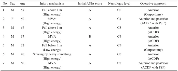
Table 1. Demographics of patients who had CSF leaks
No. Sex Age Injury mechanism Initial ASIA score Neurologic level Operative approach
1 M 57 Fall above 1 m A C6 Anterior
(High energy) (Corpectomy)
2 F 50 MVA
aA C4 Anterior and posterior
(High energy) (ACDF
bwith PSF
c)
3 M 67 Fall above 1 m A C5 Anterior
(High energy) (ACDF)
4 M 17 MVA B C4 Anterior
(High energy) (ACDF)
5 M 22 Fall below 1 m A C5 Anterior
(Low energy) (Corpectomy)
6 M 40 Striking by heavy something A C6 Anterior
(High energy) (ACDF)
7 M 60 MVA A C5 Anterior and posterior
(High energy) (ACDF with PSF)
MVA
a: motor vehicle accident, ACDF
b: anterior cervical discectomy and fusion, PSF
c: posterior screw fixation
were men and the mean age at the time of operation was 49.12±18.15 years (range, 17~67). The injury mechanism was mostly from a high energy source:
motor vehicle accident for 3 patients, a fall above 1 m for 2 patients, and being struck by something heavy for 1 patient. Only 1 patient had a low energy injury mechanism: a fall below 1 m (Table 1).(18) The initial American Spinal Injury Associate (ASIA) score was A in 6 patients and B in 1 patient. The neuro- logical level was C4 in 2 patients, C5 in 3 patients and C6 in 2 patients.
1.
Preoperative imaging
All patients were preoperatively evaluated using X-ray, computed tomography (CT) and magnetic resonance imaging (MRI). The cervical lateral X-ray
and CT showed a facet fracture and dislocation in 5 patients and a vertebral body fracture in 2 patients.
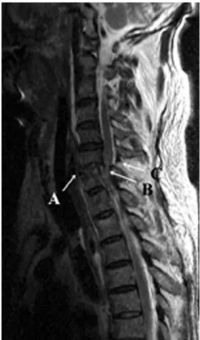
All patients showed disruption or displacement of the anterior longitudinal ligament (ALL) and poste- rior longitudinal ligament (PLL), a high signal intensity of the intervertebral disc and spinal cord, and disruption of the ligamentum flavum on the ini- tial cervical spine MRI (Fig. 1).
2.
Intraoperative findings
After the standard anterior cervical approach, ALL injury was found in 4 patients and intervertebral disc injury was found in 7 patients. During discecto- my, PLL injury and simultaneous dural tear were found in 7 patients. A resulting CSF leak from the dura tear was noted in all patients. Primary repair of the dural tear was notattempted because of severe laceration of the dura. Fibrin glue (Green plast�, Green Cross, Seoul, Korea) was applied to the dura in all cases. Among these cases, 2 of them received the TachoComb� in addition to fibrin glue (Torii Pharmaceutical, Tokyo, Japan). There were no confirmed persistent CSF leaks on the operated site.
Then, one level anterior cervical discectomy and fusion (ACDF) and corpectomy using autologous iliac bone graft and anterior plating was performed in 5 patients and 2 patients, respectively. At closure, one submuscular Jackon-Pratt (JP) drainage was insert- ed and the wound was closed layer by layer.
Additionally, 2 patients who did not achieved reduc- tion of the facet dislocation in the anterior approach had lateral mass screw fixation performed.
Postoperative lumbar CSF drains were not inserted in all the patients.
3.
Perioperative management
After the operation, the patients kept their heads in an elevated position and were allowed to do any activities they could manage. The JP drainage was removed within the first postoperative day and the drainage amount was 70.6±51.7 cc (range, 31~160).
Rehabilitation started as soon as possible.
Soo Eon Lee, et al.: Management of Cerebrospinal Fluid Leak after Traumatic Cervical Spinal Cord Injury
Fig. 1. Preoperative MRI of a traumatic cervical spine at C6-7
Sagittal T2-weighted image shows the disruption of the
ALL (A) and intervertebral disc. The disruption of the
PLL is shown (B) as well as the disruption of the liga-
mentum flavum (C). This patient’s dural tear and
resulting CSF leak were identified during the operation
4. Hospital course
The perioperative complications developed includ- ing pneumonia in 4 patients, urinary tract infection (UTI) in 1 patient, and bacteremia and sepsis in 1 patient. None of the patients developed complica- tions related to their CSF leak such as wound dehis- cence, meningitis, and pseudomeningocele. ASIA score at discharge was A in 4 patients, B in 2 patients and C in 1 patient.
IV. Discussion
Traumatic cervical SCI is frequently accompanied by dural tears and the resulting CSF leak after anterior cervical spine surgery can be troublesome, delay rehabilitation and increase morbidity.
Although direct repair is the optimal treatment modality in most cases of dural tears in all spinal regions, primary dural repair is difficult in trauma cases.(11-13) Moreover, although various techniques have been described to manage CSF leak related complications after surgery for degenerative spinal diseases,(11-13) there are no reports on cervical spine trauma. Thus, the authors of this study sug- gest a simple, effective method to manage CSF leaks during the perioperative period.
The incidence of cervical dural tears and CSF leaks after anterior decompression procedures have ranged from 0.5% to 3%.(13) For ossification of the posterior longitudinal ligament (OPLL), the inci- dence is much higher, ranging from 4.3% to 32%.(11,13) The most common cause of CSF leaks is injury to the dura while resecting the posterior lon- gitudinal ligament. Thus, OPLL itself is a risk factor for dural tears and as a consequence, CSF leaks.(19) However in trauma cases, dural tears mostly devel- oped at the moment of injury. In the present study, 7 patients(13.2%) had dural tears. The incidence after cervical spine trauma was as frequent as that in OPLL. All patients who had a CSF leak showed a disruption or displacement of the ALL, PLL and lig- amentum flavum and high signal intensity of the intervertebral disc and spinal cord on the initial MRI. Hence, a dural tear by spine trauma could be predicted on the preoperative MRI when it shows
injury of the ligament, intervertebral disc and spinal
cord. Additionally, the surgeon should predict the
possibility of an intraoperative CSF leak in these
cases. If injury to the dura is observed during the
surgery, primary repair with microsurgical sutures
could be attempted. Although primary repair fre-
quently is not technically feasible, various repair
techniques with gelatin foam, fibrin glue, collagen
matrix, fat and fascia grafts, biological grafts, syn-
thetic materials, blood patch or microdural stapling
have been described to manage dural tears intraop-
eratively. Additionally, wound drain, lumbar drain,
lumboperitoneal or wound-peritoneal shunts, or
ventriculostomy have been described to manage CSF
leaks during the postoperative period.(13,20-22) The
authors’policy for managing CSF leaks consisted of
first primary repair of the identified dura tear in
spinal surgery for degenerative spinal disease and
spinal tumors. However, if primary dural repair was
not feasible, duraplasty was attempted and then
fibrin glue was applied over the surgical site. Wound
drains were not routinely inserted, especially in sur-
gical cases with small incisions or intradural
surgery. Although a wound drain was inserted at
closure, the drain was removed if CSF appeared in
the drain tube or bag, and the tube was clamped and
removed regardless of the drainage amount.(23)
Additionally, lumbar drainage was not inserted after
the operation. Instead, the patients were not allowed
bed rest and encouraged to ambulate. If CSF collec-
tion under a surgical wound did develop, it would be
observed unless the symptoms were related to an
airway obstruction. Over time, CSF collection grad-
ually disappears.(23) Besides trauma cases, dural
tears and the resulting CSF leaks already existed
before the operations accompanied simultaneously
by PLL tears. However, it was not an iatrogenic
result. PLL is believed to provide stability to the
cervical spine and the removal of the PLL could lead
to a decrease in cervical stability after surgery.(24)
However, the removal of the PLL was helpful in
getting more decompression in the anterior
approach for cervical spondylotic myelopathy (CSM)
although more technically demanding.(25) In the
operative procedure, the further resection of the
PLL was not meaningful in the trauma cases
because the purpose of the operation was to stabilize the unstable spine. Therefore, if CSF leak was observed during the discectomy, we did not try fur- ther resection of the PLL to avoid further dural defect. Primary dural repair was not possible and duraplasty was also not successful due to a wide, rough defect of the dura. Application of fibrin glue was the most useful method. At closure, wound drainage was inserted to evacuate hematoma from the edematous prevertebral soft tissue after cervical trauma. However, it was stopped within the first 24 hours after operation. The most important manage- ment protocol was to encourage early ambulation and rehabilitation. During the hospital course, none of the patients developed complications related to their CSF leak such as wound dehiscence, meningi- tis, and pseudomeningocele. Therefore, we recom- mend head elevation and early ambulation to decrease intracranial pressure and early removal of wound drains to increase the pressure of the subcu- taneous space. The use of CSF diversion through lumbar drainage for the treatment of CSF leaks is well documented.(13,19,20,26) Although effective in treating CSF leaks, the use of lumbar drains is associated with infection, over-drainage of CSF and pneumocephalus with brainstem compression.(26- 28) While the standard position of patients with lumbar drainage is bed rest to prevent over- drainage of the CSF, bed rest may increase the risk of postoperative morbidity due to the development of deep venous thrombosis and pulmonary embolism after prolonged immobilization.(29) The present study had a relatively small number of cases and a short follow-up period. However, the authors feel it is important to report on the management of CSF leaks after trauma to the cervical spine.
V. Conclusion
The incidence of CSF leaks after surgery for cer- vical spinal trauma is relatively higher than that of cervical spinal stenosis. In trauma cases, the sur- geon can predict the possibility of dural tears on the preoperative MRI. Intraoperatively, if a dural tear and the resulting CSF leak are observed, then fur- ther resection of the PLL may not be attempted to
prevent further damage to the dura. Thus, applying fibrin glue on the operated site is the most useful method. Moreover, the patients should be positioned with their heads elevated and early ambulation should be encouraged as well as rehabilitation with- out any postoperative lumbar drains.
REFERENCES