Veterinary Science
http://dx.doi.org/10.4142/jvs.2014.15.2.187
Received: 22 Mar. 2013, Revised: 11 Jun. 2013, Accepted: 9 Jul. 2013
Original Article
*Corresponding author: Tel: +82-2-880-1265; Fax: +82-2-880-8662; E-mail: [email protected]
†The first two authors contributed equally to this work.
ⓒ 2014 The Korean Society of Veterinary Science.
This is an Open Access article distributed under the terms of the Creative Commons Attribution Non-Commercial License (http://creativecommons.org/licenses/by-nc/3.0) which permits
unrestricted non-commercial use, distribution, and reproduction in any medium, provided the original work is properly cited.
Computed tomographic evaluation of cervical vertebral canal and spinal cord morphometry in normal dogs
Eunjeong Seo
1,†, Jihye Choi
2,†, Mincheol Choi
1, Junghee Yoon
1,*
1
College of Veterinary Medicine and the Research Institute for Veterinary Science, Seoul National University, Seoul 151-742, Korea
2
College of Veterinary Medicine, Chonnam National University, Gwangju 500-757, Korea
The height, width, and cross-sectional area of the vertebral canal and spinal cord along with the area ratio of spinal cord to vertebral canal in the cervical vertebra were evaluated in images obtained using computed tomography (CT).
Measurements were taken at the cranial, middle, and caudal point of each cervical vertebra in eight clinically normal small breed dogs (two shih tzu, two miniature schnauzers, and four mixed breed), 10 beagles, and four German shepherds. CT myelography facilitated the delineation of the epidural space, subarachnoid space, and spinal cord except at the caudal portion of the 7th cervical vertebra. The spinal cord had a tendency to have a clear ventral border in the middle portion of the vertebral canal and lateral borders near both end plates. The height, width, and area of the vertebral canal and spinal cord in the cervical vertebra were increased as the size of dog increased. However, the ratio of the spinal cord area to vertebral canal area in the small dogs was higher than that of the larger dogs. Results of the present study could provide basic and quantitative information for CT evaluation of pathologic lesions in the cervical vertebra and spinal cord.
Keywords: cervical vertebra, computed tomography, dog, spinal cord, vertebral canal
Introduction
The vertebra consists of a bony portion including the vertebral body, laminae, pedicles, and processes along with soft tissues including the spinal cord, nerves, vessels, and ligaments [8]. Most vertebral disorders are caused by damages to the spinal cord, which make the spinal cord compressed or swollen [24]. Wobbler’s syndrome,
intervertebral disk disease, hydromyelia, syringomyelia, intramedullary neoplasia, myelomalacia, and spinal cord atrophy are examples of spinal disorders [10,13,23,26].
Radiography is the primary diagnostic modality used to diagnose vertebral diseases in practice [18,21]. While basic radiography and standard myelography provide important information regarding the subarachnoid space, spinal cord, and vertebral canal, the complex shape of the vertebra as well as overlapping bony and soft tissue structures make radiographic evaluation of the vertebral column difficult [16]. Diagnostic imaging techniques used to visualize transverse planes such as computed tomography (CT) and magnetic resonance imaging (MRI) can reduce confusion encountered when evaluating vertebrae and the spinal cord by eliminating the superimposition of normal structures and pathological lesions. CT images can be displayed in different gray scale formats using various window setting according to the region of interest, and provide reformatted images that can enhance the visualization of specific structures without additional movement of the patient or further exposure to radiation [4,7,25].
Quantitative evaluation of spinal cord size along with the relationship between the spinal cord and vertebral canal can provide important data for distinguishing different diseases and predicting a patient’s prognosis. Previous studies have evaluated the ratios of the diameter or area between spinal cord and vertebral canal in specific vertebral regions in dogs according to body size [5-6,9,12].
Data from these studies supported the conclusion that
evaluation of the spinal cord, subarachnoid space, epidural
space, and vertebral canal should performed based on the
variety of breed-specific characteristics because there are
significant differences in the component proportion ratio
C3 Height Width Area C4 Height Width Area C5 Height Width Area C6 Height Width Area C7 Height Width Area
6.3 ± 1.1 9.0 ± 1.0 47.0 ± 8.9 6.4 ± 0.6 10.1 ± 1.3 51.0 ± 7.4 7.0 ± 0.5 10.0 ± 0.9 57.4 ± 7.1 7.4 ± 0.8 10.8 ± 0.6 64.0 ± 10.8
7.5 ± 0.9 10.6 ± 0.8 62.3 ± 10.3
7.0 ± 0.7 8.7 ± 0.9 48.4 ± 7.8 7.5 ± 0.7 8.8 ± 0.6 53.4 ± 5.4 8.2 ± 0.7 9.5 ± 0.7 63.2 ± 5.1 8.3 ± 0.7 9.9 ± 0.8 70.1 ± 7.5 7.5 ± 1.2 10.4 ± 0.8 66.5 ± 9.9
6.7 ± 0.9 8.9 ± 1.0 53.8 ± 11.8
7.2 ± 0.5 9.2 ± 1.0 59.8 ± 8.2 7.7 ± 0.4 9.8 ± 0.7 67.4 ± 5.5 8.3 ± 0.8 10.4 ± 0.6 74.8 ± 7.6 7.5 ± 1.0 10.4 ± 0.8 65.4 ± 9.6
7.2 ± 0.5 11.2 ± 1.2 62.9 ± 8.0 6.8 ± 0.4 11.7 ± 0.3 63.1 ± 5.9 7.4 ± 0.5 11.9 ± 0.6 70.2 ± 4.5 8.4 ± 0.3 12.0 ± 0.9 85.7 ± 3.3 8.7 ± 0.5 13.1 ± 0.7 93.1 ± 3.9
8.4 ± 0.6 9.6 ± 1.0 61.6 ± 4.4 8.6 ± 0.4 9.8 ± 1.1 64.8 ± 6.5 9.6 ± 0.7 10.2 ± 0.6 76.1 ± 5.7 10.3 ± 0.6 11.5 ± 0.6 92.2 ± 7.1 8.9 ± 0.7 12.9 ± 0.6 93.7 ± 3.5
7.8 ± 0.7 11.1 ± 1.3 72.8 ± 13.6
8.4 ± 0.3 11.6 ± 1.0 81.7 ± 7.7 9.4 ± 0.5 11.6 ± 0.8 95.6 ± 10.6
9.8 ± 0.4 12.7 ± 0.5 104.4 ± 3.5 10.1 ± 0.6 13.0 ± 0.8 105.7 ± 5.2
9.3 ± 1.3 13.1 ± 0.8 102.5 ± 20.4
9.3 ± 0.7 15.3 ± 1.3 114.0 ± 17.1
9.6 ± 1.0 14.8 ± 0.7 119.4 ± 15.6
9.5 ± 0.8 16.1 ± 1.3 133.2 ± 17.1
9.7 ± 1.4 16.6 ± 1.2 140.1 ± 20.6
11.1 ± 1.5 12.4 ± 0.4 109.8 ± 16.0
11.7 ± 1.0 12.4 ± 0.7 121.7 ± 13.1
13.3 ± 1.2 13.4 ± 1.0 142.1 ± 19.5
12.5 ± 2.1 14.7 ± 1.1 152.2 ± 28.5
12.3 ± 2.2 16.1 ± 1.4 164.8 ± 44.9
10.8 ± 1.6 13.2 ± 0.6 128.5 ± 23.8
10.4 ± 1.5 12.8 ± 0.4 133.0 ± 20.6
11.7 ± 0.5 13.6 ± 1.4 149.7 ± 27.8
11.3 ± 1.0 14.3 ± 1.6 164.8 ± 22.5
12.3 ± 1.9 15.2 ± 1.5 168.3 ± 42.5
Data are expressed as the mean ± standard deviation (SD). Units of measurement are mm for height and width, and mm
2 for area. C, cervical
vertebra; Cranial point, immediately caudal to cranial end-plate; Middle point, the narrowest portion of the individual vertebral body; Caudal
point, immediately cranial to the caudal end plate.
vertebral canal diameter is significantly greater than that in the normal control with a widened spinal cord [14].
Therefore, delineation between the bony structure and spinal cord in vertebrae is important to establish reliable differential diagnostic techniques.
CT and MRI can define the bony and soft tissue structures more clearly compared to radiographic myelography. MRI can provide a higher contrast within soft tissues compared to CT [2]. However, MRI is less available than CT in veterinary medicine. In human medicine, the normal range of cross-sectional diameter and spinal cord area has been established, and correlation between alterations of the spinal cord area and clinical signs has been assessed with CT myelography in the cervical vertebra [20,22]. In dogs, the normal range of the cervical spinal cord to vertebral canal area ratio was proposed using radiographic myelography in small and large animals [6]. The vertebral canal and body ratio as well as morphologic features of the cervical vertebra have been investigated in studies of Doberman pinschers focused on cervical spondylomeylopathy [1,3]. However, no investigation has been performed using trans-sectional imaging modalities such as CT or MRI to determine the
spinal cord to vertebral canal area ratio in the cervical region of normal dogs.
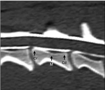
CT myelography can help delineate the spinal cord, particularly in small dogs. This can improve the visualization of anatomic details in cases of spinal diseases [11]. Margins of the spinal cord, subarachnoid space, and epidural space were clearly observed using CT myelography in the present study. The height, width, and areas of the spinal cord and vertebral canal were evaluated at three points located in the cranial, middle, and caudal regions of the cervical vertebra body. The space in the ventral region at the middle point of each vertebra and in both lateral sides near the end plates tended to be greater even though these differences were not statistically significant.
Variations of the vertebral and spinal cord structures
according to body size have been reported in the literature
[6,12,17]. In small dogs, the subarachnoid space is
relatively narrow and conforms closely to bones forming
the vertebral canal in the thoracolumbar region. In contrast,
large breeds tend to have relatively thinner spinal cords and
a uniform, parallel-sided subarachnoid space [12]. In the
present study, the height, width, and area measured in the
Table 3. Spinal cord to vertebral canal area ratio for the cervical vertebra in normal dogs
Group 1 Group 2 Group 3
Cranial point
Middle point
Caudal point
Cranial point
Middle point
Caudal point
Cranial point
Middle point
Caudal point C3
C4 C5 C6 C7
0.54 ± 0.05 0.56 ± 0.05 0.55 ± 0.05 0.56 ± 0.04 0.53 ± 0.06
0.55 ± 0.05 0.56 ± 0.02 0.55 ± 0.04 0.57 ± 0.06 0.55 ± 0.08
0.55 ± 0.04 0.52 ± 0.03 0.51 ± 0.04 0.53 ± 0.02 0.48 ± 0.02
0.46 ± 0.06 0.5 ± 0.08 0.5 ± 0.09 0.51 ± 0.06 0.45 ± 0.07
0.48 ± 0.08 0.5 ± 0.07 0.49 ± 0.05 0.49 ± 0.05 0.44 ± 0.05
0.42 ± 0.1 0.41 ± 0.06 0.41 ± 0.07 0.43 ± 0.06 0.35 ± 0.05
0.32 ± 0.02 0.31 ± 0.02 0.38 ± 0.04 0.38 ± 0.04 0.3 ± 0.04
0.31 ± 0.03 0.3 ± 0.03 0.34 ± 0.04 0.34 ± 0.05 0.31 ± 0.04
0.27 ± 0.04 0.31 ± 0.03 0.34 ± 0.05 0.33 ± 0.01 0.28 ± 0.04
Data are expressed as the mean ± SD.
Table 4. Mean ratio of the spinal cord to vertebral canal area of the cervical vertebra regardless of measuring point in normal dogs
Group 1 Group 2 Group 3
C3 C4 C5 C6 C7
0.55 ± 0.01 0.55 ± 0.02 0.54 ± 0.02 0.55 ± 0.02 0.52 ± 0.04
0.45 ± 0.03 0.47 ± 0.05 0.47 ± 0.05 0.48 ± 0.04 0.41 ± 0.06
0.30 ± 0.03 0.31 ± 0.01 0.35 ± 0.02 0.35 ± 0.03 0.3 ± 0.02
Data are expressed in the mean ± SD.
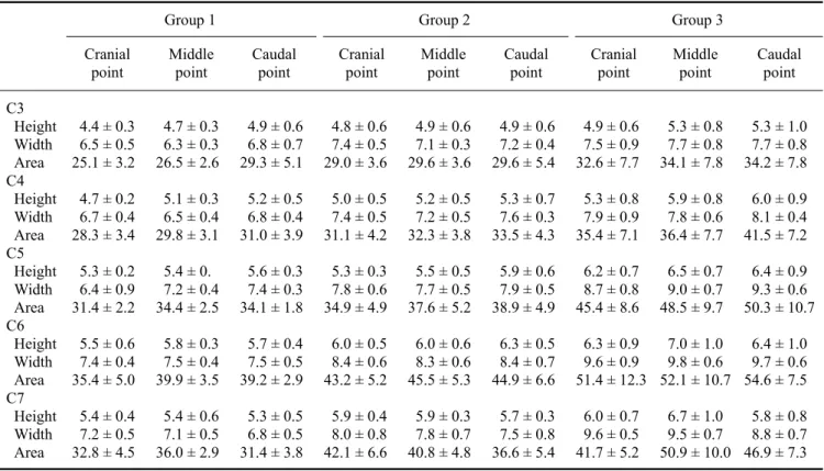
Table 2. Spinal cord height and width, and cervical vertebral area in normal dogs
Group 1 Group 2 Group 3
Cranial
point Middle
point Caudal
point Cranial
point Middle
point Caudal
point Cranial
point Middle
point Caudal point C3
Height Width Area C4 Height Width Area C5 Height Width Area C6 Height Width Area C7 Height Width Area
4.4 ± 0.3 6.5 ± 0.5 25.1 ± 3.2 4.7 ± 0.2 6.7 ± 0.4 28.3 ± 3.4 5.3 ± 0.2 6.4 ± 0.9 31.4 ± 2.2 5.5 ± 0.6 7.4 ± 0.4 35.4 ± 5.0 5.4 ± 0.4 7.2 ± 0.5 32.8 ± 4.5
4.7 ± 0.3 6.3 ± 0.3 26.5 ± 2.6 5.1 ± 0.3 6.5 ± 0.4 29.8 ± 3.1 5.4 ± 0.
7.2 ± 0.4 34.4 ± 2.5 5.8 ± 0.3 7.5 ± 0.4 39.9 ± 3.5 5.4 ± 0.6 7.1 ± 0.5 36.0 ± 2.9
4.9 ± 0.6 6.8 ± 0.7 29.3 ± 5.1 5.2 ± 0.5 6.8 ± 0.4 31.0 ± 3.9 5.6 ± 0.3 7.4 ± 0.3 34.1 ± 1.8 5.7 ± 0.4 7.5 ± 0.5 39.2 ± 2.9 5.3 ± 0.5 6.8 ± 0.5 31.4 ± 3.8
4.8 ± 0.6 7.4 ± 0.5 29.0 ± 3.6 5.0 ± 0.5 7.4 ± 0.5 31.1 ± 4.2 5.3 ± 0.3 7.8 ± 0.6 34.9 ± 4.9 6.0 ± 0.5 8.4 ± 0.6 43.2 ± 5.2 5.9 ± 0.4 8.0 ± 0.8 42.1 ± 6.6
4.9 ± 0.6 7.1 ± 0.3 29.6 ± 3.6 5.2 ± 0.5 7.2 ± 0.5 32.3 ± 3.8 5.5 ± 0.5 7.7 ± 0.5 37.6 ± 5.2 6.0 ± 0.6 8.3 ± 0.6 45.5 ± 5.3 5.9 ± 0.3 7.8 ± 0.7 40.8 ± 4.8
4.9 ± 0.6 7.2 ± 0.4 29.6 ± 5.4 5.3 ± 0.7 7.6 ± 0.3 33.5 ± 4.3 5.9 ± 0.6 7.9 ± 0.5 38.9 ± 4.9 6.3 ± 0.5 8.4 ± 0.7 44.9 ± 6.6 5.7 ± 0.3 7.5 ± 0.8 36.6 ± 5.4
4.9 ± 0.6 7.5 ± 0.9 32.6 ± 7.7 5.3 ± 0.8 7.9 ± 0.9 35.4 ± 7.1 6.2 ± 0.7 8.7 ± 0.8 45.4 ± 8.6 6.3 ± 0.9 9.6 ± 0.9 51.4 ± 12.3
6.0 ± 0.7 9.6 ± 0.5 41.7 ± 5.2
5.3 ± 0.8 7.7 ± 0.8 34.1 ± 7.8 5.9 ± 0.8 7.8 ± 0.6 36.4 ± 7.7 6.5 ± 0.7 9.0 ± 0.7 48.5 ± 9.7 7.0 ± 1.0 9.8 ± 0.6 52.1 ± 10.7
6.7 ± 1.0 9.5 ± 0.7 50.9 ± 10.0
5.3 ± 1.0 7.7 ± 0.8 34.2 ± 7.8 6.0 ± 0.9 8.1 ± 0.4 41.5 ± 7.2 6.4 ± 0.9 9.3 ± 0.6 50.3 ± 10.7
6.4 ± 1.0 9.7 ± 0.6 54.6 ± 7.5 5.8 ± 0.8 8.8 ± 0.7 46.9 ± 7.3
Data are expressed as the mean ± SD. Units of measurement are mm for height and width, and mm
2 for area.
three groups of dogs tended to increase as the size of the dog increased. This result concurs with the finding that vertebral canal height in German shepherds is greater throughout the length of the vertebral column compared to that of dachshunds in the thoracolumbar region [17] even though no data was obtained from the cervical region.
In the present study, the ratio of the spinal cord area to
vertebral canal area was greater in small dogs compared to
large dogs in this study. This result coincides with findings
from a previous study in which radiographic myelography
in cervical vertebra was performed to evaluate similar