Journal of Trauma and Injury Vol. 26, No. 3, September, 2013
[ J Trauma Inj 2013;26:63-73 ]
� Address for Correspondence : Sang Ryong Jeon, M.D., Ph.D.
Department of Neurological Surgery, Asan Medical Center, University of Ulsan College of Medicine, 388-1 Pungnap-2dong, Songpa-gu, Seoul 138-736, Korea
Tel : 82-2-3010-3550, Fax : 82-2-476-6738, E-mail : [email protected] Submitted : February 6, 2013 Revised : April 19, 2013 Accepted : July 1, 2013
본 연구는 한국연구재단을 통해 교육과학기술부의 미래유망 융합기술 파이오니어사업으로부터 지원받아 수행되었습니다 (2012-0000447).
I. 서 론
척수 손상은 기원전 2000~3000년 전 경, Edwin Smith 는 파피루스에“치료되지 않는 병”으로 기록되어 있으며, 환 자의 감각, 운동 마비와 소변 기능 상실 등 비교적 자세한 기 록이 남아 있다. 그러나 수천 년이 흐른 현대의학에서도 응 급처치, 내과 및 외과적 치료, 재활치료에서 상당한 발전이 있었음에도 불구하고 많은 환자들이 상당한 신경학적 결손으
로 인해 삶의 질의 저하를 감수해야 하는 실정이다.(1) 척수 손상의 치료는 수술적 치료, 이차 손상을 막는 약물 의 개발, 신경 기능의 보존 및 합병증 발생을 막기 위한 재활 치료로 발전해 왔다. 그러나, 손상된 신경의 복원과 재생을 위한 치료법은 아직 개발 되지 못했다. 척수 외상에 의한 척 수 손상은 대부분 생산력이 왕성한 젊은 사람에게 발생하므 로 사회적인 손실이 크고 이들 환자는 신경학적 장애가 영구적 으로 발생하여 의료비 부담 등 사회경제적 손실이 큰 질환이다.
척수손상 치료 약제의 현재와 미래: 체계적 고찰
울산대학교 의과대학 서울아산병원 신경외과학교실 최 일, 하진경, 전상용
- Abstract -
Current Concept and Future of the Management of Spinal Cord Injury: A Systematic Review
Il Choi, M.D., Jin Gyeong Ha, M.D., Sang Ryong Jeon, M.D., Ph.D.
Department of Neurological Surgery, Asan Medical Center, University of Ulsan, College of Medicine, Seoul, Korea
Spinal cord injury (SCI) is a serious condition associated with social and familial burden, as well as significant neurologic deficit. Despite the many advances in the treatment of spinal cord injury, a fundamental treatment for neurologic functional recovery has not yet been developed. In this article, we review two directions of devel- opment for spinal cord injury treatment: neuroprotective pharmacological agents and axon-regenerating cell therapy. We expect developments in these two to lead to improve functional recovery in patients with spinal cord injuries and to reduce burdens on society, as well as the patients’ families.
Key Words: Cell therapy, Pharmacologic therapy, Spinal cord injuries, Stem cell
일반적으로 중추 신경계 손상의 하나인 척수 손상은 비가 역적인 것으로 손상 후 재생은 매우 어려운 것으로 알려졌 고, 현재, 척수 손상에 대한 치료 및 재활 요법의 괄목할 발 전에도 불구하고 손상의 근본 원인이 되는 신경 조직의 재생 이 이루어 지지 않아 근본 치료가 불가능한 상태이다. 최근 의 임상에서 사용되는 치료법으로는 손상된 척수에 대한 근 본 치료보다는 이차적인 척수 손상을 막기 위한 수술적 치료 와 약물 치료만이 임상적으로 이용되는 실정이다. 따라서 기 존의 치료법에 덧붙여 줄기세포 치료(stem cell treatment) 을 비롯한 신경재생(neuroregeneration)치료에 대한 연구 가 활발히 진행 중이다.
이 글은 척추 손상의 치료에 있어 세포 치료를 포함한 약 물치료를 중심으로 고찰하고자 한다. 또한 기존약제의 현재 이용 현황과 최근에 각광 받고 있는 세포 치료를 중심으로 이룬 성과와 앞으로 기대되는 결과에 대해 다루어 보기로 한다.
II. 본 론
1. 척수 손상의 독특한 병리학적 측면
척수손상은 외상 시 발생한 물리적 손상을 의미하는 1차 손상과 1차 손상을 증폭시키는 분자, 세포 단위의 일련의 연 쇄 반응인 2차 손상으로 구분한다.(2) 1차 손상은 최초의 척 수 손상에 의한 결과로서 즉각적인 축삭 돌기의 절단과 척수 세포들의 손상을 일으키며. 2차 손상은 첫 척수 손상 후 수 주, 수개월 또는 길게는 수년에 걸쳐 진행되는 조직의 손상 을 말한다.(3,4) 1차 손상은 상위 운동 신경원과 하위 운동 신경원을 손상시키고 운동, 감각, 자율 신경기능에 대한 직 접적인 조직손상이므로 현재는 치료의 대상이 되지 못한다.
이에 반해 2차 손상은 1차 손상 직후 수 분에서 수 주일에 걸 쳐 야기되는 병리과정이다. 2차 손상은 물리적 손상의 최소 화라는 긍정적 측면도 있으나 동시에 조직 손상을 악화시키 고 수초재형성(remyelination)을 통한 복원, 재생과정에 억 제환경을 제공하는 부정적인 측면이 있다. 구체적 병리 기전 으로 염증(inflammation), 허혈(ischemia), 유리기(free radicals)형성, 이온채널의 파괴, 축삭 돌기의 수초 탈락 (axonal demyelination), 신경 교 반흔 형성(glial scar- ring), 괴사(necrosis), 세포 자멸 사(programmed cell death) 등이다. 특히 2차 손상기에는 신생혈관을 통해 혈류 가 재 관류되고 세포잔해가 제거됨과 동시에 손상된 신경회 로(neural circuits)가 재구성되는 현상도 발생한다. 이러한 현상이 연구와 치료의 주 대상이 된다.(5)
2. 현재까지의 척수 손상 치료 전략
척수 손상 치료의 방향은 이차적인 척수의 손상을 최대 한
도로 줄이고 신경학적 기능을 최대한 회복하는 데 있다. 척 수손상의 현재 치료는 다음과 같이 요약 할 수 있다.(6) 추가 적 척수 손상 방지, 척수 혈류 량의 유지, 척수 압박의 해소, 이동과 재활을 위한 척추 구조물의 안정성 제공이다. 척수 손상의 치료는 사고 현장에서부터 척수 손상의 가능성을 염 두에 두고 치료를 시작하는 것이 매우 중요하다.(6) 사고 현 장에서 환자의 적절한 고정 및 이송 과정의 견고함은 다시 말 할 나위 없이 중요하다. 척수 손상의 상당수의 경우 다발 성 장기의 손상을 동반하기 때문에 반드시 다른 출혈 성 쇼 크나 장기의 손상을 확인하여야 한다.(7) 혈압과 산소 분압을 포함한 적절한 활력징후의 안정이 이루어 지면 척추의 정복 과 고정이 이루어 져야 한다. 빠르고도 정확히 신경학적 검 사와 방사선학적인 검사를 시행하여 척수 손상이 어느 부위 에서 어느 정도 인지를 진단하고 치료 방향을 결정하여야 한 다.(8)
수술적 치료는 손상된 신경을 근본적으로 재생시키는 것이 아닌 남아 있는 신경 기능의 보호 개념의 치료 법이다.(9) 수 술적인 치료는 다음과 같은 단계로 생각해 볼 수 있다. 척추 의 전위가 정복이 안되어 척수 압박이 계속되는 경우, 비록 정복은 되었으나 골편이나 파열된 추 간판 탈출 증으로 척수 의 압박이 계속 남아 있는 경우 혹은 척추가 불안정하여 추 가적인 척수 손상 가능성이 남아 있는 경우 시행한다. 급성 기 수술적 치료가 끝난 후 신경학적으로 안정된 상태에 다다 른 환자에게서의 다음 단계 치료는 재활 및 만성 기 합병증 의 예방 및 관리이다. 우리가 이 글의 후반부에서 다룬 세포 치료를 포함한 약물 치료는 급성 기의 이차적 신경손상을 방 지하고, 만성 기에서 신경재생을 촉진하는 역할로 실험적인 방법이라고 볼 수 있다.(10)
3. 급성 척수 손상에 관한 약물 치료 및 무작위 임상 시험들
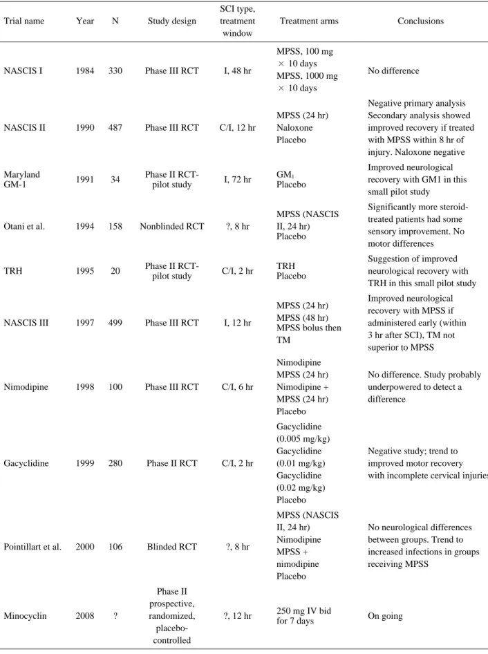
척추 손상의 약물 치료는 주로 이차적 손상 기전을 차단하 여 손상의 파급과 악화를 방지하는 개념의 치료로 지난 20여 년간 많은 약물들이 실험적으로 신경 기능의 보호 기능이 있 다고 발표 되었지만 실제로 임상적 적용이 가능했던 약은 고 용량 스테로이드 요법(methylprednisolone)이 유일하 다.(11,12)
고 용량 스테로이드는 염증 성 반응 및 부종을 줄임과 동 시에 지질 과산화(lipid peroxidation), 흥분독성(exci- totoxicity)에 대한 억제 작용이 있는 것으로 보고 되고 있 다.(13) 용량은 척수 손상 후 8시간 이내에 30 mg/kg의 메 틸프레드니솔론(methylprednisolone)을 정맥 내 급속 투여 하고, 이후 24시간 동안 5.4 mg/kr/hr을 지속적으로 정맥 주사 한다. 이 경우 불완전 마비 환자에서는 일부 효과가 있 는 것으로 보고 되어 있으나 완전마비에 대한 효과는 아직
Table 1. Prospective completion and ongoing study for spinal cord injury SCI type,
Trial name Year N Study design treatment Treatment arms Conclusions
window
MPSS, 100 mg
NASCIS I 1984 330 Phase III RCT I, 48 hr × 10 days
No difference MPSS, 1000 mg
× 10 days
Negative primary analysis MPSS (24 hr) Secondary analysis showed NASCIS II 1990 487 Phase III RCT C/I, 12 hr Naloxone improved recovery if treated
Placebo with MPSS within 8 hr of injury. Naloxone negative
Maryland Phase II RCT- GM
1Improved neurological
1991 34 I, 72 hr recovery with GM1 in this
GM-1 pilot study Placebo
small pilot study
MPSS (NASCIS Significantly more steroid- Otani et al. 1994 158 Nonblinded RCT ?, 8 hr II, 24 hr) treated patients had some
sensory improvement. No Placebo
motor differences
Phase II RCT- TRH Suggestion of improved
TRH 1995 20 C/I, 2 hr neurological recovery with
pilot study Placebo
TRH in this small pilot study MPSS (24 hr) Improved neurological MPSS (48 hr) recovery with MPSS if
NASCIS III 1997 499 Phase III RCT I, 12 hr administered early (within
MPSS bolus then
3 hr after SCI), TM not
TM superior to MPSS
Nimodipine
MPSS (24 hr) No difference. Study probably Nimodipine 1998 100 Phase III RCT C/I, 6 hr Nimodipine + underpowered to detect a
MPSS (24 hr) difference Placebo
Gacyclidine (0.005 mg/kg)
Gacyclidine Negative study; trend to Gacyclidine 1999 280 Phase II RCT C/I, 2 hr (0.01 mg/kg) improved motor recovery
Gacyclidine with incomplete cervical injuries (0.02 mg/kg)
Placebo MPSS (NASCIS
II, 24 hr) No neurological differences Pointillart et al. 2000 106 Blinded RCT ?, 8 hr Nimodipine between groups. Trend to
MPSS + increased infections in groups
nimodipine receiving MPSS
Placebo Phase II
prospective,
250 mg IV bid
Minocyclin 2008 ? randomized, ?, 12 hr On going
placebo- for 7 days
controlled
Continue
입증되지 못하고 있는 상태이다.(14)
포유류 신경세포막 구성성분이며, 척수 손상에서 신경 유 연성(plasticity)과 축삭의 회복과정에 중요한 역할을 하는 강글리오시드(GM-1,ganglioside.Sygen)는 최초 신경손상 에 대한 예방 및 신경의 재생에 효과가 있는 것으로 보고 되 어 실험되었으나 임상적으로 그 효과를 입증하는데 실패하였 다.(15) 이차적 염증반응을 억제하는 역할을 하는 갑상선 자 극 호르몬 방출 호르몬(Thyrotrophic-Releasing Hormone),(16) 중추신경계의 대표적인 신경 흥분 전달 물 질 인 글 루 탐 산 염 (Glutamate)의 경 쟁 적 억 제 자 인 Gacyclidine (GK-11), 흥분 시 세포 내 증가되는 칼슘 이온 (Ca++) 등을 억제하는 니모디핀(Nimodipine),(17) 그리고 척수 손상 시 분비되어 신경독성작용을 하는 아편 유사 제 수용체(opioid receptor)를 억제하는 아편 유사 억제 제 (opioid antagonism) 등이 척수 손상의 이차적 손상을 줄일 목적으로 한동안 사용되었으나, 임상적으로 의의를 나타내지 는 못했다.(14) 이처럼 다양한 시도에도 불구하고, 아직 약물 요법도 손상된 중추 신경의 재생에 효과가 있다고 알려진 약 제는 아직 없는 실정이다. 현재까지 알려진 연구들 및 진행 중인 연구들에 대해 Table 1에 표시하였다.
4. 척수손상에서 약물 치료의 향 후 연구
현재 척수 손상에 대한 70여 개의 임상실험들이 www.
Clinicltrials.gov에 등록되어 있으며, 다양한 치료 약제 들 이 실험실 안 밖에서 검증되고 있다.(12) 치료 목표에 따라 다음과 같이 나누어 볼 수 있다. 첫째, 축삭 재생 억제 인자 의 차단(Targeting myelin-associated inhibitors of
regeneration)에 관한 연구이다. 척수 손상 후 손상 부위내 의 신경 축삭의 재생은 매우 어렵다. 중추 신경계는 말초 신 경과 달리 염증 반응이 낮으며 대식 세포(macrophage) 등 의 침윤이 떨어져 손상된 조직의 부산물이 많이 남아 있게 된다. 이 중에 축삭의 성장을 억제하는 대표적인 물질이 유 수 연관 단백질(myelin associated protein)인데 이는 손상 된 유수(myelin)이나 희 돌기 세포(oligodendrocyte)의 사 멸 과정에서 생성된다고 알려져 있다. 이 단백질에 대한 중 화 항체가 개발되어 동물 실험에서 항체 투여 시에 축삭의 재생이 촉진된다고 보고 있다. 이 중 대표적인 것이 chon- droitin sulfate proteoglycans (CSPGs)이다. CSPG의 투 여가 척수손상 후 기능회복에 도움이 된다고 보고 되고 있 다.(18) 또한 실험적으로 염증과정에서 신경재생을 막는 요 소인 NOGO에 대해 알려지고 이 과정을 목표로 하는 BA- 210(Cethrin), ATI-355 등이 임상 실험 중에 있다.(19) 둘 째는 신경 세포 촉진인자(Neurotrophins)들이다. 신경세포 촉진인자들은 몇몇 경험적 연구에서 척수손상 후 신경기능을 호전 시킨다고 되어 있다: 예를 들어 표피 성장 인자(epi- dermal growth factor, EGF), 섬유 모세포 성장 인자 (fibroblast growth factor-2, FGF-2)의 척수 액 내의 주 입은 손상된 신경의 생존을 촉진시키고, 손상조직의 혈관 재 생을 촉진한다.(20) 그 밖에도 다양한 아이디어들이 연구되 었다. 미노사이클린(Minocycline)은 척수 손상의 동물 모델 에서 이차적 척수 손상을 줄여주며, 기능적 회복을 증진 시 켜 주는 것으로 알려져 있다.(21) 작용 메커니즘으로는 미세 아교세포(microglia cell) 활성을 줄여주며, 세포 자멸 과정 (apoptosis)을 줄여 주는 것으로 되어 있다.(22) Riluzole은 현재 근위축성 축삭 경화증(일명 루게릭 병, amyotrophic
Table 1. Prospective completion and ongoing study for spinal cord injury
SCI type,
Trial name Year N Study design treatment Treatment arms Conclusions
window
Phase I Na+ channel
Riluzole 2007 36 multicenter SCI C/I, 12 hr blockade, On going
antiglutamatergic trial
50 mg PO bid
Phase I infused into the
ATI-355 2006 52 multicenter open- C, ? subarachnoid No posted
label phase I/IIA space for 28 days Phase I/II
nonrandomized a dose of 0.3 to
27% ASIA impairment grade
Cethrin 2005 48 open-label C, ? 9 mg was mixed
multicenter with Tisseel conversion rate
clinical trial
GM: monosialotetrahexosylganglioside , MPSS: methylprednisolone, NASCIS: National Acute Spinal Cord Injury Study, RCT: ran-
domized controlled trial, SCI: spinal cord injury, TM: tirilazad mesylate, TRH: thyrotropin-releasing hormone
lateral sclerosis)의 치료에 있어 이용된 지 10년 가량 된 benzothiazole 항 경련 제 계열 약물이다.(23) 흥미롭게도, 척수 손상에 있어서는 메틸프레드니솔론(methylpred- nisolone)과 시너지 효과를 갖는 것으로 보이며, 척수 손상 의 동물모델에서 일부 신경 보호 역할을 제공하는 것으로 알 려져 있다.(24)
5. 척수 손상에서 세포 치료 연구
척수 손상은 결국 신경 조직의 손실을 가져 오기 때문에, 척수로 조직 이식에 대한 관심은 꾸준히 증가해 왔다. 이 이 식된 세포들은 신경 보호 및 재생에 관여하여, 신경 세포 촉 진 인자 같은 물질 의 분비를 촉진하며, 기능적으로 잃어 버 린 신경세포를 대신하고, 축삭의 재생 등을 촉진하여, 잃어 버린 신경망을 대체할 새로운 연결을 이루어 줄 것으로 기대 하고 있다. 최근 주목 받고 연구 되는 이식세포는 줄기세포, 신경줄기 세포 계열, 골수 유래 줄기 세포, 슈반 세포 (Schwann cell), 후각 세포(olfactory ensheathing cell, OEC, OEG)가 그것이다. 후각 세포(olfactory ensheating cell)를 척수에 직접 주입하는 방법에 대한 효능이 여러 연구 를 통해 시도되었고(25-28) 척수 실질 내 슈반 세포 이식은 안정성은 확인할 수 있었으나 운동기능개선 및 삶의 질 개선 효능은 만족스럽지 못했다.(29)
후각 세포, 슈반 세포 등의 세포치료 이후 인간 발생단계 의 태생기 배 세포(blastocyte)에서 유래하는 배아줄기세포 (embryonic stem cell)와 성인의 골수줄기세포(bone marrow stem cell)로 대표되는 줄기세포 치료가 시도되었 다.(30) 성인 골수줄기세포는 혈액세포로부터 유래되는 조혈 모세포(hematopoietic stem cell)와 골수 기질에서 주로 배 양조직을 얻는 골수 중간 엽 줄기세포(bone marrow mes- enchymal stem cell)로 다시 구분할 수 있다. 현재 척수 손 상에서 세포 치료 임상연구는 대부분이 1상 혹은 2상 임상연 구로 아직 출발점에 있다고 볼 수 있다.(31-35) 줄기세포의 근원(source)과 분화능력에 따라 다양한 줄기세포가 있다.
배아줄기세포는 세가지 배엽(germ layer)으로 분화 할 수 있는 능력 때문에 전능(pluripotent) 줄기세포이며,(36,37) 성체 줄기세포는 보다 제한적인 분화 능으로 인해 다 분화능 (multipotent) 줄기세포이다.(38-41) 성체 줄기세포의 대표 적인 예가 조혈모세포와 중간 엽 줄기세포로 양분되는 골수 유래 줄기세포이다. 배아줄기세포는 거의 모든 형태의 세포 로 분화 할 수 있으나 기형 종(teratoma) 및 암 형성의 위험 이 있어 안전성에 문제가 있다.(42-44) 또한 배아줄기세포 에서 유도된 신경세포에 대해서도 안전성에 대한 추가 연구 가 필요한 상태로 배아줄기세포를 이용한 임상연구는 현재 진행되지 않는 상태이고,(43) 윤리적 논쟁으로부터 자유로울 수 없다는 단점 또한 있다. 이에 반해 골수 유래 중간 엽 줄
기세포는 골수 천자, 흡인을 통해 비교적 쉽게 추출 할 수 있 고 이를 배양 후 다시 자가 이식 편(autograft)으로 인체 내 로 주입할 수 있으므로 거부반응 등의 이상반응이 발생할 가 능성이 없다는 점이 가장 큰 장점이다. 중간 엽 줄기세포는 체외 배양이 용이하고 연골세포(chondrocyte), 골아 세포 (osteoblast), 지방세포(adipose cell) 등으로 분화 할 수 있 다.(45) 척수 손상 동물실험에서 중간 엽 줄기세포의 이식은 척수공동(cavity) 형성과 조직손상을 감소시키며 신경학적 기능의 회복이 향상됨이 보고되고 있고,(46-53) 다수의 임 상연구에서도 중간 엽 줄기세포 치료는 안전성 및 유효성이 보고 되고 있다.(34,46,47,54-62) 현재까지 보고된 주요 연 구들을 Table 2에 나열하였다.
6. 척수손상에서 줄기세포의 치료 방향
척수손상에서 줄기세포치료분야의 발전적인 방향은 진화 된 영상기법과 영상 기반 기술(image guided technique) 을 통해 정위적 능력(stereotactic capability)를 높이고 술 기와 연관된 합병증을 줄이는 것이다. 또한 접근이 어려운 중추신경계에서 접근 성을 해결하여 줄기세포의 반복적 주입 을 가능하게 하는 것이 중요하다.
줄기세포 연구에서 또 하나의 중요한 점은 이식된 줄기세 포의 상태를 체내에서 감시 할 수 없다는 점이다. 즉 주입된 줄기세포의 정확한 위치, 생존, 이동(migration) 등을 확인 하기가 어렵다. 따라서 체내 줄기세포의 상태를 확인하게 하 는 세포 표지기법(cell label technique)이 주목 받고 있다.
세포는 주입되기 전에 생물지표(biomarker) 혹은 조영제 (contrast agent)로 표지를 달아 체내 가시화(in vivo visualization)를 할 수 있다. 대표적인 예들이 자기공명영 상으로 확인 가능한 super paramagnetic iron oxide (SPIO) particles, PET (positron emission tomogra- phy) 혹은 SPECT (single-photon emission computed tomography)로 확인 가능한 방사성 핵 종(radionuclide) 등이다. super paramagnetic iron oxide (SPIO) parti- cles로 표지화 된 줄기세포를 추적하는 연구는 많은 동물 실 험, 임상시험에서 논의된 바 있다.(63-68) 이들 연구는 표지 기법이 생체 내 확인을 가능하게 한다는 점과 안전성을 규명 하여 향후 연구표준이 될 가능성이 있다.
마지막으로 유전자 조작을 통해 줄기세포의 성장인자, 영 양인자 등 분비물질이 향상되는 소위 스마트 줄기 세포 (smart stem cell)를 개발하여 치료효과를 향상시키는 방법 을 생각할 수 있다. 이 경우 유전자 주입을 위한 전달 체 (vector)의 안전성을 확보하는 것이 중요 할 것이다.
Table 2. Clinical trials of cell therapy for SCI Year Authors (reference) Inclusion criteria cells transplant Follow-up and outcome total 10 patients: 8× 10
6cells directly injected 4 patients with ASIA A, into the spinal cord, 6 with ASIA B, and 4 × 10
7cells injected into Although 6 of the 10 patients showed motor power 2012 Jeon SR at al.(31) 4 patients (3~12 months BM MSCs the subdural space, improvement of the upper extremities at 6-month follow-up, after SCI ) and After 4 and 8 weeks, an 3 showed gradual improvement in ADL, and changes on MRI. 6 patients (> 12 months), additional 5 × 10
7cells were No permanent complications all cervical injected into each patient through lumbar tapping. total 5 patients: Follow-up duration was initially 6 months. 3 patients with ASIA A, One patient with ASIA B and another with ASIA C 2012 Saito et al.(32) 1 with ASIA B, BM MSCs 3~5 × 10
7cells given via lumbar has been improved to ASIA D respectively. 1 with ASIA C, puncture The patients were further followed up for 1 to 4 years. all patients (< 21 days after No serious complications SCI), all cervical total 13 patients with ASIA A, One dose (1 × 10
6~4 × 10
6Follow-up duration ranged from 6 to 38 months. 6 patients (3~12 months cells/kg) was given directly at One patient had improvement in motor power. after SCI ) and the site of injury followed by Two patients had a patchy improvement in pinprick 2011 Bhanot Y et al.(33) 7 patients (> 12 months),
BM MSCs two doses (1 × 10
6~2 × 10
6sensation below the level of injury 5 cervical and 8 thoracic cells/kg) given via lumbar No serious complications puncture within a span of 21 days Subjects were evaluated at entry and at 12 months after completing the 6-months intervention. total 64 patients: Forty-four subjects received Although a higher percentage of the MSC group 2010 Kirshk NA et al.(57) case group-44 patients/ BM MSCs monthly cells (5 × 10
6~10 × 10
6increased motor scores by 1-2 points and changed control group-20 patients, cells/kg) for 6 months via LP from ASIA A to B, no significant between-group at a mean of 3.6 years after SCI. and 20 subjects served as controls. improvements were found in clinical measures One subject with a history of post-infectious myelitis developed encephalomyelitis after her third injection. total 30 patients: 24 patients with ASIA A, Patients were transplanted with Follow-up duration ranged from 1 to 3 years. 6 with ASIA C, 2~3 doses of 1 × 10
6cells per No significant improvement in neurological 2009 Pal et al.(34) 20 patients (< 6 months after BM MSCs kilogram of bodyweight at 1- ASIA scores were reported. However, patients with SCI) and week interval via LP. less than 6 months of thoracic level injury showed 10 patiets (> 6 months after SCI), No control group some improvement in Barthel’s index score. 7 cervical and 23 thoracic No serious complications Continue
Table 2. Clinical trials of cell therapy for SCI Year Authors (reference) Inclusion criteria cells transplant Follow-up and outcome case report: Follow-up duration of 6 months. Motor and sensory one patient with ASIA A, scores (SNCSCI) gradually improved at 1 and 3 months. 2008 Saito et al.(35) Acute stage BM MSCs This patient was transplanted A slight improvement to motor not sensory score was (13 days after SCI), with 3.1 × 10
7cells via LP. observed at 6months compared with that at 3 months. cervical No serious complications total 8 patients: Follow-up duration of 2 years. 5 patients with ASIA A and Patients were transplanted with Three of the four acute patients showed improved neurological 1 with ASIA B and a mean population of 4 × 10
8cells, function (ASIA A to C) and 3 of the four chronic patients 2008 Geffner et al.(54) 2 with ASIA C. 4 patients BM MCPs injected at multiple sites directly also improved(one patient improved from ASIA A to C, (5 days to 7 months after SCI) into the cavity, via LP one from ASIA B to C, one from ASIA C to D). and 4 patients (5~21 years and intravenously. All transplanted patients demonstrated increased bladder after SCI), No control group control/sensation and had improved quality of life scores. all thoracic No serious complications Patients were transplanted with Follow-up duration of 1 year. total 9 patients with ASIA A between 20 × 10
6and 67 × 10
6cells All of the patients showed improved neurological 08 Deda et al.(69) (> 6 months after SCI), BM MCPs injected at multiple sites directly function (1 patient improved from ASIA A to B and 6 cervical and 3 thoracic into the lesion and intravenously. eight improved from ASIA A to C). No control group No serious complications four patients aged, chronic autologous 3~4.5 million Schwann cells in (28~80 months) sources 300 μ l cell suspension was Of the four patients, only one patient with incomplete 2008 Saberi et al.(70) spinal cord injury for Schwann injected into the syrinx using SCI showed motor and sensory improvement 1 year (ASIA A-C) cells insulin syringe connected to after transplantation a 30.5 gauge needle total 48 patients: Patients were transplanted with case group-35 patients/ 2× 10
8cells injected directly into control group- 13 patients. the lesion. After surgery a total of Mean follow-up duration was 10 months, 29.5% of acute, case group 35 patients five cycles (daily for the first 33.3% of subacute, 0% of chronic and 7.7% of control with ASIA A. 5 days of each month over patients showed improved neurological function. 2007 Yoon et al.(55) 17 acute (within 14 days of SCI), BM MCPs 5 months) of GM-CSF was injected Although GM-CSF administration induced fever, 6 subacute (between 14 days subcutaneously (250 mg/m
2of body facial rashes/flushing, and headaches. and 8 weeks after SCI) surface area). Thirteen control Some patients in both the treatment and control groups and 12 chronic (more than patients were treated with also experienced neuropathic pain, 8 weeks after SCI), conventional decompression and no serious complications 23 cervical and 12 thoracic fusion surgery Continue
Table 2. Clinical trials of cell therapy for SCI Year Authors (reference) Inclusion criteria cells transplant Follow-up and outcome total 20 patients: Follow-up duration ranged from 3 to 12 months. 15 patients with ASIA A and Patients were transplanted with All four acute/subacute and one of the two chronic patients 4 with ASIA B and 1 ASIA C. 104.0 × 10⁸ ± 55.3 × 10⁸ that received intra-arterial MCP delivery showed improved 006 Sykova et al.(71) Seven acute/subacute BM MCPs autologous MCPs injected either neurological function. (10~30 days after SCI) and intra-arterially (n=6) or Of the 14 patients who received intravenous MCP 13 chronic (2~17 months intravenously (n=14). transplantations only one acute/subacute patient showed after SCI), 12 cervical No control group an improved ASIA score. and 8 thoracic No serious complications Patients were transplanted with 2006 Callera and do Ten patients with SCI, Chronic BM MCPs 1.98 × 10
9cells injected via LP No neurological assessments were reported. Naseimento(72) (mean 3 years after SCI) into the CSF. No serious complications No conrtol group Seven patients with ASIA A Transplanted into lesions ranging 2006 Lima et al(28) Cervical and thoracic OEC from 1 to 6 cm that were present Every patient had improvement in ASIA motor scores. at C4-T6 neurological levels Patients were transplanted with 1.98 × 10
9cells injected directly into the lesion After surgery, Follow-up duration ranged from 6 to 18 months. Six patients with ASIA A. a total of five cycles (daily for One patient improved from ASIA A to B and 2005 Park et al.(62) Acute (7 days after SCI), BM MCPs the first 5 days of each month four improved from ASIA A to C. GM-CSF adminis tration 5 cervical and 1 thoracic over 5 months) of GM-CSF was induced fever, myalgic pain, and leukocytosis, injected subcutaneously No serious complications. (250 mg/m
2of body surface area). No control group 8 patient, ASIA A, with Activated The resulting incubated autologous The resulting incubated autologous macrophages were 2005 Knoller et at.(73) 14 days of injury Autologous macrophages injected into the injected into the patien’s spinal cord immediately caudal to Macrophages patient’s spinal cord immediately the lesion within 14 days of injury ADL: activities of daily living, ASIA: American Spinal Injury Association, BM: bone marrow, CSF: colony-stimulating factor, SCI : spinal cord injury, GM: granulocyte- macrophage, LP: lumbar puncture, MCP: mononuclear cell preparations, MSC: mesenchymal stem cell, OEC: Olfactory ensheathing cel l, SNCSCI: scoring for standard neurologic classification of spinal cord injury
III. 결 론
지금까지 살펴 본 바와 같이 척수손상의 치료 약제는 크게 2차 손상을 감소 시켜 주는 약물 치료와 축삭의 재생을 목표 로 하는 세포 치료로 진행되고 있다. 두 가지 발전 방향을 통 하여, 손상된 척수 기능 회복이 충분히 이루어 져서 환자의 장애가 최소화 되고, 사회적 부담이 현격히 개선될 수 있는 날이 오기를 기대한다.
REFERENCES