CASE REPORT
비장 혈관종과 직장 정맥류를 보인 Klippel-Trenaunay Syndrome 1예
최윤정, 지삼룡, 박관식, 류충헌, 서효림, 하승인, 이상헌, 옥경선
인제대학교 의과대학 부산백병원 내과학교실
Involvement of Splenic Hemangioma and Rectal Varices in a Patient with Klippel - Trenaunay Syndrome
Youn Jung Choi, Sam Ryong Jee, Kwan Sik Park, Choong Heon Ryu, Hyo Rim Seo, Seoung In Ha, Sang Heon Lee and Kyung Sun Ok Department of Internal Medicine, Busan Paik Hospital, Inje University College of Medicine, Busan, Korea
Klippel - Trenaunay syndrome (KTS) is characterized by a cutaneous vascular nevus of the involved extremity, bone and soft tissue hypertrophy of the extremity and venous malformations. We present a case of KTS with splenic hemangiomas and rectal varices. A 29-year-old woman was referred for intermittent hematochezia for several years. She had history with a number of operations for cutaneous and soft tissue hamangiomas since the age of one year old and for increased circumference of her left thigh during the last few months. Abdominal CT revealed multiple hemangiomas in the spleen, fusiform aneurysmal dilatation of the deep veins and soft tissue hemangiomas. There was no evidence of hepatosplenomegaly or liver cirrhosis.
Colonoscopy revealed hemangiomatous involvement in the rectum. There were rectal varices without evidence of active bleeding.
Upon venography of the left leg, we also found infiltrative dilated superficial veins in the subcutaneous tissue and aneurysmal dilatation of the deep veins. The patient was finally diagnosed with KTS, and treated with oral iron supplementation only, which has been tolerable to date. Intervention or surgery is not required. When gastrointestinal varices or hemangiomatous mucosal changes are detected in a young patient without definite underlying cause, KTS should be considered. (Korean J Gastroenterol 2011;58:157-161)
Key Words: Klippel-Trenaunay-Weber syndrome; Hemangioma; Varicose veins
Received December 9, 2010. Revised March 5, 2011. Accepted March 5, 2011.
CC This is an open access article distributed under the terms of the Creative Commons Attribution Non-Commercial License (http://creativecommons.org/licenses/
by-nc/3.0) which permits unrestricted non-commercial use, distribution, and reproduction in any medium, provided the original work is properly cited.
교신저자: 지삼룡, 614-735, 부산시 부산진구 개금동 633-165, 인제대학교 의과대학 내과학교실
Correspondence to: Sam Ryong Jee, Department of Internal Medicine, Inje University College of Medicine, 633-165, Gaegeum-dong, Busanjin-gu, Busan 614-735, Korea. Tel: +82-51-890-6536, Fax: +82-51-892-0273, E-mail: [email protected]
Financial support: None. Conflict of interest: None.
INTRODUCTION
Klippel - Trenaunay syndrome (KTS) is a rare but relatively well-documented congenital malformation characterized by the triad of capillary and venous malformations, in addition to bony or soft tissue hypertrophy of the affected limb.1 The clinical features of KTS are variable and have been reported worldwidely. Vascular malformations involving the gastro- intestinal tract have also been reported rarely and can be a
source of significant morbidity and even mortality. Thus, dif- ferent approaches to manage the bleeding sources are needed.2 KTS with splenic hemangiomas has not been re- ported in Korea yet. We report here a case of KTS in a 29-year-old woman who had rectal varices and multiple splenic hemangiomas. A literature review on KTS is also presented.
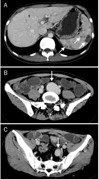
Fig. 1. An abdominal CT scan revealed multiple hemangiomas in the spleen (A), and fusiform aneurysmal dilatation of the left common (B) and internal iliac veins (C).
Fig. 2. Colonoscopy showed hemangiomatous involvement in the rectum; there were rectal varices, but no evidence of active bleeding.
CASE REPORT
A 29-year-old woman was referred to our institution with re- current hematochezia. She had suffered from intermittent hematochezia for several years. She had a gait disturbance and operative scars on the left shin. She had had a number of operations for hemangioma of the abdomen at age 1, the left ankle at age 7 and the left lower leg in adolescence. She had used lightweight orthosis on the left ankle because of the development of foot drop 7 years ago. The circumference of the left thigh had increased during the last several months.
up and slapping her foot down onto the floor. We could not find any signs of family history of hepatitis, liver cirrhosis, limb deformity or hemangioma.
On physical examination, the liver and the spleen were not palpable and the abdomen was flat and soft. At the time of admission, basic metabolic analyses, liver function tests, and complete blood count analyses were performed. All pa- rameters were within normal limits with the exception of Hb 10.4 g/dL (MCV 80.7fL, MCH 23.9 pg), Fe 15 μg/dL, and ferri- tin 3.8 ng/mL. An abdominal CT scan showed multiple he- mangiomas in the spleen and fusiform aneurysmal dilatation of the left common and internal iliac veins without proximal obstructive lesions (Fig. 1). Additionally, there were diffuse mural thickenings with multifocal calcifications involving rec- tum compatible with rectal varices and phleboliths, and mul- tifocal delayed enhancing nodular lesions in the left buttock and gluteus muscle with several calcifications suggesting soft tissue hemangioma with phleboliths. There was no evi- dence of hepatosplenomegaly, liver cirrhosis or ascites. The outcome of an esophagogastroduodenoscopic investigation was unremarkable. However, colonoscopy showed the loss of the normal submucosal vascular pattern. There were he- mangiomatous involvement and variceal change in the rec- tum without evidence of active bleeding (Fig. 2). The blood vessels in the lower sigmoid colon were also dilated.
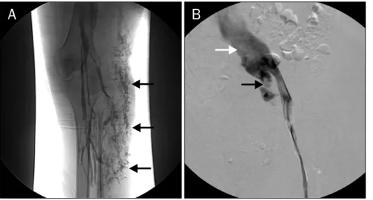
Venography of the left leg showed dilated superficial veins in the subcutaneous tissues and aneurysmal dilatation of the
Fig. 3. Venography of the left leg revealed dilated superficial veins in the subcutaneous tissues (A) and aneurysmal dilatation of the left common (white arrow) and internal iliac veins (black arrow) (B).
left common and internal iliac veins (Fig. 3).
Given the degree of anemia, the age of the patient, and her current quality of life, she has been treated with oral iron sup- plementation only. No additional interventions or additional surgery have so far been necessary.
DISCUSSION
KTS was originally described by the French physicians Klippel and Trenaunay in 1900. While it is characterized by a cutaneous vascular nevus of the involved extremity (capillary malformation), bone and soft tissue hypertrophy of the extremity and venous malformations or varicose veins, KTS can be diagnosed on the basis of any two of these features.1
Although KTS is a relatively well-documented disease, with some large-scale studies reported,1,3 there are few case re- ports in Korea. Recently, rectal involvement, esophageal and gastric varices, and sigmoid varices were reported in Korea.4 The present case involves a patient who presented with mul- tiple cutaneous hemangioma, soft tissue hemangioma and hypertrophy, rectal varix, splenic hemangioma, and deep ve- nous aneurysmal dilatation, all of which are compatible with a diagnosis of KTS. Nevertheless, the fact that some of these clinical features had existed since her childhood, and that she was barely diagnosed with KTS at the age of 29, suggests the point that this syndrome could be underdiagnosed.
Gastrointestinal involvement occurs in 1% to 12.5% of KTS patients.2 The most common involvement in the gastro- intestinal tract is diffuse cavernous hemangiomas of the dis-
tal colon and the rectum.5,6 In one of the largest published studies of KTS patients, hematochezia was observed in six of the 588 patients.3 Although rare, bleeding from jejunal he- mangiomas and esophageal and gastric varices also have been reported.2,4,7 The clinical spectrum of the gastrointes- tinal bleeding may vary from asymptomatic occult bleeding to life-threatening massive bleeding. Gastrointestinal hem- orrhage usually presents in the first decade of the patient’s life and tends to be intermittent.5 In the present case, the pa- tient had suffered from recurrent hematochezia for several years and was diagnosed with rectal varix and iron deficiency anemia. In general, hematochezia may be caused by the pos- terior or compensatory venous pathways of the extremity, which drain into the internal vein.2,8 If these veins can no lon- ger drain efficiently into the dilated internal iliac vein, rectal bleeding may occur. Increased venous pressure on defeca- tion or mucosal trauma resulting from the passage of feces can worsen the recurrent hematochezia.9
Treatment plans for colorectal hemangiomas in KTS should be individualized on the basis of the extent and se- verity of blood loss. Supportive management and iron supple- ments can be sufficient for those patients who only experi- ence the occasional insignificant bleeding. The need for transfusion after massive bleeding or worsening of the qual- ity of life can suggest surgical treatment or an intervention to the involved bowel segment is required. Conservative man- agements such as endoscopic therapy with sclerosis, band ligation, and laser are usually ineffective due to the extensive nature of the lesion.10 Thus, endoscopic therapeutic modal- ities are generally used for postoperative residual sites or lo-
laser for residual lesions and argon laser photocoagulation for hemangiomas involving the distal 7 cm of the anorectum were effective. Our patient was treated with oral iron supple- ment only because of no evidence of active bleeding.
In addition to the gastrointestinal tract, other visceral or- gan involvements such as the liver, bladder, spleen, kidney, heart and lung have also been reported.1 CT of the abdomen and pelvis is a simple, noninvasive method by which visceral hemangiomas can be found.12
Phleboliths are also known as pathognomonic features of venous malformations in very young patients and may be manifestations of previous hemorrhage or thrombus.13
The involvement of the spleen is rare and can be due to the presence of hemangioma or lymphangioma.14 Percutane- ous biopsy of spleen is not advisable because of the high risk of hemorrhage. Noninvasive imaging with ultrasonography, MRI, or CT, and lymphoscintigraphy is used for diagnosis of these lesions. Complications of splenic hemangiomas in- clude hypersplenism, rupture and malignant degenera- tion.11 Fortunately, since recent reviews of adult patients have revealed that asymptomatic patients with a small splen- ic hemangioma (<4 cm) can be treated conservatively with observation,15 we also decided to follow this approach with our patient. KTS with splenic hemangiomas has not been re- ported in Korea yet.
Our patient had deep venous aneurysmal malformations.
This abnormality can lead to thrombotic episodes in the ex- tremities and multiple recurrent pulmonary emboli.16 One study reported that 2.2% of the KTS patients who were en- rolled in the study experienced a venous thrombo-embo- lism.17 In these conditions, oral contraceptives should be strongly avoided. The physician may have to warn their pa- tients that pregnancy can aggravate the symptoms, resulting in either lower extremity swelling, venous malformations, varicosities or venous insufficiency.1 In addition, a popliteal venous aneurysm can induce foot drop because of common peroneal nerve compression,18 although we failed to detect such an aneurysm in our patient. Since the foot drop can be reversed by proper surgical intervention, when a patient with KTS complains of being unable to turn the ankle and toes up- ward, the physician has to consider performing venography to rule out the possibility of a popliteal venous aneurysm.
While many suggestions regarding the underlying cause of
obscure. However, it is most likely to be a generalized meso- dermal development abnormality.19 In addition, several ge- netic mutations including the recently reported E133K muta- tion, may be at least partly responsible for the heterogeneous mesodermal defect.20
We report here a case of a patient with Klippel - Trenaunay syndrome who had rectal varix and multiple splenic he- mangiomas, yet had not been diagnosed until the age of 29 years. While this patient has had a benign clinical course to date, the disease can sometimes show fatal gastrointestinal complications such as massive hemorrhage. Therefore, when endoscopists encounter gastrointestinal varix or he- mangiomatous mucosal change in young patients in the ab- sence of a definite underlying cause, it is important to consid- er the possibility of KTS and to evaluate the patient further by at least performing CT and carefully obtaining the medical history of the patient.
REFERENCES
1. Klippel M, Trenaunay P. Du nævus variqueux ostéo-hyper- trophique. Arch Gen Med 1900;185:641-672.
2. Wilson CL, Song LM, Chua H, et al. Bleeding from cavernous an- giomatosis of the rectum in Klippel-Trenaunay syndrome: re- port of three cases and literature review. Am J Gastroenterol 2001;96:2783-2788.
3. Servelle M. Klippel and Trenaunay's syndrome. 768 operated cases. Ann Surg 1985;201:365-373.
4. Kim JH, Kim CW, Son DK, et al. A case of Klippel-Trenaunay- Weber syndrome presenting with esophageal and gastric vari- ces bleeding. Korean J Gastroenterol 2004;43:137-141.
5. Servelle M, Bastin R, Loygue J, et al. Hematuria and rectal bleeding in the child with Klippel and Trenaunay syndrome.
Ann Surg 1976;183:418-428.
6. Schmitt B, Posselt HG, Waag KL, Müller H, Bender SW. Severe hemorrhage from intestinal hemangiomatosis in Klippel-Tre- naunay syndrome: pitfalls in diagnosis and management. J Pediatr Gastroenterol Nutr 1986;5:155-158.
7. Brown R, Ohri SK, Ghosh P, Jackson J, Spencer J, Allison D. Case report: jejunal vascular malformation in Klippel-Trenaunay syndrome. Clin Radiol 1991;44:134-136.
8. Capraro PA, Fisher J, Hammond DC, Grossman JA. Klippel- Trenaunay syndrome. Plast Reconstr Surg 2002;109:2052- 2060.
9. Natterer J, Joseph JM, Denys A, Dorta G, Hohlfeld J, de Buys Roessingh AS. Life-threatening rectal bleeding with Klippel- Trenaunay syndrome controlled by angiographic embolization and rectal clips. J Pediatr Gastroenterol Nutr 2006;42:581- 584.
10. Goenka MK, Kochhar R, Nagi B, Mehta SK. Rectosigmoid vari- ces and other mucosal changes in patients with portal hypertension. Am J Gastroenterol 1991;86:1185-1189.
11. Kocaman O, Alponat A, Aygün C, et al. Lower gastrointestinal bleeding, hematuria and splenic hemangiomas in Klippel-Tre- naunay syndrome: a case report and literature review. Turk J Gastroenterol 2009;20:62-66.
12. Yeoman LJ, Shaw D. Computerized tomography appearances of pelvic haemangioma involving the large bowel in childhood.
Pediatr Radiol 1989;19:414-416.
13. Azouz EM. Hematuria, rectal bleeding and pelvic phleboliths in children with the Klippel-Trenaunay syndrome. Pediatr Radiol 1983;13:82-88.
14. Jindal R, Sullivan R, Rodda B, Arun D, Hamady M, Cheshire NJ.
Splenic malformation in a patient with Klippel-Trenaunay syn- drome: a case report. J Vasc Surg 2006;43:848-850.
15. Willcox TM, Speer RW, Schlinkert RT, Sarr MG. Hemangioma of
the spleen: presentation, diagnosis and management. J Gastrointest Surg 2000;4:611-613.
16. Aggarwal K, Jain VK, Gupta S, Aggarwal HK, Sen J, Goyal V.
Klippel-Trenaunay syndrome with a life threatening throm- boembolic event. J Dermatol 2003;30:236-240.
17. Baskerville PA, Ackroyd JS, Lea Thomas M, Browse NL. The Klippel-Trenaunay syndrome: clinical, radiological, and haemo- dynamic features and management. Br J Surg 1985;72:232- 236.
18. Jang SH, Lee H, Han SH. Common peroneal nerve compression by a popliteal venous aneurysm. Am J Phys Med Rehabil 2009;
88:947-950.
19. Baskerville PA, Ackroyd JS, Browse NL. The etiology of Klippel-Trenaunay syndrome. Ann Surg 1985;202:624-627.
20. Tian XL, Kadaba R, You SA, et al. Identification of an angiogenic factor that when mutated causes susceptibility to Klippel-Tre- naunay syndrome. Nature 2004;427:640-645.