J o u r n a l o f R h e u m a t i c D i s e a s e s V o l . 1 9 , N o . 5 , O c t o b e r 2 0 1 2
http://dx.doi.org/10.4078/jrd.2012.19.5.302 □ Clinical Im age □
302
<Received:June 21, 2012, Revised:July 10, 2012, Accepted:August 14, 2012>
Corresponding to:Wan-Hee Yoo, Division of Rheumatology, Department of Internal Medicine, Chonbuk National University Medical School and Research Institute of Clinical Medicine of Chonbuk National University Hospital-Chonbuk National University, San 2-20, Geumam-dong, Deokjin-gu, Jeonju 561-180, Korea. E-mail:[email protected]
pISSN: 2093-940X, eISSN: 2233-4718
Copyright ⓒ 2012 by The Korean College of Rheumatology
This is a Free Access article, which permits unrestricted non-commerical use, distribution, and reproduction in any medium, provided the original work is properly cited.
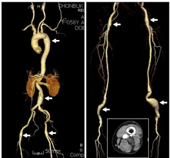
Figure 1. Multiple arterial aneurysmal dilatations on computed tomography angiogram. There were aneurysms on the aortic arch, infrarenal aorta, both common femoral and popliteal arteries (white arrows), and thrombosis in the left popliteal vein (white arrow) on the CT scan of lower extremity.
Multiple Arterial Aneurysms in Behcet’s Disease
Seol A Jang, Wan-Hee Yoo
Division of Rheumatology, Department of Internal Medicine, Chonbuk National University Medical School and Research Institute of Clinical Medicine of Chonbuk National University
Hospital-Chonbuk National University, Jeonju, Korea
Case Report
Patient: 58-year-old female
Chief complaint: Pulsatile swelling on the posterior aspect of left knee for 3 years.
Physical examination: At admission, blood pressure of 115/85 mm Hg, body temperature of 36.8oC, pulse of 67/min and res- piratory rate of 19/min were checked. On presentation, phys- ical examination revealed an about 4×7 cm-sized, non-tender, pulsating mass on the left popliteal area. Pathergy test per- formed using a 20-gauge needle was positive.
Past medical history: She had recurrent oral and genital ulcers for 7 years.
Laboratory tests: Laboratory test revealed white blood cell (WBC) of 8.1 103/mL, Hb of 12.8 g/dL and platelet of 18.3×
103/mL. A high erythrocyte sedimentation rate of 58 mm/hr (normal <20/mm/hr), C-reactive protein of 132.47 mg/L (normal <5 mg/L) and positive HLA-B51 were noted. Blood chemistries, autoantibodies and tests for viral infections were within normal limit or all negative.
Radiologic findings: Computed tomography (CT) angiogram (Panel) revealed aneurysmal dilatation of aortic arch, in- frarenal aorta, both common femoral and popliteal arteries (white arrows), and thrombosis in the left popliteal vein (white arrow) on CT scan of left lower extremity (Figure 1).
Diagnosis and treatment: A diagnosis of Behcet’s disease with arterial and venous complications was made with character- istic clinical symptoms, including recurrent oral and genital ul- cers ischemic pain and positive Pathergy test. She denied to operative treatment and has been treating with glucocorticoid,
colchicines, anti-platelet drug and azathioprine.
Discussion
We here present an unusual case of a Behcet’s disease with multiple arterial aneurysms and deep venous thrombosis.
Behcet’s disease is an inflammatory disorder of unknown cause, characterized by recurrent oral ulcers, genital ulcers,
Multiple Arterial Aneurysms in Behcet’s Disease 303
uveitis, positive Pathergy test and cutaneous and vascular le- sions (1). Vasculitis accounts for most of the pathologic proc- esses in Behcet’s disease and can affect both veins and ar- teries of any sizes (2).
The arterial lesions of Behcet’s disease (vasculo-Behcet’s disease) include aneurysms, stenosis, and occlusions. The ar- tery most often affected is the aorta followed by the pulmo- nary arteries, femoral artery, subclavian artery, and popliteal artery. In most reports, arterial lesions are isolated; rarely, they can be multiple and frequently coexist with venous thrombosis (3). The arterial inflammation is acute and destruc- tive to the vessel wall and results in rapid formation of aneur- ysms with an increased incidence of rupture and bleeding.
Acute arterial involvement in Behcet’s disease should be re- garded as a medical emergency and should be treated with pulsed intravenous corticosteroids and cyclophosphamide fol- lowed by maintenance oral corticosteroids (4). Systemic arte-
rial aneurysms in Behcet’s disease should be surgically cor- rected because of the risk of aneurismal rupture. Thus, early diagnosis and early institution of immunosuppressive therapy and surgical correction will help in preventing formation and progression of this life threatening complication.
References
1. Kastner DL. Arthritis and allied conditions: a textbook of rheumatology. 13st ed. p 1279. Baltimore, Williams &
Wilkins, 1997.
2. Sakane T, Takeno M, Suzuki N, Inaba G. Behçet's disease. N Engl J Med 1999;341:1284-91.
3. Calamia KT, Schirmer M, Melikoglu M. Major vessel in- volvement in Behçet disease. Curr Opin Rheumatol 2005;17:1-8.
4. Barnes CG. Treatment of Behcet's syndrome. Rheumatol- ogy (Oxford) 2006;45:245-7.