Korean J Gastroenterol Vol. 66 No. 3, 168-171 http://dx.doi.org/10.4166/kjg.2015.66.3.168 pISSN 1598-9992 eISSN 2233-6869
CASE REPORT
Korean J Gastroenterol, Vol. 66 No. 3, September 2015 www.kjg.or.kr
위-비장 누공을 형성한 비장 결핵 1예
이경주, 유진세, 전호성, 조승국, 이지현, 하성삼, 조미연1, 김재우
연세대학교 원주의과대학 내과학교실, 병리학교실1
A Case of Splenic Tuberculosis Forming a Gastro-splenic Fistula
Kyong Joo Lee, Jin Sae Yoo, Hosung Jeon, Sung Kook Cho, Ji Hyun Lee, Sung Sam Ha, Mee Yon Cho1, and Jae Woo Kim Departments of Internal Medicine and Pathology1, Yonsei University Wonju College of Medicine, Wonju, Korea
We report a case of a 61-year-old man who presented with a cough and abdominal discomfort. CT scan of the chest showed two lesions across both lungs, and an abdominal CT scan revealed multiple hypodense lesions in the spleen with cystic lesions on the splenic hilum. Upper gastrointestinal tract endoscopy found creamy yellowish discharge through a fistula between the stomach and splenic hilum. Under fluoroscopic guidance, forceps was inserted into the fistula tract, and forcep biopsy was done. The pathology was consistent with tuberculosis, and a nine-month anti-tuberculosis medication regimen was started.
Imaging performed three months after finishing medication indicated improvement of splenic lesions, and the gastro-splenic tract was sealed off. This case is a very rare clinical example of secondary splenic tuberculosis with a gastro-splenic fistula formation in an immunocompetent patient. (Korean J Gastroenterol 2015;66:168-171)
Key Words: Tuberculosis, splenic; Gastro-splenic fistula; Granuloma
Received March 4, 2015. Revised April 3, 2015. Accepted April 7, 2015.
CC This is an open access article distributed under the terms of the Creative Commons Attribution Non-Commercial License (http://creativecommons.org/licenses/
by-nc/4.0) which permits unrestricted non-commercial use, distribution, and reproduction in any medium, provided the original work is properly cited.
Copyright © 2015. Korean Society of Gastroenterology.
교신저자: 김재우, 26426, 원주시 일산로 20, 연세대학교 원주의과대학 내과학교실
Correspondence to: Jae Woo Kim, Department of Internal Medicine, Yonsei University Wonju College of Medicine, 20 Ilsan-ro, Wonju 26426, Korea. Tel: +82-33-741- 1222, Fax: +82-33-741-1228, E-mail: [email protected]
Financial support: None. Conflict of interest: None.
INTRODUCTION
Splenic tuberculosis is a rare form of abdominal tuber- culosis.1 Isolated splenic involvement is very unusual with only a few cases having been reported.2-4 Diagnosis of splen- ic tuberculosis is often delayed due to its nonspecific clinical presentation.5 Gastrointestinal perforation and fistula is a rare, severe complication that can occur in patients with gas- tric tuberculosis.6-8 There are no case reports available on the main manifestation of gastro-splenic fistula formation caused by splenic tuberculosis in an immunocompetent patient. Here, we report a case of secondary splenic tuberculosis that formed a gastro-splenic fistula in a 61-year-old man.
CASE REPORT
A 61-year-old man presented with cough and abdominal discomfort for one month at the outpatient department of pulmonology. On physical examination, blood pressure, pulse rate, and body temperature were within normal range.
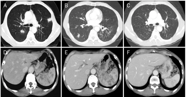
Initial chest CT scan (Fig. 1A) showed two 1.5-cm lobular con- solidations in the posterior segment of the right upper lung and the anterior segment of the left upper lung, compatible with lung abscess and bronchiectasis. The patient was treat- ed for bronchiectasis and lung abscess four years prior, which resolved after antibiotic treatment. Sputum cultures, AFB and bronchial washing cytology for Mycobacterium tuber- culosis were negative. He had no significant past history of
Lee KJ, et al. Splenic Tuberculosis Forming a Gastro-splenic Fistula 169
Vol. 66 No. 3, September 2015
Fig. 1. CT images of chest and abdomen. (A) First chest scan shows two newly-developed 1.5-cm lobular consolidations in posterior segment of right upper lung and anterior segment of left upper lung. (B) Chest scan after one month of antibiotics shows mild improvement of initial chest lesion, but newly developed focal consolidation lesion on right lower lobe. (C) Final chest scan after six months of antituberculous treatment shows disappearance of focal consolidation on both upper lobe and right lower lobe. (D) First abdominal scan shows multiple focal hypodense lesions in spleen with cystic lesion on splenic hilum. (E) Abdominal scan after one month of antibiotics shows enlargement of multiple hypodense lesions on spleen. (F) Final abdominal CT scan after twelve months of antituberculous treatment shows splenic abscesses nearly healed.
Fig. 2. Sonographic and fluoroscopic images. (A) Abdominal ultrasonography shows multiple low echoic nodules in spleen. (B) When dye was injected through ulceration site using gastro- scope under fluoroscopic guidance, there was a fistular tract between the stomach and spleen.
pulmonary tuberculosis or contact with tuberculosis pa- tients, and serum ELISA test for HIV test was negative. Blood biochemical profiles, including liver and kidney function tests, were within normal range. The respiratory specialist who first saw the patient believed the lung lesions to be lung abscesses because they appeared different from typical tu- berculosis presentation, and because sputum cultures and bronchial washing cytology test for tuberculosis were negative.
Therefore, the patient was started on typical oral antibiotics including third-generation cephalosporin and metronidazole.
The chest lesions improved after one month of antibiotic
therapy. However, the initial chest CT scan also revealed mul- ti-focal hypodense lesions in the spleen with cystic lesions on the splenic hilum (Fig. 1D). While follow-up chest and abdomi- nal CT scans showed the chest lesions to be improving, a new- ly developed focal consolidation lesion on the right lower lobe was detected (Fig. 1B), with enlargement of multiple hypo- dense lesions on the spleen (Fig. 1E). The patient also started to complain of poor oral intake and abdominal discomfort.
Therefore, he was transferred to the gastroenterology clinic of our hospital for further evaluation. Abdominal ultrasono- graphy showed multiple low echoic nodules in the spleen (Fig.
170 이경주 등. 위-비장 누공을 형성한 비장 결핵
The Korean Journal of Gastroenterology
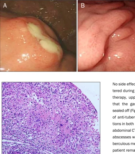
Fig. 3. Endoscopic findings. (A) Initial gastroscopy shows creamy yellowish discharge on ulceration of subepithelial mass from fundus to upper body, similar to an abscess. (B) After two months of antitubercular therapy, follow-up gastroscopy shows gastro- splenic fistular tract sealed off.
Fig. 4. Microscopic finding of spleen biopsy specimen shows well- defined granuloma with epithelial cells and central caseous necrosis (H&E, ×100). Finding is compatible with chronic granulomatous inflammation with caseous necrosis, suggestive of tuberculosis.
2A). Upper gastrointestinal tract endoscopy revealed a creamy yellowish discharge from an ulceration of the sub- epithelial mass, from the fundus to the upper body, similar to an abscess (Fig. 3A). Under fluoroscopic guidance, a dye was injected through the ulceration site and a fistula tract be- tween the stomach and spleen was observed (Fig. 2B). The forceps were inserted more than 3 cm deep into the fistula tract, and forcep biopsy was done. The Ziehl-Nielsen stain for AFB and PCR for tuberculosis were negative. However, the pathologist suggested tuberculosis as the main diagnosis, as the forcep biopsy showed a well-defined granuloma with epi- thelial cells and central caseous necrosis (Fig. 4). The patient was started on four anti-tuberculous agents (isoniazid 300 mg/day, rifampin 600 mg/day, ethambutol 1,200 mg/day, pyrazinamide 1,500 mg/day) for nine months, as splenic tu- berculosis is usually associated with disseminated disease.
No side effect from the anti-tuberculous drugs was encoun- tered during therapy. After two months of anti-tuberculous therapy, upper gastrointestinal tract endoscopy revealed that the gastro-splenic fistula tract had spontaneously sealed off (Fig. 3B). A follow-up chest CT scan after six months of anti-tuberculous treatment showed multiple consolida- tions in both lungs to have disappeared (Fig. 1C). A follow-up abdominal CT scan at nine months indicated that the splenic abscesses were also nearly healed (Fig. 1F), so the anti-tu- berculous medication was discontinued at nine months. The patient remains well for one year without recurrence.
DISCUSSION
Gastro-splenic fistula formation due to secondary splenic tuberculosis is a very rare manifestation. Splenic tuberculosis is thought to be increasing in frequency because of the in- creasing number of immunocompromised patients.9 The clinical presentation of splenic tuberculosis is nonspecific.
Splenic tuberculosis is usually confirmed by involvement of other organs. Therefore, invasive diagnostic modalities are not needed in the workup of splenic lesions in most tuber- culosis patients.10 However, the diagnosis of splenic tuber- culosis is difficult if tubercular foci are not observed in other locations, including the lung. In our case, although there was a visible lung lesion and primary pulmonary tuberculosis was suspected, several tuberculosis diagnostic tests were nega- tive. Furthermore, the initial chest lesions improved with typi- cal antibiotic treatment, obscuring the diagnosis of pulmo- nary tuberculosis. However, the lung lesion that simulta- neously presented with splenic lesion disappeared after anti- tuberculous treatment, leading to our suspecting pulmonary
Lee KJ, et al. Splenic Tuberculosis Forming a Gastro-splenic Fistula 171
Vol. 66 No. 3, September 2015
tuberculosis as the primary disease in this patient. Since the disease was progressing without treatment, gastro-splenic fis- tula formation was expressed as a main manifestation of sec- ondary splenic tuberculosis.
Splenic involvement by tuberculosis is usually associated with disseminated tuberculosis. The tuberculous abscess formation in the spleen likely resulted from the over-reaction of the host immune system, with formation of caseating necrosis. Splenic tuberculosis occurs in two forms. The first form is encountered with miliary tuberculosis, especially in immunocompromised patients. The second form is primary involvement of the spleen, which is extremely rare, especially in an immunocompetent person.11,12
Gastrointestinal perforation and fistula caused by tuber- culosis are rare, and can cause serious complication than obstruction.6-8 However, the fistula that occurred in our pa- tient did not result in serious complications because it was contiguously formed between the posterior wall of stomach and the splenic hilum. Since creamy yellowish exudate was discharged through the fistula, biopsies were performed through the tract, and splenic tuberculosis was diagnosed.
Splenic abscess due to M. tuberculosis can be treated ini- tially with antibiotics, limiting splenectomy to those cases that do not respond to medical treatment.9 As most patients with splenic tuberculosis may have disseminated disease, a few controlled trials of treatment in patients with extra-pulmonary tuberculosis strongly suggest a 12-month regimen, with pro- longed treatment if deemed necessary.13,14 In contrast, one study showed very slight improvement between imaging studies at 12 and 24 months although anti-tuberculous treat- ment was continued for over 24 months due to unresolved lesions.13 The optimal duration of treatment for splenic tuber- culosis is undetermined: in our case, further treatment was discontinued after nine months of anti-tuberculous medi- cation since most of the lesions had disappeared. Imaging studies taken one year after cessation of treatment showed healed splenic abscesses and pulmonary lesions without evi- dence of recurrence. This case is a very rare clinical example
of secondary splenic tuberculosis with a gastro- splenic fistula formation in an immunocompetent patient.
REFERENCES
1. Pramesh CS, Tamhankar AP, Rege SA, Shah SR. Splenic tuber- culosis and HIV-1 infection. Lancet 2002;359:353.
2. Ho PL, Chim CS, Yuen KY. Isolated splenic tuberculosis present- ing with pyrexia of unknown origin. Scand J Infect Dis 2000;
32:700-701.
3. Kim HH, Park SJ, Park MI, Moon W. Primary tuberculous abscess of the spleen in an immununocompetent patient diagnosed by biochemical markers and radiologic findings. J Clin Med Res 2012;4:149-151.
4. Raul SK, Tiwari S, Raina V, Sharma R, Joseph M. Tubercular splenic abscess in immunocompetent patients: a rare entity.
Indian J Surg 2009;71:213-215.
5. Tung CC, Chen FC, Lo CJ. Splenic abscess: an easily overlooked disease? Am Surg 2006;72:322-325.
6. Gill RS, Gill SS, Mangat H, Logsetty S. Gastric perforation asso- ciated with tuberculosis: a case report. Case Rep Med 2011;
2011:392769.
7. Nagi B, Lal A, Kochhar R, Bhasin DK, Thapa BR, Singh K.
Perforations and fistulae in gastrointestinal tuberculosis. Acta Radiol 2002;43:501-506.
8. Sharma D, Gupta A, Jain BK, et al. Tuberculous gastric perfo- ration: report of a case. Surg Today 2004;34:537-541.
9. Llenas-García J, Fernández-Ruiz M, Caurcel L, Enguita-Valls A, Vila-Santos J, Guerra-Vales JM. Splenic abscess: a review of 22 cases in a single institution. Eur J Intern Med 2009;20:537-539.
10. Sharma SK, Smith-Rohrberg D, Tahir M, Mohan A, Seith A.
Radiological manifestations of splenic tuberculosis: a 23-patient case series from India. Indian J Med Res 2007;125:669-678.
11. Ray S, Kundu S, Goswami M, Sarkar D, Saha M. Isolated tu- bercular splenic abscess: can we defer splenectomy? Our single experience with anti-tuberculous therapy alone. Indian J Med Microbiol 2012;30:101-103.
12. Kundu PR, Mathur SK, Singh S, Duhan A, Aggarwal G, Sen R.
Isolated tuberculous splenic abscess in an immunocompetent individual. Asian Pac J Trop Med 2011;4:81-82.
13. Hamizah R, Rohana AG, Anwar SA, Ong TZ, Hamzaini AH, Zulkarnaen AN. Splenic tuberculosis presenting as pyrexia of un- known origin. Med J Malaysia 2007;62:70-71.
14. Dede F, Doğan E, Demir M, et al. Unusual presentation of tuber- culosis as a splenic mass. Tohoku J Exp Med 2006;210:79-82.