수근관증후군은 수근관내 압력의 증가로 생기는 정중신경 의 압박신경병증으로 정중신경의 분포 영역에 작열통, 무감 각, 지각 이상, 무지구부 근육의 위축, 그리고 심한 경우에는 어깨로의 방사통을 일으키는 비교적 흔한 만성질환이다(1, 2).
여자에서 많이 발생하여 남:녀 발생비는 1 : 2 - 5이며, 40-60세에 서 호발한다( 1 ) .
진단은 주로 임상증상을 기초로 하여 근전도 검사에 의해 이루어지나, 정중신경의 압박정도와 만성도에 따라 수근관증 후군 증상을 호소하고 있는 환자들에서도 근전도 소견이 모 호하거나 음성으로 나올 수가 있다(3, 4).
M R I는 연조직 대조도가 우수하여 수근관증후군을 진단하 는데 유용한 방법으로 알려져 있지만(3, 5, 6-8), 비용이 비싸 고 시간이 많이 드는 단점 때문에 보편적으로 사용되는 초기 의 영상장치는 아니며, 최근 적은 비용과 짧은 시간에 검사할 수 있는 고해상 초음파가 수근관증후군의 진단에 유용하다는
보고들이 있다(1, 3, 6).
이에 저자들은 근전도 검사로써 진단된 수근관증후군 환자 와 정상대조군을 대상으로 고해상 초음파를 실시하고, 수근관 증후군의 진단에 도움을 줄 수 있는 초음파 소견 및 그 진단 적 유용성을 알아보고자 본 연구를 시행하였다.
대상 및 방법
1 9 9 7년 8월부터 1 9 9 8년 9월까지 임상적으로 수근관증후근을 시사하는 소견을 호소하여 내원한 환자들중 각각 근전도 및 초음파 검사를 시행한 후 근전도로 진단된 수근관증후군 환 자 2 6명의 4 4예와 정상대조군 1 5명의 3 0예를 대상으로 하였다.
대상자는 모두 여자였고, 환자군의 평균 연령은 5 2세( 3 5 - 6 7 세)였으며 그 중 1 9명은 양측에 병변이 있었다. 정상대조군의 평균 연령은 4 8세( 3 3 - 6 2세)였다.
사용된 초음파 기기는 Acuson 128XP(Acuson, Mountain view, California, U.S.A.)였고 7.5MHz 선형 탐촉자를 사용 하였다. 손목관절의 초음파검사는 손목을 중립위로 위치한 상 목적: 수근관증후군의 초음파 소견 및 그 진단적 유용성을 알아보고자 하였다.
대상 및 방법:수부의 저림을 호소하여 내원한 환자들 중 근전도로 진단된 수근관증후군 환
자 2 6명의 4 4예(평균연령 5 2세, 35-67세)와 정상대조군 1 5명의 3 0예(평균연령 4 8세, 33-62세) 를 대상으로 하였다. 초음파 촬영은 원위부 요골, 두상골 및 유구골 선상에서 각각 수근관내 정중신경의 단면적 및 편평도를 측정하였고, 유구골 선상에서 굴근지대의 굽힘정도(수장전 위)를 측정하였다. 또한 인지의 위치를 7 0°굴곡위 및 7 0°신전위로 변화시키면서 두상골 선상 에서 척골동맥으로부터의 정중신경의 이동성을 측정하였다.
결과:정중신경의 단면적은 원위부 요골에서 환자군은 9 . 2 9±2 . 63m m2( m e a n±S.D.), 대조군은 5 . 4 5±1 . 98m m2였고, 두상골 선상에서 각각 1 0 . 6 8±3 . 38m m2, 6.55±2 . 01 m m2였으며, 유구골 선상 에선 각각 1 0 . 8 8±2 . 78 m m2, 6.34±2 . 00m m2으로, 세 위치 모두 환자군에서 대조군에 비해 유의 하게 증가되었다(p=0.0001). 정중신경의 편평도는 유구골 선상에서만 환자군 2 . 3 7±0 . 5 6으로 대조군 2 . 0 6±0 . 3 6에 비해 유의하게 증가되었다(p=0.0064). 굴근지대의 수장전위는 환자군 3 . 4 4±0 . 90mm, 대조군 2 . 2 0±0 . 55 m m로 전자에서 유의하게 증가되었다(p=0.0001). 인지의 수 동 굴곡과 신전시에 정중신경의 이동거리는 환자군 0 . 9 8±1.03 mm, 대조군 1 . 6 5±1 . 22 m m로 환자군에서 유의하게 감소하였다(p=0.0180).
결론:고해상 초음파는 수근관증후군을 진단하는데 유용한 검사이며, 정중신경의 단면적 증 가, 수근관 원위부에서의 편평도 증가, 굴근지대의 수장전위 증가 및 인지의 굴곡과 신전시 정중신경의 이동거리 감소 등이 유의한 소견이었다.
1가톨릭대학교 의과대학 대전성모병원 진단방사선과
2가톨릭대학교 의과대학 대전성모병원 재활의학과
이 논문은 1 9 9 9년 5월 2 4일 접수하여 1 9 9 9년 8월 1 9일에 채택되었음.
수근관증후군의 초음파 소견
1이연수・최은석2・민태규・김지창・이은자・김 현・강시원
태에서 축상면 및 시상면 스캔을 실시하였다.
수근관증후근을 초음파로 진단하기 위한 정량적 분석 방법 으로 축상면상 원위부 요골(distal radius), 두상골( p i s i f o r m ) , 그리고 유구골(hamate) 선상의 세 위치에서 각각의 정중신경 단면적을 측정하였고, 정중신경의 전후 및 좌우 직경을 측정 하여 신경의 편평도를 계산하였다(좌우 직경/전후 직경).
굴근지대(flexor retinaculum)의 굽힘 정도을 측정하기 위해 굴근지대의 양측 부착 부위인 대능형골 융기부(tubercle of the t r a p e z i u m )와 유구골 갈고리(hook of the hamate)사이에 직선 을 긋고 이 직선에서부터 굴근지대의 수장첨(palmar apex)까 지의 거리를 측정하였다(수장 전위)(Fig. 1).
또한 정중신경의 이동성을 알아보기 위하여 대상자의 인지 를 7 0°굴곡위와 7 0°신전위로 움직이면서, 두상골 선상에서 척골동맥으로부터의 정중신경의 거리를 측정하였으며, 인지 의 굴곡과 신전시 정중신경과 척골동맥과의 거리의 차이를 정중신경의 활주거리(sliding distance)로 정의하였다.
자료분석은 unpaired t-test를 사용하여 환자군과 대조군사 이에 정중신경의 단면적, 편평도, 수장전위 및 정중신경의 활 주거리를 비교분석하였으며 통계적 유의수준은 p value 0.05 미만으로 하였다.
결 과
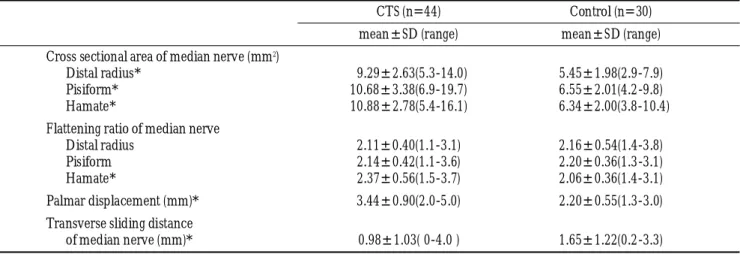
정상대조군과 비교한 수근관증후군의 초음파 소견은 T a b l e 1과 같다.
환자군과 대조군에 있어서 정중신경의 단면적은 원위부 요 골에서 각각 9 . 2 9±2 . 63 m m2(range, 5.3-14.0m m2), 5.45±1 . 98m m2 (range, 2.9-7.9 m m2), 두상골 선 상 에 서 각각 1 0 . 6 8±
3 . 38 m m2(range, 6.9-19.7 m m2) , 6.55±2.01 mm2(range, 4.2-
9 .8 m m2) 유구골 선상에서 1 0 . 8 8±2.78 mm2(range, 5.4- 1 6 .1 m m2), 6.34±2 . 00m m2 (range, 3.8-10.4m m2)로, 세 측정부위 모두 환자군에서 대조군에 비해 유의하게 증가되었다 (p=0.0001) (Figs. 2, 3).
정중신경의 평균 편평도는 원위부 요골과 두상골선상에서는 두 군간에 유의한 차이를 보이지 않은 반면, 유구골 선상의 경 우 환자군에서 2 . 3 7±0.56(range, 1.5-3.7)으로 대조군 2 . 0 6±
0.36(range, 1.4-3.1)에 비해 유의하게 증가하였다( p = 0 . 0 0 6 4 ) (Fig. 4).
굴근지대의 평균 수장 전위는 환자군 3 . 4 4±0.90 mm (range, 2.0-5.0mm), 대조군 2 . 2 0±0 . 55mm(range, 1.3-3.0m m )로 환자군에서 대조군에 비해 유의하게 증가되었다( p = 0 . 0 0 0 1 ) (Fig. 5).
인지를 굴곡과 신전시키면서 측정한 정중신경의 평균 활주 거리는 환자군에서 0 . 9 8±1 . 03mm(range, 0-4.0m m )로 대조군 1 . 6 5±1 . 22mm(range, 0.2-3.3m m )에 비해 유의하게 감소하였다 (p=0.0180) (Figs. 6, 7).
고 찰
수근관증후군은 잘 알려진 질환이지만 병인론은 아직 확실 하지 않다. 원인은 크게 수근관 내용물의 부피를 증가시키는 경우와 수근관의 크기를 감소시키는 경우로 나눌 수 있으나 (3, 9, 10), 원인과 상관없이 수근관의 좁아진 공간 내에서 정 중신경이 압박되어 수근관증후군이 발생된다. 즉, 수근관 내 에 증가된 간질액 압이나 직접적인 정중신경의 압박을 일으 키는 압력이 정중신경의 미세혈관을 변화시켜 허혈 및 혈류 장애를 야기하며(11, 12), 혈류장애에 의한 반응으로 신경외막 의 부종에 의해 신경종창이 일어나고(13), 만성 압박 부위에 서 수초의 변형 및 신경의 편평화를, 그리고 압박 근위부에선 신경의 종창을 유발시킨다. 일부 환자에서 수근관증후군의 진 단은 고전적 병력, Tinel 징후나 Phalen 검사와 같은 이학적 검사, 그리고 정중신경 분포 지역의 지각감퇴 등과 같은 임상 적 소견을 기초로 하여 쉽게 이루어질 수 있다(3, 12). 그러나 오늘날 대부분의 수근관증후군 환자들은 심한 근위축 또는 감각의 저하가 발생하기 전인, 병의 초기에 내원하기 때문에, 진단이 어려운 경우가 많다.
근전도 검사는 수근관을 통한 정중신경 전도의 지연을 특 징적으로 보여줄 뿐 아니라, 미만성 말초신경병증, 경수신경 근병증, 그리고 단일신경병증과 같은 다른 신경질환들과 감별 할 수 있으므로 수근관증후군을 진단하는데 매우 유용한 검 사이지만, 수근관증후근 환자의 5 - 10%에서, 특히 경한 증상 을 가진 환자에서는 정상 소견을 나타낼 수 있다( 1 4 ) .
정상적인 수근관의 해부와 병리적 변화를 영상화하기 위해 많은 시도가 있었으나, 단순 방사선촬영은 전위 골절, 이상배 열, 가골형성 또는 비후성 변화 등의 수근골 변형을 보여 주 는 것 외에 별 도움을 주지 못하며, CT는 연조직 대조도가 낮 기 때문에 그 유용성이 제한되어 있다(1, 15). 반면, MRI는 뛰 어난 연부조직 대조도 때문에 수근관의 정상적인 해부와 병 Fig. 1. Schematic diagram of palmar displacement (PD) of the
flexor retinaculum shows that a straight line is drawn between the tubercle of the trapezium and the hook of the hamate, and the distance from this line to the palmar apex of the flexor reti- naculum is measured.
H:hook of hamate, C:capitate, Td:Trapezoid, Tm:trapezium, PD:palmar displacement, Fr:flexor retinaculum.
변을 진단하는데 가장 훌륭한 검사이나(7, 8, 16, 17), 시간이 오래 걸리고 고비용 등의 단점이 있어 보편적으로 이용되지 는 못하고 있다.
1 9 8 8년 고해상 초음파가 손의 연부조직 병변의 진단에 사용 된 이후(18), Fornage(19)에 의해 정중신경을 포함한 다양한 사지의 말초신경에 대한 초음파 소견이 발표되었고, 수근관증 후군의 초음파 소견에 대해서도 많은 연구가 이루어지고 있 다(1, 3, 6).
수근관은 수장측의 굴근지대와 배측의 수근골 사이에 위치 하는 제한된 공간으로(Fig. 8), 공통의 활액초에 의해 둘러싸 인 4개의 심부 수지굴건(flexor digitorum profundus tendon)과 4 개의 표재성 수지굴건(flexor digitorum superficialis tendon), 독 립된 활액초(synovial sheath)에 의해 둘러싸인 장무지굴건 (flexor pollicis longus tendon), 그리고 정중신경을 포함하고 있 다( 1 0 ) .초음파상 정중신경은 굴근지대 바로 아래에 위치하며
음속이 신경면에 수직일 때 고에코, 음속이 약간 경사질 때 저 에코로 나타나고, 신경 주위 섬유조직을 의미하는 좁은 고에 코의 테두리에 의해 둘러싸여 있다. 고에코의 굴근지대는 정 상적으로 직선 또는 약간 볼록한 형태이고 굴건들은 초음파 음속(ultrasound beam)의 각에 따라 에코가 다양하며, 각 건들 의 활액막을 나타내는 저에코의 구조에 의해 싸여 있다(1, 3).
Buchberger 등(1, 6)은 수근관증후군의 고해상 초음파 진단 에 있어서 원인과 관계없이 근위부 수근관에서 정중신경의 종창, 원위부 수근관에서 정중신경의 편평화, 그리고 굴근지 대의 증가된 굽힘(bowing) 등의 3가지 객관적 소견을 기술하 였는데, 이러한 초음파 소견은 Middleton 등( 7 )과 M e s g a r - zadeh 등( 8 )이 발표한 MRI 소견과 유사하였다.
저자들의 연구에서 정상대조군의 수근관내 정중신경의 단 면적은 두상골 선상에서 6 . 5 5±2 . 01 m m2로 가장 컸으며, 환자 군의 경우 대조군에 비해 세 측정 위치 모두에서 정중신경의
Fig. 2. A 35-year-old woman with normal control.
Axial sonogram of wrist at pisiform level shows median n- erve(arrow) with normal size and shape. Cross sectional area of the median nerve is measured as 5.0 m m2.
Fig. 3. A 51-year-old woman with carpal tunnel syndrome.
Axial sonogram of wrist at pisiform level shows enlarged hy- poechoic median nerve. Cross sectional area of the median n- erve is measured as 15.3 m m2.
MN:median nerve.
Table 1.Comparison of Ultrasonographic Findings between Patient and Control
CTS (n=44) Control (n=30)
m e a n±SD (range) m e a n±SD (range) Cross sectional area of median nerve (mm2)
Distal radius* 09 . 2 9±2 . 6 3 ( 5 . 3-14.0) 5 . 4 5±1 . 9 8 ( 2 . 9-7 . 9 ) Pisiform* 1 0 . 6 8±3 . 3 8 ( 6 . 9-19.7) 6 . 5 5±2 . 0 1 ( 4 . 2-9 . 8 ) Hamate* 1 0 . 8 8±2 . 7 8 ( 5 . 4-16.1) 6 . 3 4±2 . 0 0 ( 3 . 8-10.4) Flattening ratio of median nerve
Distal radius 2 . 1 1±0 . 4 0 ( 1 . 1-3.1) 2 . 1 6±0 . 5 4 ( 1 . 4-3.8) Pisiform 2 . 1 4±0 . 4 2 ( 1 . 1-3.6) 2 . 2 0±0 . 3 6 ( 1 . 3-3 . 1 )
Hamate* 2 . 3 7±0 . 5 6 ( 1 . 5-3.7) 2 . 0 6±0 . 3 6 ( 1 . 4-3 . 1 )
Palmar displacement (mm)* 3 . 4 4±0 . 9 0 ( 2 . 0-5.0) 2 . 2 0±0 . 5 5 ( 1 . 3-3.0) Transverse sliding distance
of median nerve (mm)* 0 . 9 8±1 . 0 3 ( 0-4 .0 ) 1 . 6 5±1 . 2 2 ( 0 . 2-3 . 3 ) CTS: carpal tunnel syndrome
*: p<0.05
단면적이 유의하게 증가되었고 특히 두상골과 유구골 선상에 서 현저하게 증가되었다(두상골 1 0 . 6 8±3 . 38 m m2, 유구골 1 0 . 8 8
±2 . 78 m m2). 이는 두상골선 상에서 단면적이 가장 컸던 Buchberger 등( 6 )의 보고와 다소 차이가 있었는데, 그 이유는 가신경종으로 생각되는 수근관 근위부의 국소적인 정중신경 의 종창보다는 전반적으로 정중신경이 커진 경우가 많았기 때문으로 생각된다(20, 21).
저자들은 환자군에서 정중신경의 편평도가 유구골 선상에 서만 유의하게 증가됨을 관찰하였는데, 이는 주로 굴건과 횡수 근인대 사이에서 정중신경이 압박되는 것을 시사하며, Buch- berger 등(1, 6)의 연구에서도 같은 소견을 보고한 바 있다.
굴근지대의 수장전위는 환자군에서 유의하게 증가하였는데,
이러한 수장전위의 증가는 수근관내 내용물의 부피 증가 또는 수근관내의 압력 증가의 결과로 해석된다(14, 22). 한편, Mesgarzadeh 등( 8 )은 굴근지대의 수장전위가 유구골 선상에서 가장 잘 관찰된다고 하였으며, 그 이유는 이 부위의 굴근지대 가 가장 굵어 측정이 용이하기 때문이라고 기술한 바 있다.
본 연구의 결과에서 m e a n±2 SD를 적용한 수근관내 측정 지수들의 정상범위는 단면적의 경우, 원위부 요골에서 1 . 4 9 - 9.41 mm2, 두상골 선상에서 2 . 5 3 - 1 0 . 57 m m2, 유구골 선상에서 2 . 3 4 - 1 0 . 34 m m2였으며, 유구골 선상에서의 정중신경 편평도의 정상범위는 1.34-2.78, 그리고 수장전위는 1 . 1 - 3 .3 m m로 측정되 었다.
즉, 고해상 초음파 검사상 수근관내 정중신경의 단면적이
Fig. 4. A 52-year-old woman with carpal tunnel syndrome.
Axial sonogram at hamate level shows increased flattening of the median nerve (arrow). Flattening ratio of the median nerve is measured as 3.7.
Fig. 5. A 52-year-old woman with carpal tunnel syndrome.
Axial sonogram at hook of hamate level shows increased pal- mar bowing of flexor retinaculum. Palmar displacement of flexor retinaculum is measured as 4.1 mm.
PD:palmar displacement.
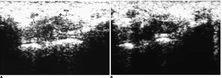
Fig. 6. Transverse sliding of median nerve in a 38-year-old woman with normal control.
A . Axial sonogram of wrist during flexion of index finger.
B . Axial sonogram of wrist during extension of index finger shows that median nerve moves toward ulnar aspect of wrist. Sliding distance of the median nerve is measured as 3.1 mm.
MN:median nerve, UA:ulnar artery.
A B
1 0 .6 m m2 이상이거나, 편평도가 2 . 8이상이며, 수장전위가 3 .3mm 이상이면, 수근관증후군을 강력하게 시사하는 것으로 생각된다.
한편, 본 연구에서 인지의 수동적인 굴곡 및 신전시에 정중 신경의 활주거리가 환자군에서 유의하게 감소하였는데, Nakamichi 등( 2 3 )도 같은 소견을 보고한 바 있다. 즉 정상인에 서 인지의 수동 신전시 생리적인 현상으로 심지굴건과 표재굴 건이 정중신경의 배측 및 요골 측으로 압력을 가하여 정중신 경을 척골측으로 밀어내는 이동현상이 관찰되나, 수근관증후 군 환자에서는 이러한 정중신경의 이동이 감소되는데, 이는 수 근관 내용물의 부피증가나 공간의 상대적인 감소로 인한 만성 적인 신경압박 또는 주변 조직과의 유착에 기인한 것으로 해 석될 수 있으며, 이러한 소견은 초음파가 M R I와 달리 정중신 경의 이동성을 관찰할 수 있는 역동적인 검사임을 나타낸다.
본 논문의 제한점으로 모든 연구대상이 근전도로 진단되었 으나 수술로써 그 해부학적 원인을 확인하지 못하였는데, Pha- l e n ( 2 0 )은 수근관증후근의 수술적 소견에 대한 연구에서 가장 많은 원인으로 굴건초의 비후 또는 섬유화를 보고하였고,
Werner 등( 1 1 )과 Resnick 등( 9 )도 굴건의 비후 또는 염증을 보 고한 바 있다. 수근관증후군의 병인을 확인하는데는 초음파보 다 M R I가 우수한 것으로 보고되며(6), 특히 초음파로 건초의 비후를 평가하는 것은 쉽지 않은 것으로 알려져 있다(1, 12).
Nakamichi 등( 2 4 )은 초음파로 활막염의 진단이 가능하다고 하 였으나, 저자들의 경우 활막염의 유무를 판단하기는 어려웠으 며 향후 수술소견과 비교한 연구가 필요할 것으로 사료된다.
또한 M R I는 초음파와 비교하여 정중신경의 신호강도를 알 수 있고, 수장전위의 측정이 더 정확하며, 초음파에서 놓칠 수 있는 경한 신경압박도 진단할 수 있다는 보고가 있으나( 6 ) , 본 연구는 MRI 검사를 시행하지 않아 두 검사의 비교는 이 루어지지 않았고, 향후 두 검사방법에 의한 소견의 비교연구 가 필요하다고 생각된다.
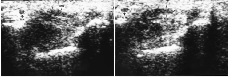
결론적으로 다른 영상장치에 비해 적은 비용과 짧은 시간 에 검사할 수 있는 장점을 가지고 있으며, 정중신경의 횡활주 와 같은 신경조직의 역동적 성질을 평가할 수 있는 고해상 초 음파는 수근관증후근 진단에 유용한 영상장치로 생각되며, 특 히 정중신경 단면적의 증가, 원위부 수근관에서 정중신경 편 Fig. 7. Transverse sliding of median nerve in a 48-year-old woman with carpal tunnel syndrome.
A . Axial sonogram of wrist during flexion of index finger.
B . Axial sonogram of wrist during extension of index finger shows little difference in transverse sliding of median nerve as com- pared with flexion view. Sliding distance of the median nerve is measured as 0.2 m m .
MN:median nerve, UA:ulnar artery.
A B
Fig. 8. Schematic diagram of normal carpal tunnel at hamate level shows that median nerve lies underneath flex- or retinaculum and courses ventral to the flexor tendons.
H:hamate, C:capitate, Td:trapezoid, Tm:trapezium,
Fr:flexor retinaculum, Fds:flexor digi- torum superficialis tendon,
Fdp:flexor digitorum profundus ten- don, Fpl:flexor pollcis longus tendon, MN:median nerve, UA:ulnar artery, UN:ulnar nerve.
평도의 증가, 굴근지대의 수장전위 증가 및 정중신경의 횡활 주거리 감소 등이 유의한 소견으로 사료된다.
참 고 문 헌
1 . Buchberger W, Schon G, Strasser K, Jungwirth W. High-resolution ultrasonography of the carpal tunnel. J Ultrasound Med 1 9 9 1 ; 1 0 ; 5 3 1 - 5 3 7
2 . Phalen GS. The carpal tunnel syndrome: seventeen year’s experi- ence in diagnosis and treatment of six hundred fifty-four hands. J Bone Joint Surg 1 9 6 6 ; 4 8 A : 2 1 1 - 2 2 8
3 . Chen P, Maklad N, Redwine M, Zelitt D. Dynamic high resolution sonography of the carpal tunnel. AJR 1 9 9 7 ; 1 6 8 : 5 3 3 - 5 3 7
4 . Dawson DM. Entrapment neuropathies of the upper extremities.
N Engl J Med 1 9 9 3 ; 3 2 9 : 2 0 1 3 - 2 0 1 8
5 . Brahme SK, Hodler J, Braun RM, Sebrechts C, Jackson W, Resnick D. Dynamic MR imaging of carpal tunnel syndrome. Skeletal Radiol 1 9 9 7 ; 2 6 : 4 8 2 - 4 8 7
6 . Buchberger W, Judmaier W, Birbamer G, Lener M, Schmidaur C.
Carpal tunnel syndrome: diagnosis with high-resolution sonogra- phy. AJR 1 9 9 2 ; 1 5 9 : 7 9 3 - 7 9 8
7 . Middleton WD, Kneeland JB, Kellman GM, et al. MR imaging of the carpal tunnel: normal anatomy and preliminary findings in the carpal tunnel syndrome. AJR 1 9 8 7 ; 1 4 8 : 3 0 7 - 3 1 6
8 . Mesgarzadeh M, Schneck CD, Bonakdarpour A, Mitra A, Conaway D. Carpal tunnel: MR imaging. II. carpal tunnel syn- drome. Radiology 1 9 8 9 ; 1 7 1 : 7 4 9 - 7 5 4
9 . Resnick D, Kang HS. Internal derangement of joints;Emphasis on MR I m a g i n g . Philadelphia;Saunders 1997;149-151
1 0 . Stoller DW, Brody GA. The wrist and hand. In stoller DW. M a g n e - tic resonance imaging in orthpedics & sports medicine. P h i l a d e l - pia:Lippincott-Raven 1997:956-961
1 1 . Werner RA, Armstrong TJ. Carpal tunnel syndrome: Ergonomic risk factors and intracarpal canal presure. Phys Med Rehabil Clin North Am 1 9 9 7 ; 8 : 5 5 5 - 5 6 9
1 2 . Akelman E, Weiss AP. Carpal tunnel syndrome: Etiology and en- doscopic treatment. Orthop Clin North Am 1 9 9 5 ; 2 6 : 7 6 9 - 7 7 8 1 3 . Omer GE. Median nerve compression at the wrist. Hand Clin 1 9 9 2 ;
8 : 3 1 7 - 3 2 4
1 4 . Rosenbaum RB. The role of imaging in the diagnosis of carpal tun- nel syndrome. Invest Radiol 1 9 9 3 ; 2 8 : 1 0 5 9 - 1 0 6 2
1 5 . John V, Nau HE, Nasher HC, Reinhardt V, Venjakob K. CT of carpal tunnel syndrome. AJNR 1 9 8 3 ; 4 : 7 7 0 - 7 7 2
1 6 . Zeiss J, Skie M, Ebraheim N, Jackson WT. Anatomic relations be- tween the median nerve and flexor tendons in the carpal tunnel:
MR evaluation in normal volunteers. AJR 1 9 8 9 ; 1 5 3 : 5 3 3 - 5 3 6 1 7 . Mesgarzadeh M, Schneck CD, Bonakdarpour A. Carpal tunnel:
MR imaging. I. Normal anatomy. Radiology 1 9 8 9 ; 1 7 1 : 7 4 3 - 7 4 8 1 8 . Fornage BD, Rifkin MD. Ultrasound examination of the hand and
f o o t . Radiol Clin North Am 1 9 8 8 ; 2 6 : 1 0 9 - 1 2 9
1 9 . Fornage BD. Peripheral nerves of the extremities: imaging with US. Radiology 1 9 8 8 ; 1 6 7 : 1 7 9 - 1 8 2
2 0 . Phalen GS. The carpal tunnel syndrome:clinical evaluation of 598 h a n d s . Clin Orthop 1 9 7 2 ; 8 3 : 2 9 - 4 0
2 1 . Rietz KA, Omne L. Analysis of 65 operated cases of carpal tunnel syndrome. Acta Chir Scan 1 9 6 7 ; 1 3 3 : 4 4 3 - 4 4 7
2 2 . Gelberman RH, Hergenroeder PT, Hargens AR, Lundborg GN, Akeson WH. The carpal tunnel syndrome: A study of carpal canal pressures. J Bone Joint Surg 1981;63A:380-383
2 3 . Nakamichi K, Tachibana S. Restricted motion of the median nerve in carpal tunnel syndrome. J Hand Surg 1995;20B:460-464 2 4 . Nakamich K, Tachibana S. The use of ultrasonography in detection
of synovitis in carpal tunnel syndrome. J Hand Surg 1 9 9 3 ; 1 8 B : 1 7 6 - 1 7 9
J Korean Radiol Soc 1999;4 1:9 99- 1 0 0 5
U l t ra s o n o g raphic Findings of Carpal Tunnel Sy n d ro m e
1Yeon Soo Lee, M.D., Eun Seok Choi, M.D.2, Tae Kyu Min, M.D., Ji Chang Kim, M.D., Eun Ja Lee, M.D., Hyun Kim, M.D., Si Won Kang, M.D.
1Department of Radiology, Taejon St Mary’s Hospital, The Catholic University of Korea
2Department of Rehabilitation Medicine, Taejon St Mary’s Hospital, The Catholic University of Korea
Purpose :To describe the ultrasonographic (US) findings of carpal tunnel syndrome (CTS) and to evaluate the diagnostic value of US in CTS.
Materials and Methods :Forty-four wrists of 26 patients aged 35 to 67 (mean, 52) years with CTS who were electrophysiologically diagnosed, and 30 wrists of 15 normal control subjects aged 33-62(mean, 48 years) were studied using US with a 7.5MHz linear transducer. Axial images of these wrists in the neutral position were obtained at the level of the distal radius, pisiform, and hook of hamate. The following measurements were tak- en: at each level, cross sectional area (CSA) and flattening ratio (FR) of the median nerve; at the hamate level, bowing of the flexor retinaculum (palmar displacement: PD); during passive flexion and extension of the index finger, transverse sliding of the median nerve.
Results :CSA at each level was significantly higher in patients than in controls (p=0.0001): 9.29±2 . 63 m m2 ( m e a n±S.D.) vs 5.45±1.98 mm2at the distal raidus; 10.68±3.38 mm2vs 6.55±2.01 mm2at the pisiform;
1 0 . 8 8±2.78 mm2, vs 6.34±2.00 mm2at the hamate. FR was significantly higher in patients(2.37
±0.56) than in controls (2.06±0.36) only at the level of the hamate (p=0.0064). In addition, PD of the flexor retinaculum was also significantly higher in patients (3.44±0.90 mm) than in controls (2.20±
0.55mm) (p=0.0001). The sliding distance of median nerve during passive flexion and extension of the index finger was, however, significantly lower in patients (0.98±1.03 mm) than in controls (1.65±1 . 22 m m ) ( p = 0 . 0 1 8 0 ) .
Conclusion : For the diagnosis of CTS, US proved useful. Significant ultrasonographic findings in CTS were swelling of the median nerve, increased flattening ratio of the median nerve at the distal carpal tunnel, in- creased bowing of the flexor retinaculum, and decreased mobility of the median nerve during motion of the in- dex finger.
Index words : Wrist, US
Wrist, abnormalities Wrist, injury
Address reprint requests to : Yeon Soo Lee, M.D., Department of Radiology, Taejon St. Mary’s Hospital, The Catholic University of Korea
#520-2 Taehung-dong, Chung-ku, Taejon, 301-723, Korea.
Tel. 82-42-220-9625 Fax. 82-42-257-0511 E-mail : [email protected]