ISSN 2234-3806 • eISSN 2234-3814
https://doi.org/10.3343/alm.2017.37.6.563 www.annlabmed.org 563
Ann Lab Med 2017;37:563-565
https://doi.org/10.3343/alm.2017.37.6.563
Letter to the Editor
Diagnostic Genetics
Phenotype of a Patient With a 1p36.11-p35.3 Interstitial Deletion Encompassing the AHDC1
Hae-Yeon Park, M.D.1, Myungshin Kim, M.D.2,3, Woori Jang, M.D.2,3, and Dae-Hyun Jang, M.D.1
Department of Rehabilitation Medicine1, College of Medicine, The Catholic University of Korea, Incheon St. Mary’s Hospital, Incheon; Department of Laboratory Medicine2, College of Medicine, The Catholic University of Korea, Seoul; Catholic Genetic Laboratory Center3, College of Medicine, The Catholic University of Korea, Seoul, Korea
Dear Editor,
1p36 deletion syndrome is the most common terminal chromo- somal deletion in humans, affecting 1 in 5,000 newborns [1-3].
Common clinical features include developmental delay, hypoto- nia, seizures, learning difficulties, feeding difficulties, cardiomy- opathy, hearing loss, and visual defects. In most cases, the ter- minal end or the distal critical region of the chromosome is missing, and most breakpoints occur within the distal 10.5 Mb of 1p36 [2]. In 2007, five cases of children with interstitial dele- tions in a more proximal region, 1p36.23-1p36.11, were re- ported [3]. Unlike most cases of 1p36 deletion, these children exhibited frontal and parietal bossing, epicanthal folds, and hir- sutism. We recently identified a patient with a 1p36.11-p35.3 interstitial deletion in a region more proximal than previously re- ported. Here, we described the phenotype of this patient and reviewed the possible genotype related to the clinical features.
To our knowledge, this is the first case of de novo 1p36.11- p35.3 interstitial deletion, encompassing AHDC1.
A 12-month-old boy presented at Incheon St. Mary’s Hospital, Korea, with global developmental delay. He was born after 39 weeks and 6 days of gestation by spontaneous vaginal delivery, with a birth weight of 3.48 kg (25–50th percentile). The patient was a firstborn child; the pregnancy was uncomplicated, and
the family history was noncontributory. At presentation, the pa- tient’s height was 73 cm (5–10th percentile), body weight was 10.6 kg (50–75th percentile), and head circumference was 48 cm (90–95th percentile). The patient exhibited hypotonia; he could roll over and sit without arm support but could not creep or crawl. He possessed a simian crease, but a Down syndrome evaluation showed no abnormalities. Bayley Scales-based de- velopmental evaluation indicated an approximately 5-month de- lay in receptive language cognitive ability and fine and gross motor domain scores and approximately 10-month delay in ex- pressive language score. Brain magnetic resonance imaging (MRI) showed corpus callosum thinning (Fig. 1). Nerve conduc- tion and electromyography evaluations were normal. A Denver Development Screening Test II administered at 28 months of age revealed personal-social, fine-motor adaptive, gross-motor, and language skill equivalents of 20, 17, 12, and 7 months, re- spectively.
The patient was referred to the genetic clinic for further evalu- ation. Written informed consent was obtained from the patient’s parents after they have been briefed about the study. This study was approved by the institutional review board of the Catholic University of Korea, Incheon St. Mary’s Hospital. Chromosome analysis revealed 46,XY at the 550 band level, and an array
Received: January 6, 2017 Revision received: March 8, 2017 Accepted: July 6, 2017
Corresponding author: Dae-Hyun Jang
Department of Rehabilitation medicine, Incheon St. Mary’s Hospital, College of Medicine, The Catholic University of Korea, 56 Dongsu-ro, Bupyeong-gu, Incheon 21431, Korea
Tel: +82-32-280-5207, Fax: +82-32-280-5040 E-mail: [email protected]
© Korean Society for Laboratory Medicine.
This is an Open Access article distributed under the terms of the Creative Commons Attribution Non-Commercial License (http://creativecommons.org/licenses/by-nc/4.0) which permits unrestricted non-commercial use, distribution, and reproduction in any medium, provided the original work is properly cited.
1 / 1 CROSSMARK_logo_3_Test
2017-03-16 https://crossmark-cdn.crossref.org/widget/v2.0/logos/CROSSMARK_Color_square.svg
Park H-Y, et al.
1p36.11-p35.3 deletion including the AHDC1
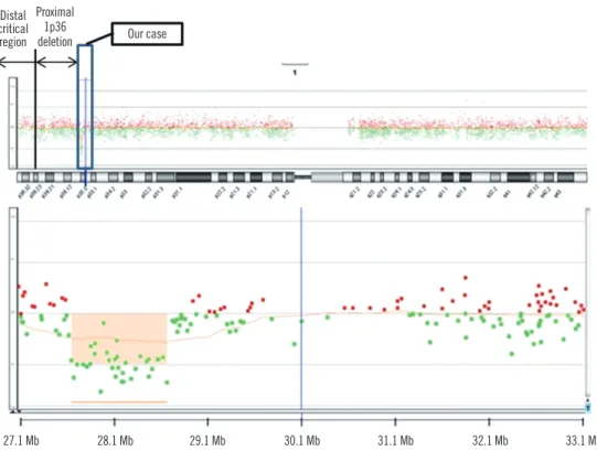
564 www.annlabmed.org https://doi.org/10.3343/alm.2017.37.6.563 comparative genomic hybridization (CGH) revealed a de novo
1p36.11-p35.3 interstitial deletion. The deletion size was esti-
mated to be 1 Mb, and the deleted base pairs of chromosome 1 spanned 27,750,155–28,754,771 (hg18) (Fig. 2). The array CGH of both parents revealed no abnormalities, confirming the de novo origin of the deletion. Approximately 20 known genes were included in this region, among which 13 genes were regis- tered in the Online Mendelian Inheritance in Man (OMIM) data- base. However, according to a search of the Developmental Dis- orders Genotype-Phenotype Database (DDG2P), only AHDC1 has been confirmed as a morbid gene responsible for a devel- opmental disorder. The other 12 genes (ATPIF1, EYA3, FGR, IFI6, MED18, PHACTR4, PPP1R8, PTAFR, RPA2, SESN2, STX12, and WASF2) were found to have no relationship with the specific developmental phenotype of the patient. Therefore, we could exclude the involvement of other genes in the abnormal phenotype of the patient. AHDC1 (OMIM 615790) is a protein- coding gene located on the short arm of chromosome 1 (1p36.1-p35.3). In 2014, Xia et al [4] reported four unrelated children with mental retardation exhibiting clinical features con- sistent with developmental delay, congenital hypotonia, mildly dysmorphic features, and sleep apnea. All four exhibited a de- velopmental delay in expressive language and hypoplasia of the corpus callosum on brain MRI. Mutations in AHDC1 were con- firmed by whole-exome sequencing [4].
Fig. 1. Brain magnetic resonance imaging revealing corpus callo- sum thinning (marked as an arrow).
Fig. 2. Comparative genomic hybridization of chromosome 1 in the patient. Breakpoints were revealed in 1p36.11-p35.3 (27,750,155–
28,754,771).
Our case
27.1 Mb 28.1 Mb 29.1 Mb 30.1 Mb 31.1 Mb 32.1 Mb 33.1 Mb
Distal critical region
Proximal 1p36 deletion
Park H-Y, et al.
1p36.11-p35.3 deletion including the AHDC1
https://doi.org/10.3343/alm.2017.37.6.563 www.annlabmed.org 565
This patient possessed an interstitial deletion in 1p36.11- p35.3, where AHDC1 is located, and showed congenital hypo- tonia and delay in development of gross motor skills and expres- sive language. Brain MRI showed corpus callosum thinning. Al- though the patient’s clinical features were typical of those seen in 1p36 deletion and proximal 1p36 interstitial deletion, his phenotype more closely resembled that of cases with de novo truncated mutations in AHDC1. Many cases of 1p36 deletion have been reported, but to our knowledge, this is the first report of a de novo 1p36.11-p35.3 interstitial deletion with loss of AHDC1. Molecular studies are needed to identify the genotype- phenotype relationship of this deletion and to understand the effects of AHDC1 expression.
Authors’ Disclosures of Potential Conflicts of Interest
No potential conflicts of interest relevant to this article were re- ported.
Acknowledgments
This study was supported by the Research Fund of Seoul St.
Mary’s Hospital, The Catholic University of Korea.
REFERENCES
1. Jordan VK, Zaveri HP, Scott DA. 1p36 deletion syndrome: an update.
Appl Clin Genet 2015;8:189-200.
2. Heilstedt HA, Ballif BC, Howard LA, Lewis RA, Stal S, Kashork CD, et al.
Physical map of 1p36, placement of breakpoints in monosomy 1p36, and clinical characterization of the syndrome. Am J Hum Genet 2003;
72:1200-12.
3. Kang SH, Scheffer A, Ou Z, Li J, Scaglia F, Belmont J, et al. Identifica- tion of proximal 1p36 deletions using array-CGH: a possible new syn- drome. Clin Genet 2007;72:329-38.
4. Xia F, Bainbridge MN, Tan TY, Wangler MF, Scheuerle AE, Zackai EH, et al. De novo truncating mutations in AHDC1 in individuals with syn- dromic expressive language delay, hypotonia, and sleep apnea. Am J Hum Genet 2014;94:784-9.
![외국인 PCR검사 가능 기관 [Referral Laboratories : 11sites]](data:image/gif;base64,R0lGODlhAQABAIAAAP///wAAACH5BAEAAAAALAAAAAABAAEAAAICRAEAOw==)