VEGF 조절을 통한 육계 추출물의 신생혈관 형성 촉진 작용
고정민․이상훈․백용현․최도영 경희대학교 한의과대학 침구학교실
목적 : 한의학 분야에서 육계(CCE: Cinnamomum cassia)는 혈류 순환이 저하된 상태에 널리 쓰이는 약 재이다. 이 연구에서는 CCE와 그 활성 혼합물인 cinnamic acid가 in vitro와 in vivo에서 발휘하는 신생혈관 형성 작용에 대해 알아보고자 한다.
방법 : CCE와 CA의 신생혈관형성 작용을 알아보기 위하여 Human unbilical endothelial cells(HUVECs) 와 Bovine arterial endothelial cells(BAECs) 그리고 Matrigel assay를 이용하였다.
결과 : In vitro에서 CCE와 그 활성 혼합물은 HUVEC 증식, 이동, 모세혈관-유사 관 형성을 유도한다.
CCE에서의 25-50% 증가(1 또는 10ng/ml 용량에서)는 CA에서 증식과 유사하게 나타났다. HUVEC에서는 증식이 뚜렷하게 나타났으나 BAEC에서는 증식이 뚜렷하게 나타나지 않아 증식 효과는 내피세포의 기원에 따라 다른 것으로 나타났다. BAEC는 CCE와 CA에서 증식은 보이지 않았지만 이동과 모세혈관-유사 관 형 성 또는 분화를 보였다. 또한 CCE와 CA는 HUVEC에서 VEGF 발현과 Flk-1/KDR 발현을 각각 2.2배와 2.5 배 증가시켰다.
결론 : CCE와 그 활성 혼합물은 in vivo와 in vitro에서 혈관 내피세포의 VEGF 발현을 통하여 신생혈관 형성 작용을 유도하며 이는 앞으로 다른 한약 추출물의 신생혈관형성 촉진 작용 연구에 모델을 제시할 것으 로 보인다.
핵심단어 : Cinnamomum cassia, Cinnamic acid, Angiogenesis, VEGF; Flk-1/KDR
1)
Stimulatory Efect of Cinnamomum cassia Extract on Angiogenesis Through Regulation of VEGF
Ko Jeong-min, Lee Sang-hoon, Baek Yong-hyeon and Choi Do-young Department of Acupuncture and Moxibustion, College of Oriental Medicine,
Kyung Hee University
․Acceptance : 2009. 2. 5. ․Adjustment : 2009. 2. 9. ․Adoption : 2009. 2. 10.
․Corresponding author : Choi Do-young, Department of Acupuncture & Moxibustion, College of Oriental Medicine, Kyung Hee University, 1 Hoegidong, Dongdaemungu, Seoul 130-701, Korea
Tel. 02-958-9196 E-mail : [email protected]
국문초록
Original Article
Ⅰ. Introduction
Angiogenesis, the formation of new capillaries from preexisting blood vessels
1-5)is crucial in physiological processes such as tissue repair and female reproductive cycle in adults
2,6). It is also required in pathophysiological processes such as the progression of tumor growth
7,8). It is a multi-step process which involves endothelial cell (EC) acti- vation; breakdown of the basement membrane;
migration, proliferation, organization and tube for- mation of EC; and maturation of the newly formed vessels
9-11).
Cinnamomum cassia has been used in conditions with Blood deficiency. It contains relatively large amount of coumarin, a naturally occurring com- pound that acts as a blood thinner.
There are reports from studies on animals and human that Cinnamon lowers blood glucose and LDL cholesterol
12). Recently reported Cinnamomum
cassia (CCE) possesses significant stimulatoryeffect on bone formation in osteoblastic cells and may contribute to the prevention of osteoporosis and inflammatory bone diseases
13).
In this study, we evaluated the distinct effects of CCE and its active compound cinnamic acid (CA) in in vivo and in vitro. We used animal model to study infiltration of endothelial cells in the Matrigel assay. Also, we observed whether CCE and CA stimulate endothelial cell proliferation, migration, and differentiation in vitro, and regulate the expression of the VEGF and Flk-1/KDR in Human umbilical vein endothelial cells (HUVEC).
Ⅱ. Materials and methods
1. Materials
Human recombinant VEGF165 were purchased from R&D systems (Minneapolis, MN, USA). Cell culture reagents were from Gibco (Invitrogen, Grand
Island, NY, USA). The 5-bromo-2’-deoxyuridine (BrdU) colorimetric assay kit was purchased from Roche (Stanhofer, Mannheim, Germany). The polyester membranes (12m pores) and the 48 well chemotaxis chamber were purchased from Nuero Probe, Inc. (Cabin John, MD, USA). Matrigel was purchased from Becton Dickinson (San Jose, CA, USA). Drabkins reagent kit 525, heparin, gelatin, CA and other chemicals were purchased from Sigma chemical (St. Louis, MO, USA). The chemiluminescence (ECL) detection system and nitrocellulose membrane were purchased from Amersham Life Science (Arlington Heights, IL, USA). The rabbit anti-Von Willebrand factor ABC kit and DAB substrate kit were from DAKO (Glostrup, Denmark).
2. Preparation of CCE and standardization
CCE was purchased from Medicine stuffs depart- ment of Kyung Hee University Medical center (Seoul, Korea), and voucher specimen was depos- ited at the herbarium at Pharmacy of Oriental medicine of Kyung Hee Medical Center, Kyung Hee University (KH04-20). Material was extracted with 50% ethanol in boiling water under pressure for 8h and filtered and lyophilized for a complete removal of the residual solvent to yield dark-brown powder.
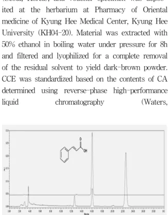
CCE was standardized based on the contents of CA determined using reverse-phase high-performance
liquid chromatography (Waters,
Fig. 1. Representative HPLC chromatogram of CA and the extract of CCE
CA was detected at round 14 min in this system.
Milford, MA, USA) equipped with a photodiode array detector (PAD). Separation was carried out using YMC Hydrosphere C
18(S-5㎛, 4.6x250mM;
Shisheido, Tokyo) at 30°C under following conditions:
acetonitrile: water (A 15 : 85, v/v or 25 : 75, v/v) as mobile gradient as a flow rate of 1.0ml/min at 250 nm. CAwas found in the CCE at a mean level of 1.03±0.02%(n=3)(Fig. 1).
3. Mouse Matrigel angiogenic assay
In vivo angiogenesis was assayed in male C57/BL6 mice (5weeks old, Jackson Lab, Japan) as the growth of blood vessels from subcutaneous tissue into a growth factor reduced Matrigel plug containing the test sample. Matrigel in liquid form at 4°C was mixed with heparin (60U/ml; Sigma) as the control group and one of CCE (100ng/ml), CA (100ng/ml) or VEGF (100ng/ml) was added to the mixture. Matrigel (0.5ml) was then injected into abdominal subcutaneous tissue along the peritoneal mid-line of mice, and sacrificed after 7 days.
Matrigel plugs were excised and processed for histological analysis. Samples were fixed in methanol at 4°C and embedded in paraffin and 5μ m-thick sample sections were stained with Hematoxylin-Eosin (H&E) stain for microscopic observation. Blood vessels, defined as those structures possessing a patent lumen and containing red blood cells, were identified blindly by two independent observers. Positive staining for anti-Von Willebrand Factor, an endothelial specific antigen, and were shown to correlate with the presence of vessels detected by light microscopy.
4. Cell culture
HUVECs were isolated with collagenase type I and cultured in 0.2% gelatin-coated dishes as previously described. They were routinely cultured in Medium 199 (M199) supplemented with 20% fetal bovine serum (FBS), 2mM glutamin, 100μg/ml penicillin/streptomycin, 15mM of HEPES, and 0.4%
ECGS. These cells were cultured in 5% CO
2at
37°C and the media were replaced at 2-days intervals.
Bovine aortic endothelial cells (BAEC) were grown in Dulbecco’s Modified Eagle Medium (DMEM) supplemented with 10% FBS, 2mM glutamin, and 100μg/ml penicillin/streptomycin.
5. Proliferation assay
Cell proliferation assays were performed using the BrdU assay (Roche Diagnostic, Germany).
HUVEC were seeded into 96-well gelatin-coated plates (1×10⁴/well). They were grown for 6h in M199 with 20% FBS, and overnight in M199 with 5% FBS at 37°C in 5% CO
2. Cells were stimulated with different concentrations of CCE and CA (1—
1000ng/ml) in M199 with 5% FBS. After 48 h, 10㎕
of BrdU were added to each well, and the samples were incubated for a further 6h at 37°C. Cells were fixed and added with anti-BrdU-POD and detected by TMB substrate reaction. This reaction was quantified by ELISA reader at 480—650nm.
6. Chemotaxis migration assay
Chemotaxis assay was carried out in 48 well Boyden chambers. Briefly, the chamber was divided into 2 compartments by a polyvinyl-pyrrolidone- free filter, 25x80mM, and 12μm pore size. The polycarbonate filter was coated with 0.2% gelatin.
The lower compartment of the chamber was filled with 30㎕ of basal medium (0.2% BSA containing M199) in the presence of CCE, CA or VEGF. Then 3x10⁵ cells suspended with control medium were loaded on the upper compartment of the chamber.
After 4hr at 37°C in 5% CO
2, the filter was fixed
and stained with Diff Quick solution (hematoxylin
and eosin). Nonmigrating cells on the upper surface
of the filter were removed by wiping with a cotton
swab, and chemotaxis was quantified by counting
the cells that migrated to the lower side of the
filter with optical microscope (×100, Nikon, Tokyo,
Japan). 10 fields were counted for each assay. Each
assay was conducted three times and similar
results came out at least 4 times.
7. Capillary-like tube formation assay
Growth factor-reduced Matrigel was added to 24-well plates with a total volume of 250㎕ in each well and was polymerized for 30min at 37°C.
HUVEC, 1x10⁵cells/well, in a final volume of 500㎕
was cultured in M199 basal medium treated with various concentrations of CCE, CAor VEGF were plated on the Matrigel. After 18h of incubation in 5% CO
2at 37°C, the area covered by the tube network was determined by using an optical imaging technique in which pictures of the tubes were scanned by Adobe Photoshop and quantitated using Image-Pro Plus (Media Cybernetics).
8. Quantitative real-time polymerase chain reaction (RT-PCR)
Confluent HUVEC (6cm Petri dishes) were cultured for 24h in M199 basal medium. For stimulation, the medium was replaced with the same M199 in the presence of test samples or VEGF for 24h. After incubation, cells were lysed for RNA isolation using a modified guanidium isothiocyanate method (TRIzol, Invitrogen), according to the recommendations of the manufacturer. Final RNA concentrations were determined by OD at 260nm and integrity was verified by ethidium bromide staining of ribosomal 18 S and 28 S bands on agarose gel. Total RNA was reverse transcribed in a final volume of 25μl containing 2.5×buffer, 40U/μl of RNAzine, 10mM of dNTP, 200U/μl ofmMLRT (Promega, Charbonnières, France) and 1 μg of total RNA. After hexanucleotides were annealed for 10 min at 70°C, cDNA synthesis was performed for 1h at 42°C, followed by an enzyme inactivation step for 5 min at 95°C. cDNA was store at -20°C until use.
All PCRs were performed by real-time fluorescence detection method using LightCycler System with a FirstStart DNA Master SYBR Green I kit (Roche Diagnostics). A reaction mixture
containing the following components at the indicated end-concentration was prepared according to the manufacturer’s instructions: 12.4μl water, 2.4μl MgCl
2(2.5mM), 0.6μl of forward primer (0.5μM), 0.6μl of reverse primer (0.5μM) and 2μl LightCycler Fast Start DNA Master SYBR Green 1. Eighteen microliters of the reaction mixture was distributed into precooled capillaries and 40ng of reverse- transcribed total RNA in a volume of 2μl was added as PCR template. To avoid amplification of contaminating genomic DNA, one of the two primers for Flk-1/KDR was chosen at the junction between two exons (10, accession number AF 063658AF063658). The primer sequences for Flk-1/
KDR were as follows: forward 5’TCTCAATGT- GGTCAACCTTCTAGG3’ reverse 5’TTTAAACGT- CTTAAGGGTGT TAGTGG3’. The primer sequences for Flt-1 were as follows: forward 5’CGACGTG- TGGTCTTACGGAGTA3’ reverse 5’CTTCCCTCA- GGCGACTGC3’. The primer sequences for VEGF were as follows: forward 5’ GGCCTC CGAA ACCATGAACTTTCTGCT3’ reverse 5’ CCTCCT- GCCCGGCTCACCGC3’.
The cycling conditions were as follow: initial denaturation at 95°C for 10 min, followed by 40 amplification cycles of 95°C for 15s, 55°C for 5s and 72°C for 20s. After amplification, the temperature was slowly elevated above the melting temperature of the PCR product to measure the fluorescence and thereby to determine the melting curve. A negative control without cDNA template was performed to assess the overall specificity.
9. Statistical analysis
All data were expressed as mean±SEM.
Statistical analysis was performed by analysis of
mean with the Mann—Whitney rank sum test. A
value of P<0.05 was considered statistically
significant.
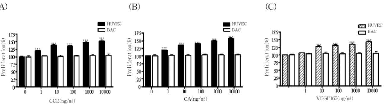
Fig. 3. CCE and CA induce proliferation of endothelial cells
Cells were seeded on 96-wells culture plates at a density of 1000 cells/well in M199 medium supplemented with 20% of FBS. The cells were cultured without (control) or with CCE (a), CA (b) or VEGF165 (c) used at 1 - 10000ng/ml. Data represent 3 independent experiments, each done in triplicate. Each value represents as mean ±SEM.
*** : p<0.001 compared to control.
Fig. 2. In vivo activation of the Matrigel infiltration of endothelial cells
(A)Matrigel plugs were sectioned and stained using the H&E method for microscopic observation (magnification ×200).
(B) Endothelial cell infiltration of the plugs was assessed using Factor VIII immunostaining. There was endothelial cell infiltration in plugs with CCE (b), CA (c) and VEGF165 (d) but not in the control plug (a). (C) Matrigel plugs were tested for hemoglobin (Hb) assay to quantify for formation of functional blood vessel. Each value represents as mean
±SEM.
** : p 0.01 and *** : p 0.001 compared with control plug.
Ⅲ. Results
1. CCE and CA induce angiogenesis in the in vivo Matrigel assay
The effect of CCE and CA on angiogenesis was investigated in vivo using subcutaneously implanted Matrigel plug assay. As compared to control (absence of VEGF), CCE or CA and VEGF165 showed infiltration of endothelial cells into Matrigel.
Histological analysis showed numerous endothelial cells well-organized into tube-like structures; red blood cells in some of these capillaries indicating their functionality(Fig. 2A). In CCE plugs, many
endothelial cells were positively stained with von Willebrand factor VIII already organized into capillary or large vascular cavities engorged with blood cells. Altogether, these experiments indicate that CCE or CA enhances in vivo angiogenesis(Fig. 2B).
Hb contents of the Matrigel plugs were also measured to quantify the functional vasculature. Control plugs showed 5.4±0.44g/dl of Hb and CCE-containing plugs showed 15.5±1.5g/dl. Also, CA significantly increased Hb levels to 13.2±1.1g/dl (Fig. 2C).
2. CCE and CA increase endothelial cell proliferation
The effect of CCE and CA on endothelial cell
(A) (B) (C)
(A) (B) (C) a
a
c
Control CCE CA VEGF
CCE(ng/㎖) CA(ng/㎖) VEGF165(ng/㎖)
HUVEC BAC
HUVEC BAC
HUVEC BAC
b
d
a
c
b
d
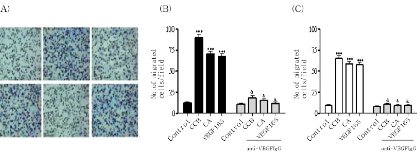
Fig 4. CCE and CA enhances migration of endothelial cells in chemotactic migration assay
(A) HUVEC were photographed and migrating cells were analyzed as described in materials and methods a; control, b;
CCE at 10ng/ml, c; CCE plus anti-VEGF neutralizing Ab, d; CA (CA), e; CA plus anti-VEGF neutralizing Ab, f; VEGF (B) Numbers of migrated cells per field in HUVEC under optical microscope (100 x). (C) Similar experiments were performed using BAEC. All experiments were conducted three times and similar results came out at least four times. Each value represents as mean ±SEM.
*** : p <0.001 compared to control, a : p<0.05 versus test sample alone.
proliferation was analyzed. Similar to CCE, CA stimulated HUVEC proliferation as shown in Fig. 1.
As compared to control, CCE at a low dose of 10ng/ml significantly increased proliferation of HUVEC by 38% (P(Fig. 3A). This effect was dose-dependent. 25 and 50% increase of HUVEC proliferation occurred in the presence of 1 and 100ng/ml CA, respectively(Fig.3B). The effect of exogenous CCE and CA on endothelial cell proliferation was also analyzed on BAEC. CCE and CA (10ng/ml) exerted a lower proliferative effect (5% increase) on BAEC(Fig. 3A, B).
3. CCE and CA increase endothelial cell migration
We then determined whether CCE and CA stimulate endothelial cell migration in chemotactic migration. As shown in Fig. 4, CCE (7.3-fold increase for 10ng/m) significantly induced the migration of HUVEC similarly to CA (5.8-fold increase for 10ng/ml). Cell migration observed with CCE or CA was completely inhibited with an excess (1μg/ml) of neutralizing anti-VEGF antibody (Fig. 4A, B). Interestingly, CCE also significantly
induced the migration of BAEC (7.7-fold increase for 10ng/ml) similarly to CA (5.6-fold increase for 10ng/ml). The specificity of this assay was also demonstrated by the inhibition with neutralizing anti-VEGF antibody(Fig. 4C).
4. CCE and CA induce endothelial cell differentiation
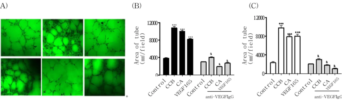
CCE and CA were tested for stimulatory potency on the differentiation of endothelial cells into tube- like structure. As shown in Fig. 5, CCE significantly induced the differentiation of HUVEC and BAEC (10780±20.5 tube area/field, 10450±112.8 tube area/
field respectively 64% and 53% increase for 10ng/ml, respectively, n4, Psimilarly to CA (10780±20.5 tube area/field, 10450±112.8 tube area/field respectively 64% and 53% increase for 10ng/ml, respectively, n4, P(Fig. 5A, B).
5. Differential expression of VEGF, Flk-1/KDR and Flt-1 mRNA in endothelial cells
In order to analyze whether differences CCE and
(A) (B) (C)
anti-VEGFIgG anti-VEGFIgG
Fig. 5. CCE and CA stimulated capillary-like tube formation of endothelial cells
(A) HUVEC was treated with CCE or CA on growth factor-reduced Matrigel (5 mg/ml) in the absence or presence of anti-VEGF Ab for 18 h. After washing and fixation, cells were observed under microscope (100x) and photographed. a;
control, b; CCE at 10ng/ml, c; CCE plus anti-VEGF neutralizing Ab, d; CA, e; CA plus anti-VEGF Ab, f; VEGF. (B) Tube area from randomly chosen field was quantified using image analysis system on HUVEC. (C) Similar experiments were performed using BAEC. All experiments were conducted three times. Each value represents as mean±SEM.
*** : p<0.001 compared to control, a : p<0.05 versus test sample alone.
VEGF
Flt-1
Flt-1/KDR
GAPDH
Fig. 6. Expressions of VEGF and its receptors in endothelial cells
Total RNA was isolated from HUVEC treated with CCE or CA (10ng/ml) for 24h. The mRNA expression of VEGF, Flt-1 and Flk-1/KDR in HUVEC is depicted on the first line and BAEC on the second line. The level of gene expression was calculated after normalizing against the GAPDH level in each sample.
CA have on endothelial cell proliferation is due to different levels of receptor expression in HUVEC and BAEC, we developed a quantitative PCR analysis for measuring both VEGF and their receptors expression(Fig 6). As shown in Fig. 6, HUVEC express high levels of VEGF, Flt-1 and Flk-1/KDR transcripts (4.8-, 1.2- and 3.7-fold respectively), while BAEC express lower levels of VEGF, Flt-1 and Flk-1/KDR (1.6-, 0.04- and 0.5-fold respectively).
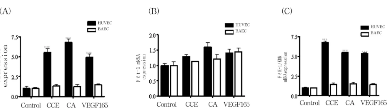
6. CCE and CA increased VEGF, Flk-1/KDR RNA but not Flt-1 expression in endothelial cells
W e then evaluated whether CCE and CA increased VEGF, Flk-1/KDR expression, the receptor involved in endothelial cell proliferation, migration and differentiation. As shown in Fig. 7, CCE significantly induced mRNA expression of VEGF and Flk-1/KDR (5.5-fold and 6.8-fold) in HUVEC, as compared to the control. Also, CA has shown significant induction of VEGF and Flk-1/KDR transcript (6.8-fold and 5.5 fold) similar to that observed with VEGF165. In contrast, CCE, CA or VEGF165 did not modify Flt-1 expression(Fig. 7).
Moreover, CCE, CA or VEGF165 (10ng/ml) did not modify VEGF, Flk-1/KDR transcripts level in BAEC(Fig. 7).
(A) (B) (C)
anti-VEGFIgG anti-VEGFIgG
Fig. 7. Quantification of real-time quantitative RT-PCR of VEGF, Flk-1/KDR and Flt-1 expression
Total RNA was isolated from HUVEC treated with CCE or CA (10ng/ml)for 24h. The levels of VEGF (A), Flt-1 (B) and Flk-1/KDR (C) transcripts were measured in the presence on its respective primers and SYBR Green I dye in real-time quantitative PCR. The level of gene expression was calculated after normalizing against the GAPDHlevel in each sample, and presented as fold induction±SEM in three independent experiments. * : p<0.05, *** : p<0.001 compared to control
Ⅳ. Discussion
In the present study, we demonstrate that CCE and its active compound CA have a novel function as angiogenic factor. CCE and CA stimulated angiogenesis by the mouse Matrigel plug assay in vivo where the ability of CCE and CA to promote neovessel formation was comparable with that of the well-established angiogenic factor VEGF165.
Vascular endothelial growth factor (VEGF), an endothelial cell-specific growth factor and the main regulatory factor involved in angiogenesis
14,15)is a critical factor for development of the vascular system in physiological and pathological angiogenesis. VEGF acts on endothelial cells through two highaffinity tyrosine kinase receptors, namely Flt-1 (or VEGF- R1) and Flk-1/KDR (or VEGF-R2)
16-19). Flk-1/KDR mediates the VEGF-dependent mitogenic effect, while Flt-1 is usually considered as a decoy receptor.
Through mRNA splicing, a single VEGF gene gives rise to several distinct isoforms which differ in their biochemical properties as well as their expression patterns
20-23). The two main human isoforms consist of 121 and 165 amino acids, and VEGF165 is the VEGF isoform most extensively studied. These isoforms are soluble, but some VEGF165 remains bound to the cell surface or
extracellular matrix (ECM).
In in vitro angiogenesis models, we have revealed that the stimulation of HUVEC with CCE and CA leads to an increase in cell proliferation as well as chemotactic motility and strong induction of tube network formation. Our data also showed that the mechanism by which CCE and CA induce angiogenesis is similar to VEGF165 which requires VEGF expression for its angiogenic activity.
The proliferative effect observed with CCE and CA appears dependent on the origin of the endothelial cell, since it was not observed with BAEC. Using the chemotactic migration assay, we observed that CCE and CA increases endothelial cell migration similarly to VEGF165 in both HUVEC and BAEC, and this effect was inhibited in the presence of anti-VEGF antibodies. Interestingly, the differentiation effect was also observed with BAEC, which express VEGF and VEGF receptors, suggesting different mechanisms involved in proliferation, and migration and differentiation. Our results suggest that CCE and CA acts as a VEGF, remains active as an endothelial cell mitogen.
The mechanisms of the mitogenic, chemotatic and differentiating effects on endothelial cells are not clear. CCE and CA could thus be active VEGF binding to Flk-1/KDR, the receptor mediating the VEGF-dependent mitogenic effect. Recent reports
(A) (B) (C)HUVEC
BAEC HUVEC
BAEC
HUVEC BAEC
Control CCE CA VEGF165 Control CCE CA VEGF165 Control CCE CA VEGF165
show that the sequence encoded by exon 6 could control growth factor release and proliferative effects through signaling pathways
24-26). One of the downstream molecular targets of VEGF action is the Flk-1/KDR receptor whose expression is increased by VEGF165. Our study demonstrates that CCE and CA also increase Flk-1/KDR expression in HUVEC. This regulation also appears dependent on the phenotype of the endothelial cell, since it is not observed in BAEC. However Flt-1 expression is not modified by CCE and CA in endothelial cells.
Ⅴ. Conclusion
In this study we have presented pharmacological evidence for the first time that CCE significantly induces angiogenesis in vivo and in vitro, latter process of which is possibly dependent on the cell origin through regulation of VEGF. This study may contribute to further studies on various herbal extracts for investigation on angiogenesis.
Ⅵ. Reference