인삼의 혈관신생(angiogenesis) 조절에 대한 음(陰)과 양(陽)
송 범 석 해태제과(주) 연구소
1. 서론
대체의학으로서 식용약초의 사용은 미국과 유럽 에서 다시 급증하고 있다. 그 예로 1997년에 미국 성인의 12.1% 이상이 식용약초를 사용한 것으로 집계되었다. 2001년에는 1,780만 달러의 영양보조 식품이 판매되었고 그 중 420만 달러가 식용약초 이었다. 흥미롭게도 미국에서 이러한 식용약초들은 1994년에 제정된 영양보조식품건강교육법 (Dietary Supplements and Health Education Act)에 의해 영양보조식품으로 규제되며 따라서 FDA의 승인 없이 유통될 수 있다.
그리스어로 pan (all)과 akos (healing)로부터 유 래된 Panax란 속(屬)명을 갖는 인삼은 중국 전통 의학에 있어 중요한 재료이며 미국에서 매년 22.
5~30만 달러가 판매될 정도로 서방국가에서 가장 광범위하게 사용되는 식용약초 중 하나이다.
인삼 추출물의 효능은 모호하게도 angiogenesis에 대 한 각기 반대되는 효과를 통해 창상치료(wound healing)를 촉진하며 암세포 성장을 저해한다고 보
고되었다. 본 연구는 이러한 인삼 효능의 혼란을 명확하게 밝히기 위해 혈관내피세포 (human umbilical vein endothelial cell)의 phenotype에 각 기 상반되는 효과를 나타내는 성분들을 동정하였 고, 각각의 성분이 angiogenesis 과정에서 미치는 영향을 밝히고자 하였다. 또한 지역별, 종별 차이 에 의한 각 성분들의 고유한 구성비가 나타내는 고유한 chemical fingerprint이 angiogenesis에 미치는 영향을 판단하고자 하였다.
2. 실험방법
2.1. 질량분석기에 의한 성분분석
Chinese and Korean ginseng (Panax ginseng), American ginseng (P quinquefolium), Sanqi ginseng (P notoginseng, Yunnan, China)의 4종 류 인삼을 수집하여 메탄올로 추출하였다. 각각의 추출액은 ESI mass spectrometer (Perkin-Elmer
Sciex API 365 Tandem spectrometer, positive scan mode, flow rate 0.6 mL/h)로 정성분석 하였다.
2.2. 화학물질에 의해 유도된 세포 투과도 측정
104 cells/well 농도의 혈관내피세포 (HUVECs)를 Matrigel-coated transwell (8-μm pore diameter, Costar)의 상위 chamber에 도말하였다. 시료와 성 장인자들을 알맞은 chamber에 주입하여 16시간 배양한 후 상위 chamber의 세포들을 제거하였다.
하위 chamber로 이동한 세포들을 10% formalin으 로 고정하고 hamatoxylin-eosin으로 염색한 후 Nikon Diaphot inverted microscope를 통해 육안 으로 관찰하였다.
2.3. 창상치료 측정
반원형의 coverslip이 들어있고 0.1% gelatin이 입 혀진 24개의 well plate에 3×10⁴ cells/well 농도의 HUVECs을 20% (vol/vol) FCS가 함유된 M199배 지와 함께 주입하였다. Plate에 세포들이 덮인 후 24시간 방치한 다음 coverslip을 제거하였다. 1%
(vol/vol) FCS가 함유된 M199배지와 시료를 첨가 한 후 48시간 배양하였고 hemocytometer를 통해 세포수를 측정하였다.
2.4. Western Blot analysis
위에서 언급된 바와 같이 처리한 세포들을 환원 제의 존재 하에 sample buffer(×3)에 용해 시켰다.
동일한 양의 단백질을 10% SDS-PAGE상에서 전 기영동하고 gel 상에 있는 단백질을 membrane으 로 전이 시켰다. 이 단백질들은 내피세포 nitric oxide synthase (NOS)에 직접 결합하는 항체 (1:250 희석액, New England BioLabs)와 반응시켰
다. Akt의 활성측정은 단백질들을 phospho-Akt에 특이 적인 항체 (1:800 희석액, Cell Signaling Technology) 와 반응시킨 후 Akt에 대한 항체로 반응시켜 관찰 하였다. 이후 Membrane들은 horseradish peroxidase 와 결합된 이차항체의 1:2000 희석액(Amersham) 에서 배양한 후 화학적 발광법에 의해 가시화하였다.
2.5. In Vivo Matrigel Angiogenesis assay
성장인자 및 시료와 혼합된 Matrigel을 C57/BL6 수컷 쥐 (Becton Dickinson)의 피하에 삽입하였다.
9일 후 CO2 방출에 의해 희생 시킨 다음 피부를 뒤집고 Matrigel을 절개하였다. 이후 면역조직화학 (immunihistochemistry) 검사를 위해 잘라서 동결시 켰다. 12 μm의 Matrigel 박편을 -20℃ methanol로 고정한 다음 von Willebrand factor (vWF)에 대 한 항체로 검색하였다. 신호는 Texas red-labeled secondary antibody에 의해 증폭되었고 Zeiss LSM510 confocal microscope에 의해 촬영되었다.
2.6.In Vivo Scaffold Implant Neovascularization Assay
Balb/c 수컷 쥐 (20~22 g body wt; Tuck, Rayleigh, UK)를 0.8 L/min 산소와 0.6 L/min의 질소 혼합물에 4% isoflurane으로 마취 시킨 후 2% isoflurane 농도로 유지하였다. 이후 등의 양쪽 으로 두 개의 피하 공기 주머니를 만들고 멸균된 polyether polyurethane sponge (170 mm3)를 각 주머니에 삽입한 후 봉합하였다. 부형약 (vehicle) 또는 시료 처리는 삽입 후 24시간이 경과 후부터 시작되었고 10일 동안 하루에 한번씩 sponge에 주 입하였다. 삽입 15일 후 쥐는 경추를 이탈시켜 희생시 켰다. 혈관생성 (neovascularization)은 granuloma 조직 내의 혈관의 성장으로써 정량분석 하였고 macro-lens (Nikon)로 촬영하였다.
2.7. 신혈관의 기능성 측정
Sponge implant로부터 방사성 동위원소인 133Xe 의 제거는 혈관의 기능성을 판단하는 근거가 된 다. Sponge 삽입 15일 후 Hypnom (Janssen Pharmaceutica)과 Hypnovel(Roche) 혼합물을 사 용하여 마취시켰다. 133Xe을 함유한 saline의 제거는 6분에 걸쳐 관찰되었다. 방사성은 microprocessor scalar rate meter (Nuclear Enterprise)로 측정하 였고 data는 전체농도에 대한 6분 동안 제거된
133Xe의 비율로 표현하였다.
2.8. 면역조직적(immunohistological) 혈관 염색
10 μm의 동결된 박편은 인간의 vWF에 대한 다 클론성 토끼 항체 (1:2000희석, Dako)와 blocking solution (1%[vol/vol] FCS in Tris- buffered saline with Tween)에서 하루 동안 incubation 하 였다. 항체 결합자리는 FITC와 결합한 goat anti-rabbit antibody (1:150희석액, Vector Labs) 를 사용하여 육안으로 관찰하였다. 형광색소는 488 nm laser line에 의해 발색되고 발색된 빛은 530/30-nm bandpass filter에 의해 포획되었으며 Leica TCS-NT confocal microscope로 촬영하였다.
2.9. 통계처리
실험 data는 mean±SEM으로 표시하였다. 통계 적 유의는 Dunnett or Friedman post hoc test에 따라 1-way ANOVA로 검증되었다. 전체적인 Dose-response effect를 검증하기 위해 Graphpad Prism 3 software 통해 Bonferroni test를 사용하 였다. P<0.05이면 유의성을 갖는 것으로 간주하였다.
3. 결과 3.1. 성분분석
지역적 분포와 품종이 다른 4가지 인삼-Chinese and Korean ginseng (Panax ginseng), American ginseng (P quinquefolium), Sanqi ginseng (P notoginseng, Yunnan, China)-을 대상으로 그 메탄 올 추출물을 mass spectrometer로 분석하였다 (Fig.
1A~D). 인삼의 활성성분들은 ginsenoside로 잘 알려진 triterpene dammarene류 이고 그들의 구 조에 따라 panaxdiol과 panaxtriol로 분류될 수 있 다. 가장 일반적인 diol, triol은 각각 Rb1, Rg1으 로 동일한 sterol 구조에 곁사슬과 수산화기 치환 정도가 다른 유사한 구조를 나타낸다 (Fig. 1E, 1F). 각각의 종은 Rb1과 Rg1을 모두 가지고 있으 며 두 성분의 구성비에 있어 고유한 mass spectrometric fingerprint를 나타내었다.
3.2. 막투과 유도 효과
활성화된 내피세포는 일련의 조화된 순차적 단 계를 거쳐 angiogenic phenotype을 나타낸다. 혈 관신생의 초기 주요 단계는 내피세포의 기저막 (basement membrane) 투과와 성장인자 또는 에 너지원 같은 적절한 신호를 향한 이동이다. 본 연 구에서는 내피세포의 독립적 활성인자로 이미 알 려진 hepatocyte growth factor/scatter factor (HGF/SF)를 chemoattractant로 사용하였다. 실험결 과 Rg1 뿐 아니라 Rb1도 chemoattractant로서의 기능을 나타내지는 않았지만 Rb1은 HGF/SF에 의 해 유도된 chemoinvasion을 저해하였다. 반대로 Rg1은 HGF/SF를 향한 내피세포의 chemoinvasion 을 촉진시키는 것으로 밝혀졌다. 더욱이 Rg1이 처 리된 세포는 생리적인 혈관과 유사한 선 모양으로 정렬되어 있는 것이 관찰되었다 (Fig. 2).
Fig. 1. Mass spectrometric compositional analysis of ginseng extract from different species and chemical structures of ginsenosides Rg1 and Rb1. Top panel shows the ESI mass spectrometric (Perkin-Elmer Sciex API 365 Tandem Spectrometer, positive scan mode, flow rate 0.6 mL/h) profile of a methanolic extract from american ginseng (A), Sanqi ginseng (B), Korean ginseng (C), and Chinese ginseng (D). E and F show the structures of Rb1 and Rg1, respectively.
Fig. 2. Effect of ginsenosides on chemoinvasion by HUVECs. Photomicrographs depict cells migrated under control conditions (A) and HGF/SF (1 nmol/L) (B)-facilitated HUVEC chemoinvasion and spreading. C, Rg1 treatment increases the HGF/SF-induced chemoinvasion and spreading of HUVECs. D, Rb1 (125 nmol/L) inhibited the HGF/SF-induced chemoinvasion. Right, Mean number of migrated cells per high-power field under different experimental conditions (mean±SEM;n=4). *P<0.05,
***P<0.001 vs HGF/SF-induced chemoinvasion. Upper and lower indicate the 2 chambers of the transwell; Veh, vehicle. Arrow depicts a typical cell that has migrated to the lower side and spread.
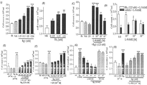
Fig. 3. Effect of Rg1 and Rb1 on the proliferation of HUVECs, after a mechanical injury to the monolayer, and modulation by NOS inhibitors. The cells were harvested and counted after 48-hour incubation in the presence of Rg1 (A) and Rb1 (B). C, The NOS inhibitor L-NAME inhibited the Rg1-induced proliferation of HUVECs in a concentration-dependent manner. At 10-3 mol/L, L-NAME did not alter the basal response. D, L-NAME inhibited the Rb1-induced proliferation at higher concentrations.
The cells were incubated in the presence of L-NAME(LN)(10-5mol/L), increasing concentrations of Rg1(E), and increasing concentrations of the NOS substrate L-arginine(L-Arg)(F). G, Effects of NOS-III inhibitor L-NIO and NOS-II inhibitor SMT on Rg-1 induced cell proliferation. H, Increasing concentrations of Rg1 can overcome the inhibitory effects of L-NIO. Values at 0 hour denote the cell count immediately after wounding. +P<0.01 vs Rb1 or Rg1 alone; *P<0.05, **P<0.01,
***P<0.001 vs vehicle(veh)-treated controls. Data are mean±SEM from 3 to 6 experiments with duplicate wells in each.
3.3. 생체 외 창상치료
내피세포는 일반적으로 접촉에 의해 성장저해를 받고 이러한 저해가 불안정해졌을 때 활동적인 표 현형 (aggresive phenotype)을 띄게 된다. 이러한 기작을 생체 외에서 표현하기 위해 monolayer injury assay를 하여 HUVECs 증식에 대한 Rg1과 Rb1의 효과를 측정하였다. Fig. 3에 나타낸 것처 럼 vehicle을 처리한 대조구와 비교적으로 두 물질 모두 농도 증가에 따라 HUVECs 증식을 촉진시
켰다. 이러한 결과는 ginsenoside는 sterol화합물이기 때문에 glucocortinoid 와 그 밖의 다른 angiostatic steroid처럼 전사단계에서 핵산수용체에 영향을 입 혀 내피세포의 증식을 저해할 것이라는 예상과는 다른 결과였다.
3.4. Ginsenosides의 생체 내 효과
생체 외 실험결과를 기초로 쥐에 polyether polyurethane scaffold implant를 삽입하여
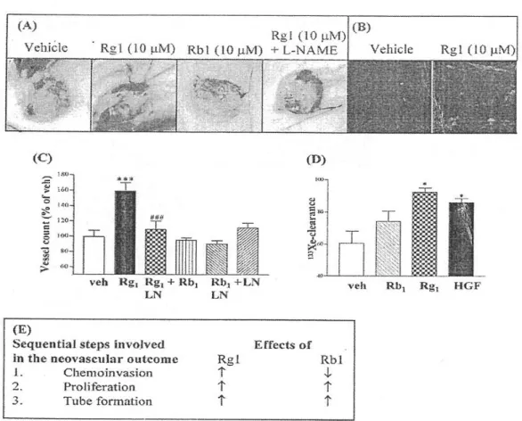
Fig. 4. Effects of Rg1 and Rb1 on neovascularization of a polyether polyurethane scaffold in mice. A, Effect of different treatments, administered for 10 days, on neovascularization into the scaffold implants, as seen on day 15 after implantation. B, Confocal images of vWF-delineated endothelial cells (labeled with FITC-coupled secondary antibody) at the same level cross section of vehicle-and Rg1-treated scaffolds. Vascularization was assessed as the ingrowth of vessels into the implant (C) and as a function of the blood flow through the implants (D), quantified by the direct injection of 133Xe-containing saline into the scaffold and with monitoring of its clearance over a 6-minute period. LN indicates L-NAME. Results were calculated from duplicate experiments with at least 3 to 4 replicates per test group. Data are expressed as mean±SEM.
*P<0.05, ***P<0.001 vs vehicle (veh)-treated controls; ###P<0.001 vs HGF/SF-treated group.
Deconstructing the neovascular response, E shows the effects of Rg1 and Rb1 at each key step in determining the final outcome. Effects of Rg1 and Rb1 on the compositional steps of angiogenesis, a possible explanation for the in vivo effects, are summarized.
angiogenesis에 대한 ginsenoside의 효과를 측정하 였다. 이 모델은 angiogenesis의 외형적 그리고 기 능적 연구를 위하여 광범위하게 사용되고 있다.
시료는 10일 동안 삽입물 내에 10 μg씩 주입되었
고 혈관생성 결과는 scaffold granuloma tissue로 의 혈관 성장으로써 정량분석 하였다. 신혈관의 기능성은 삽입물로부터 방사성 동위원소인 133Xe 의 제거에 의해 정량분석 되었으며 133Xe의 빠른
제거는 활동적인 혈액순환을 말해준다. Fig. 4에 나타 냈듯이 Rg1은 scaffold implant에 강력한 기능성 혈관을 생성시켰고 이는 vWF에 대한 immunostaining에 의해 확인되었다. 이 결과는 Rg1이 혈관생성의 모 든 순차적 단계를 자극시킨다는 생체 외 실험 결 과와 일치하는 것이다. 반대로 Rb1은 실험된 농도 에서 두드러지는 생체 내 효과를 나타내지 않았 다. 이는 fig. 4에 나타냈듯이 Rb1이 혈관형성의 초기단계 또는 내피세포의 chemoinvasion을 저해 한다는 생체 외 실험 결과와 일치한다.
인삼 품종에 따른 Rg1과 Rb1의 구성비 차이의 중요성을 평가하기 위해 Rg1과 Rb1을 규정된 비 율로 재구성하였고 Matrigel implant 실험을 통해 angiogenesis에 대한 효과를 측정하였다. Fig. 5에 나타냈듯이 Rb1보다 Rg1의 비율이 높은 추출물의 경우 vehicle과 비교하여 유효한 angiogenesis 유 도 결과를 나타내었다. 반대로 Rb1의 우세는 Rg1 에 의해 유도된 혈관생성을 저해하였다.
4. 토의
본 연구는 두 종류의 주요한 ginsenoside가 angiogenic phenotype에 미치는 상반되는 활성을 체계적으로 밝힘으로써 논쟁의 여지가 있던 창상 치료와 암세포에 대한 인삼의 기능을 설명하고 있 다. 더욱이, 종과 지역적 차이가 phenotype을 변화 시키는 구성적 지문 (compositional fingerprint)에 영향을 미친다는 것을 증명하고 있다. Rb1이 우세 한 Panax ginseng 추출물은 여러 암세포 모델에 있어 예방효과를 나타내었다. 유사하게도 American ginseng은 유방암 치료제에 상승작용을 나타낸다 고 밝혀졌으며 이는 본 연구결과에 부합하는 것이 다. 반대로 Sanqi ginseng은 우세한 ginsenoside Rg1의 proangiogenic activity로 인해 외상 치료와 모세순환계 촉진에 대한 중요한 재료로 판단된다.
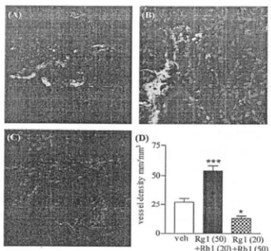
Fig. 5. Effects of extracts, reconstituted with defined ratio of Rg1 and Rb1, on angiogenesis in a murine Matrigel implant. Growth factor-reduced Matrigel was injected subcutaneously into C57/BL6 mice and allowed to gel. On day 9, the animals were euthanized, and the implants were excised, cryosectioned, and immunostained for vWF, which was detected with the use of a species-specific secondary antibody coupled with FITC. Propidium iodide was added to counterstain the nuclei.
Photomicrographs show angiogenesis in control (A), Rg1 (50㎍) plus Rb1(20㎍) (B), and Rg1 (20㎍) plus Rb1 (50㎍) (C) perimplant. The images were captured with a Zeiss LSM510 confocal microscope at a resolution of 512×512 pixels. Controls undertaken by omitting the primary antibody were imaged with the same settings for laser power and gain and showed no specific fluorescence. D, Vessel density in the different treatment groups. Veh indicates vehicle.
Data are expressed as mean±SEM; n=3. ***P<0.001 vs vehicle control; *P<0.01 vs HGF/SF treatment.
생체 내․외에서 Rb1의 활성은 Rg1의 신호전달 체계 (signal cascade)를 밝히는데 도움이 되었다.
내피세포가 phenotype을 획득하는 주요한 경로인 mitogen-activated protein kinases와 NO signaling 경로에 관한 Rg1과 Rb1의 효과를 측정하였다.
Rg1 뿐 아니라 Rb1도 mitogen-activated protein kinase 경로에 아무런 활성을 나타내지 않았다. 그 러나 흥미롭게도 Rg1이 처리된 세포에서 24시간 후 NOS-III의 강력한 발현이 발견되었다 (Fig. 6).
게다가 Rg1에 유도된 HUVECs의 증식은 NOS의 비 선택적 inhibitor인 L-NAME에 의해 저해되었 다 (Fig. 3C). 이러한 저해는 L-NAME과 활성자 리에 대해 경쟁하는 L-arginine에 의해 부분적으 로 완화 되었다 (Fig. 3E). 또한 높은 농도의 Rg1 은 그러한 억제를 완전히 반전시켰다 (Fig. 3F).
PI3K와 Akt는 신호전달체계의 상위에 있고 NOS-III를 활성화시킬 수 있다는 것이 최근에 증 명되었다. Rg1은 또한 PI3K inhibitor인 LY294002 에 의해 차단된 Akt의 인산화를 유도한다 (Fig.
6). 이러한 발견은 Rg1이 NOS 효소의 생성을 촉 진할 뿐 아니라 PI3K→phospho-Akt→NOS 경로 를 통해 활성화 시킬 수 있다는 것을 제안하고 있다.
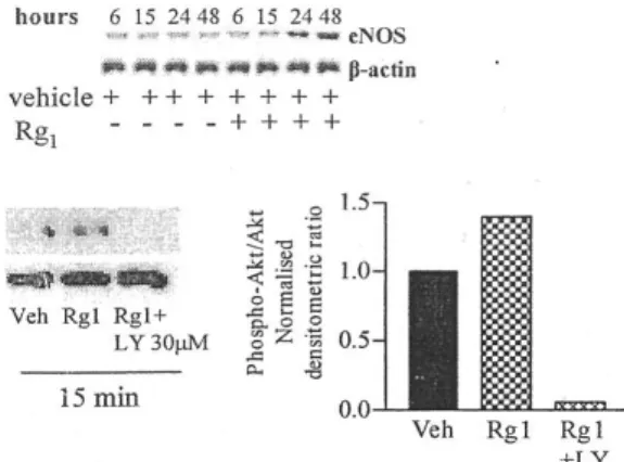
Fig. 6. Effect of Rg1 on NOS-III expression and activation of PI3K pathway. Top, Injured HUVECs were treated with vehicle or Rg1 and were lysed in sample buffer under reducing conditions at defined time points. Equal amounts of proteins were resolved in a 10%
SDS- PAGE gel. The level of NOS-II protein was detected with a species-specific anotibody and normalized to the level of β-actin expression. eNOS indicates endothelial NOS.
Bottom, Serum- starved HUVECs were incubated for 15minutes with vehicle(Veh), Rg1, or Rg1+
LY294002 (LY). The lysate was resolved on a SDS-PAGE gel and probed for phospho-Akt.
Normalization was done relative to the total Akt levels
NO는 혈관생성의 유도와 관련되어 있다. 그러 나 Pipili-Synetos 등은 NO donor가 chick chorioallantoic membrane assay에서 aggressive phenotype과 혈관생성을 저해한다고 보고하였고 이는 NOS-II의 발현과 활성의 증가와 연관되어 있다는 것을 증명하였다. 그러나 본 연구에서 NOS-II의 선택적 inhibitor인 SMT는 Rg1에 위해 유도된 세포 증식에 영향을 주지 않았으며 Western blot에서도 발견되지 않았다. 반대로 NOS-III inhibitor인 L-NIO는 Rg1에 의해 유도된 세포증식을 저해하였으나 (Fig. 3G) Rg1 농도의 증가에 의해 전환되었다 (Fig. 3H). 이는 NOS-III isoform이 Rg1에 의해 유도된 내피세포의 활성에 중요한 역할을 한다는 것을 말해준다. L-NAME과 Rg1의 병용은 생체 내에서 angiogenic effect를 차 단하였다 (Fig. 4). 이 결과는 NOS-III가 창상치료 에 관련된 혈관생성에 관련되어 있기 때문에 유의 성을 갖는다. 더욱이 NO에 의해 매개되는 Rg1의 효과는 vascular endothelial growth factor와 fibroblast growth factor-2의 tubulogenic effect에 대한 설명을 가능케 한다. 이러한 리간드들은 NO 에 의한 신호전달체계를 통해 혈관생성을 유도하 기 때문이다.
Rg1에 의해 유도된 tubulogenensis는 L-NAME 에 의해 완전히 저해되지만 Rb1의 경우에서는 유 사한 효과를 나타내지 않았다. Rb1에 의해 유도된 세포증식은 L-NAME에 의해 부분적으로 저해되 었다. 이는 Rb1 효과를 매개하는 부가적인 경로의 존재를 암시한다. 최근의 연구에서 Rb1은 tyrosin kinase를 저해한다고 보고 되었다. 이는 HGF/SF 에 의해 유도된 chemoinvasion의 저해를 설명할 수 있다. 이러한 세포내 신호전달체계의 차이는 내피세포의 막투과 단계에서 phenotype이 결정된 다는 것을 나타낸다.
이러한 ginsenoside들의 활성방식에서의 확연한 차이는 병태생리학적으로 장애가 발생한 내피세포 의 기능조절에 대한 치료적 접근을 기초로 하여 새로운 비단백질 저분자의 개발 가능성을 제시하
고 있다. 이후의 연구들은 암과 당뇨병성 망막병 증 (diabetic retinopathy)에서와 같이 혈관생성 저 해에 있어 Rb1-like molecules의 예방적 역할 (chemopreventive role)을 밝혀내는 반면 proactive modulator인 Rg1은 창상치료제에 대한 원형 (prototype)으로서 chemical scaffold의 이용가능성 을 제시할 것이다. 또한 본 연구는 규정된 비율의 Rg1과 Rb1의 첨가에 의해 재구성한 추출물이 혈 관생성 결과를 바꿀 수 있다는 발견을 통해 복용 에 대한 주의를 환기시키고 있다. 더욱이 종과 지 역적 차이는 구성비에 영향을 줄뿐만 아니라 가공 처 리되는 방법에 따라 fingerprint가 바뀔 수 있다는 것을 밝히고 있다. DSHEA에 따르면 영양보충식 품제조자는 소비자 어떠한 요구에도 정직할 의무 가 있고 요구에 대한 증거를 제시해야 한다. 그러 나 현재 요구되어지는 증거에 대한 표준을 제시하 지 못할 뿐 아니라 FDA로의 증거제시가 요구되 지도 않는다. 민간요법으로 사용되는 식물성 약물 이 안전하고 효력이 있다는 인식은 체계적인 연구 보다는 전통적 지식에 바탕을 두고 있다. 따라서 최근에 새로운 발견들은 품질, 안전, 효능보증 확 립에 박차를 가하고 있다.
본 연구는 인삼의 상반되는 작용을 하는 두 가 지 원소의 존재를 알림으로써 보다 나은 규제보다 는 구성분석을 통한 표준화의 중요성을 강조하고 있다.
5. 참고문헌
1. Marcus DM, Grollman AP. Botanical medicines: the need for new regulations. N Engl J Med. 2002;347:2073-2076.
2. Ang-Lee MK, Moss J, Yuan C-S. Herbal medicines and perioperative care. JAMA.
2001;286;208-216.
3. De Smet PA. Herbal remedies. N Engl J Med. 2002;347;2046-2056.
4. Gillis CN. Panax ginseng pharmacology: a nitric oxide link? Biochem Pharmacol.
1997;54:1-8.
5. Morisaki N, Watanabe S, Tezuka M, et al.
Mechanism of angiogenic effects of saponin from ginseng Radix rubra in human umbilical vein endothelial cells. Br J Pharmacol. 1995;115:1188-1193.
6. Sato K, Mochizuki M, Saiki I, et al.
Inhibition of tumor angiogenesis and metastasis by a saponin of Panax ginseng, ginsenoside-Rb2. Biol Pharm Bull.
1994;17:635-639.
7. Folkman J. Angiogenesis in cancer, vascular, rheumatoid and other disease. Nature Med.
1995;1:27-31.
8. Hu DE, Hiley CR, Smither RL, et al.
Correlation of 133Xe clearance, blood flow and histology in the rat sponge model for angiogenesis:further studies with angiogenic modifiers. Lab Invest. 1995;72:601-610.
9. Sengupta S. Gherardi E. Sellers LA, et al.
Hepatocyte growth factor/scatter factor can induce angiogenesis independently of vascular endothelial growth factor. Arterioscler Thromb Vasc Biol. 2003;23: 69-75.
10. Sengupta S. Sellers LA. Li RC, et al.
Targeting of mitogen-activated protein kinases and phosphatidylinositol 3 kinase inhibits hepatocyte growth factor/scatter factor-induced angiogenesis. Circulation. 2003;107:2955-61.
11. Crum R, Szabo S, Folkman J. A New class of steroids inhibits angiogenesis in the presence of heparin or a heparin fragment.
Science. 1985;230:1375-1378.
12. Sengupta S, Seller LA, Matheson HB, et al.
Thymidine phosphorylase induces angiogenesis in vivo and in vitro: an evaluation of
possible mechanisms. Br. J. Pharmacol.
2003;139:219-231.
13. Yun T, Lee Y, Lee YH, et al.
Anticarcinogenic effect of Panax ginseng C.A. Meyer and identification of active compounds. J Korean Med Sci. 2001;16 (suppl):s6-s18.
14. Duda RB, Zhong Y, Navas V, et al.
American ginseng and breast cancer therapeutic agents synergistically inhibit MCF-7 breast cancer cell growth. J Surg Oncol. 1999;72:230-239.
15. Lee K, Wang H, Itokawa H, et al. Current perspective on Chinese medicines and dietary supplements in China, Japan, and the United States. J Food Drug Anal. 2000;8:219-228.
16. Yan X, Zhou J, Xie G. Milne GWA, ed.
Traditional Chinese Medicines:Molecular Structures, Natural Sources and Application.
Hampshire, UK: Ashgate Publishing;1999.
17. Dimmeler S, Fleming I, Fisslthaler B, et al.
Activation of nitric oxide synthase in endothelial cells by Akt-dependent phosphorylation.
Nature. 1999;399:601-605.
18. Fulton D, Gratton JP, McCabe TJ. et al.
Regulation of endothelium-derived nitric oxide production by the protein kinase Akt.
Nature. 1999;399:597-601.
19. Ziche M, Morbidelli L, Choudhuri R, et al.
Nitric oxice synthase lied downstream from vascular endothelial growth factor-induced but not basic fibroblast growth factor-induced angiogenesis. J Clin Invest. 1997;99:2625-2634.
20. Pipili-Synetos E, Sakkoula E, Haralabopoulos G, et al. Evidence that nitric oxide is an endogenous antiangiogenic mediator. Br J Pharmacol. 1994;111:894-902.
21. Pipili-Synetos E, Kritikou S, Papadimitriou E, et al. Nitric oxide synthase expression, enzyme activity and NO production during angiogenesis in the chick chorioallantoic membrane. Br J Pharmacol. 2000;129:207-213 22. Lee PC, Salyapongsc AN, Bragdon GA, et
al. Impaired wound healing and angiogenesis in eNOS-deficient mice. Am J Physiol.
1999;277:H1600-H1608.
23. Dou DQ, Zhang YW. Zhang L, et al The inhibitory effects of ginsenodises on protein tyrosine kinase activated by hypoxia/
reoxygenation in cultured human umbilical vein endothelial cells. Planta Med.
2001;67:19-23.
24. Li X, Guo R, Li L. Pharmacological variations of Panax ginseng C.A. Meyer during processing. Zhongguo Zhong Yao Za Zhi. 1991;16:3-7
<출처 : Circulation 110, 1219-1225, 2004>