Veterinary Science
http://dx.doi.org/10.4142/jvs.2013.14.2.207
Received: 30 Jan. 2012, Revised: 9 May 2012, Accepted: 30 Aug. 2012
Original Article
*Corresponding author: Tel: +43-125077-6867; Fax: +43-125077-5191; E-mail: [email protected]
ⓒ 2013 The Korean Society of Veterinary Science.
This is an Open Access article distributed under the terms of the Creative Commons Attribution Non-Commercial License (http://creativecommons.org/licenses/by-nc/3.0) which permits
unrestricted non-commercial use, distribution, and reproduction in any medium, provided the original work is properly cited.
Radiation up-regulates the expression of VEGF in a canine oral melanoma cell line
Irene Flickinger
1,*, Barbara C Rütgen
2, Wilhelm Gerner
2, Ivana Calice
3, Alexander Tichy
4, Armin Saalmüller
2, Miriam Kleiter
1
1
Clinic for Internal Medicine and Infectious Diseases, Institute of Immunology, Departments for
2Pathobiology, and
4Natural Sciences, University of Veterinary Medicine, 1210 Vienna, Austria
3
Small Animal Clinic Hollabrunn, 2020 Hollabrunn, Austria
To evaluate radiosensitivity and the effects of radiation on the expression of vascular endothelial growth factor (VEGF) and VEGF receptors in the canine oral melanoma cell line, TLM 1, cells were irradiated with doses of 0, 2, 4, 6, 8 and 10 Gray (Gy).
Survival rates were then determined by a MTT assay, while vascular endothelial growth factor receptor (VEGFR)-1 and -2 expression was measured by flow cytometry and apoptotic cell death rates were investigated using an Annexin assay.
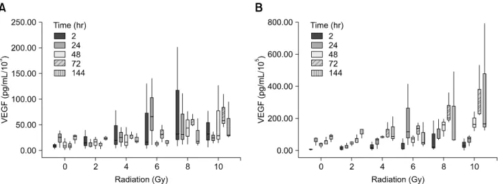
Additionally, a commercially available canine VEGF ELISA kit was used to measure VEGF. Radiosensitivity was detected in TLM 1 cells, and mitotic and apoptotic cell death was found to occur in a radiation dose dependent manner. VEGF was secreted constitutively and significant up-regulation was observed in the 8 and 10 Gy irradiated cells. In addition, a minor portion of TLM 1 cells expressed vascular endothelial growth factor receptor (VEGFR)-1 intracellularly. VEGFR-2 was detected in the cytoplasm and was down-regulated following radiation with increasing dosages. In TLM 1 cells, apoptosis plays an important role in radiation induced cell death. It has also been suggested that the significantly higher VEGF production in the 8 and 10 Gy group could lead to tumour resistance.
Keywords: cell line, radiation, TLM 1, vascular endothelial growth factor
Introduction
Melanoma is a common neoplastic disorder in dogs with variable presentation and biological behaviour. Melanomas account for 9∼20% of all skin tumours [21] and are the most common tumour of the oral cavity [13] and on the
digits [17]. Unlike cutaneous melanomas, which are often benign, melanomas within the oral cavity are highly malignant. They can be characterised by local infiltration and early metastasis to regional lymph nodes, lungs and other organs [27]. Although the pathogenesis is not completely understood, loss of functional tumour suppressor proteins such as p53, retinoblastoma susceptibility gene 1 (Rb) and phosphatase and tensin homologue deleted on chromosome 10 (PTEN) is common and may contribute to their aggressive nature [20]. Despite local control with wide surgical excisions and radiation therapy, the median survival time is only 8 months, with greater than 80% of dogs developing distant metastasis [30].
Vaccines that use immune stimulation to target melanoma cells have recently become available and have shown promising results [2]. However, other treatment options are required to further improve the long term prognosis for this common canine neoplasm. Angiogenesis represents a fundamental step in the malignant growth of tumours and metastasis. Many pro-and anti-angiogenic factors influence the formation of new blood vessels, but the central growth factor in this process is vascular endothelial growth factor (VEGF) [11].
VEGF is a heparin-binding glycoprotein with five
reported isoforms in mammals ranging from 126 to 206
amino acids [16]. VEGF binds two related receptor
tyrosine kinases, vascular endothelial growth factor
receptor (VEGFR)-1 and VEGFR-2, which are normally
found on endothelial cells [9]. In many cases, neoplastic
cells produce substantial amounts of VEGF and express
VEGF receptors, thus utilizing VEGF as an autocrine
growth stimulant [7]. Additionally, some tumour cells
have been shown to protect themselves against therapeutic