• 검색 결과가 없습니다.

Cuttlebone Shows Anti-inflammatory Activity Via Suppression of NF-κB Activation in LPS-induced RAW 264.7 Macrophages

N/A
N/A
Protected

Academic year: 2021

Share "Cuttlebone Shows Anti-inflammatory Activity Via Suppression of NF-κB Activation in LPS-induced RAW 264.7 Macrophages"

Copied!
5
0
0

로드 중.... (전체 텍스트 보기)

전체 글

(1)

DOI 10.17480/psk.2021.65.1.41

종 설(Review)

Cuttlebone Shows Anti-inflammatory Activity Via Suppression of NF-κB Activation in LPS-induced RAW 264.7 Macrophages

Geum Seon Lee* and Tae Jin Kang**

,#

*Department of Counseling Psychology, Sahmyook University

**Institute of Chronic Diseases and Department of Pharmacy, Sahmyook University

(Received January 7, 2021; Revised February 4, 2021; Accepted February 9, 2021)

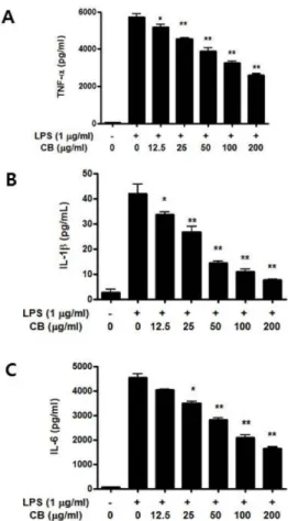
Abstract Our previous study reported that cuttlebone (CB) extract shows wound healing activity and enhances cell migration. In the present study, we examined the anti-inflammatory effect of CB in RAW 264.7 cells activated with lipopolysaccharide (LPS) for induction of inflammation. The expression of inflammatory mediators, including pro- inflammatory cytokines, was measured using enzyme-linked immunosorbent assay (ELISA) and reverse transcription polymerase chain reaction (RT-PCR) tests. The results showed that CB suppressed nitric oxide (NO) production in macrophages stimulated with LPS. Production of pro-inflammatory cytokines, such as tumor necrosis factor alpha (TNF- α), interleukin (IL)-1β, and IL-6, decreased in LPS-induced RAW 264.7 cells after treatment with CB in a dose-dependent manner. Additionally, gene expression was inhibited by CB in a dose-dependent manner in LPS-stimulated macrophages.

Furthermore, CB inhibited nuclear factor kappa B (NF-κB) p65 activation in macrophages activated with LPS. Therefore, these results suggest that CB exerts its anti-inflammatory effects via regulation of the NF-κB signaling pathway.

Keywords cuttlebone, inflammation, macrophages, cytokines, NF-κB

Introduction

Inflammation is a complex response in living tissue that neutralizes harmful pathogens and protects our bodies, without which infection and wounds cannot heal. Additionally, inflammation heals and repairs the damaged areas.

1,2)

Inflammation may be acute or chronic; acute inflammation can present in minutes to hours, whereas chronic inflammation can develop gradually. Cells involved in the inflammatory response are diverse, although macrophages play the most important role. Macrophages are antigen presenting cells and the first to recognize pathogens.

Macrophages protect healthy cells from attack by foreign substances, such as bacteria and viruses. Additionally, macrophages secrete inflammatory mediators, such as inflammatory cytokines, nitric oxide (NO), vascular amines, and prostaglandin E

2

. The various functions of macrophages help the body to maintain homeostasis through immune regulation.

3-5)

The nuclear factor kappa B (NF-κB) signaling pathway is typically associated with the inflammatory response and secretion of mediators.

6)

NF-κB is a protein complex found in cytosol,

although activated NF-κB translocates into the nucleus. Therein, NF-κB binds to the promoter region and induces the expression of a variety of genes leading to the expression of inflammatory mediators.

7,8)

Therefore, studies on the inhibition of inflammatory mediators in activated macrophages are related to the control of the inflammatory response.

Cuttlebone (CB), also known as cuttlefish bone, has been used as a traditional medicine. Our previous study shows that CB enhances wound healing and cell migration in a burned rat model, suggesting that CB aids in healing of the damaged and inflamed areas.

9-11)

Thus, we aimed to investigate the role of CB in the control of inflammatory mediators via the NF-κB pathway in lipopolysaccharide (LPS)-stimulated RAW 264.7 cells.

Methods

Reagents and kits

Cuttlebone (Gyeongdong market, Seoul, Korea), HCl, NaOH, ethyl alcohol, NaNO

2

(Daejung, Siheung, Korea), Dulbecco’s modified Eagle’s medium (DMEM), fetal bovine serum (FBS, Hyclone, Logan, UT, USA), penicillin/streptomycin (P/S, Hyclone, Logan, UT, USA), Cytokine kit (R&D systems, Minneapolis, MN, USA), total RNA purification kit (GeneAll, Seoul, Korea), cDNA synthesis kit, Taq polymerase (Bioneer, Daejeon, Korea), and NF-κB kit (Cell Signaling, Danvers, MA, USA) were used in

#

Corresponding author

Tae Jin Kang, Ph.D., Department of Pharmacy, Sahmyook University, Seoul 01795, Republic of Korea

Tel: +82-2-3399-1608, Fax: +82-2-3399-1617

E-mail: [email protected]

(2)

ethyl alcohol and dried in a vacuum oven.

Cell culture and treatment

RAW 264.7 cells, which is a mouse macrophage cell line, were grown in DMEM supplemented with 10% FBS and 1% P/S.

Macrophages were treated with CB and LPS in 24-well plates (5×10

5

cells/well) and incubated in 5% CO

2

at 37

o

C for 4 or 24 h.

Nitric oxide (NO) measurement

Nitric oxide production was measured by Griess reaction using the standard NaNO

2

curve. Briefly, the accumulation of nitrite, an estimate of nitrate (NO

3

) production, was determined colorimetrically after mixing the culture medium or sample with freshly prepared Griess reagent (0.1% NED and 1% sulfanilamide). The concentration of nitrite was estimated by comparing the absorbance readings at 540 nm (SpectraMax Microplate Reader, The Molecular Devices, San Jose, CA. USA).

Cytokine measurement

Cell culture supernatants were obtained for mouse tumor necrosis factor alpha (TNF-α), interleukin (IL)-6, and IL-1β using an ELISA kit according to the manufacturer’s instructions. Briefly, 96-well plates (Nunc, Penfield, NY, USA) were coated overnight at 4

o

C with capture antibodies for TNF-α, IL-6, and IL-1β. After washing, the plates were blocked for 1 h at 25

o

C with blocking buffer. Cytokine standards were added to wells with cell culture supernatants. Then, the detection antibody was added and incubated for 1 h at 25

o

C. Streptavidin-HRP was added for 30 min, then the plates were incubated with substrate solution for 15 min at 25

o

C. Stop solution was added and absorbance was measured at 450 nm.

RT-PCR

The attached cells were treated with RiboEx and total RNA was isolated. Reverse transcription was conducted using 1 μg of total RNA and a cDNA synthesis kit. Cyclic temperature reverse transcription (CTRT) was performed 12 times. RT-PCR was

performed for 35 cycles of denaturation (94

o

C, 30 s), annealing (each temperature, 45 s), and extension (72

o

C, 45 s) with a final extension at 72

o

C for 10 min (Table 1). The PCR products were observed by electrophoresis on 1.5% agarose gel with ethidium bromide (EtBr) under a UV-transilluminator.

Measurement of phospho-NF-κB p65 protein by ELISA The macrophages were treated with CB and LPS in 6-well plates and incubated for 4 h. The cell lysates were collected by chilled cell lysis buffer and incubated on ice for 5 min. Cell lysates were centrifuged, and the supernatants used for the ELISA. The levels of phospho-NF-κB p65 protein were assayed using a commercial kit according to the manufacturer’s instructions.

Statistical analysis

All data are expressed as the mean±SD. Statistical analysis of data was performed using Student’s t-test. Statistical significance was considered at p<0.05 or p<0.01.

Results

Effect of CB on NO production in macrophages

NO is used as a host-defense mechanism by macrophages to fight off pathogens when they enter our bodies and maintain homeostasis.

12)

Thus, to evaluate the effect of CB on NO production by macrophages stimulated with LPS, we measured NO released during inflammation. NO production decreased in a dose-dependent manner by CB in LPS-induced RAW 264.7 cells (Fig. 1).

Effect of CB on pro-inflammatory cytokine expression in macrophages

The production of pro-inflammatory cytokines was measured in

the cell supernatants. Macrophages were treated with LPS and CB

for 24 h and the supernatants were collected. The protein

production and mRNA expression were measured using ELISA

(3)

and RT-PCR, respectively. The production of TNF-α, IL-1β, and IL-6 was reduced in LPS-stimulated macrophages with CB in a

dose-dependent manner (Fig. 2). Additionally, the gene expression levels decreased in CB-treated RAW 264.7 cells (Fig. 3).

Effect of CB on the activation of NF-κB

NF-κB is a major pathway of the inflammatory response in cells and is activated by a variety of stimuli such as LPS and TNF.

6)

Components of NF-κB are IκB, p65 (RelA), and p50 (RelB). If NF-κB is activated, IκB is degraded and NF-κB subunits such as p65 and p50 are phosphorylated. The p65 and p50 proteins are translocated into the nucleus and transmit various inflammatory signals.

8)

Therefore, the reduction of phosphorylated NF-κB is associated with the control of inflammation. NF-κB expression levels were measured in the cell lysates. Phospho-NF- κB p65 protein was inhibited by CB in a dose-dependent manner in LPS-induced RAW 264.7 macrophages (Fig. 4).

Fig. 1. Effect of CB on NO production in RAW 264.7 cells. Cells were treated with LPS and CB at 37

o

C incubator. After 24 h, cell culture supernatant was collected for assay. Data are representative of at least three independent experiments in duplicate. Data are expressed as means±SD. *<0.05, **<0.01 versus LPS alone based on Student’s t-test.

Fig. 2. Effect of CB on production of pro-inflammatory cytokine in RAW 264.7 cells. Cells were treated with LPS and CB at 37

o

C incubator. After 24 h, cell culture supernatant was collected for assay.

Data are representative of at least three independent experiments in duplicate. Data are expressed as means±SD. *<0.05, **<0.01 versus LPS alone based on Student’s t-test.

Fig. 3. Effect of CB on gene expression of pro-inflammatory cytokine in RAW 264.7 cells. Cells were treated with LPS and CB at 37

o

C incubator. After 4 h, cell was harvested and total RNA was isolated. The gene expression was assayed with RT-PCR method and GAPDH was used for a control.

Fig. 4. Effect of CB on the activation of NF-κB p65 protein in RAW 264.7 cells. Cells were treated with LPS and CB and then cell lysate was collected by chilled cell lysis buffer and used for measurement of phosphorylated NF-κB p65 protein by ELISA method. Data are representative of at least three independent experiments in duplicate. Data are expressed as means±SD. *<0.05,

**<0.01 versus LPS alone based on Student’s t-test.

(4)

and IL-6 in inflammatory cells. TNF-α is involved in tissue injury and metabolic disorders and increases vascular permeability.

15)

Additionally, IL-1β and IL-6 are significant mediators in the inflammatory response. IL-1β is induced by COX-2 and iNOS and increases the expression of endothelial cell adhesion molecules.

16)

IL-6 plays a role in host defense and in the control of T and B lymphocyte functionality.

17)

Therefore, the control of inflammatory mediators is the most important factor in the inhibition of inflammation. NO production decreased in activated RAW 264.7 cells after CB treatment (Fig. 1). We measured the production of pro-inflammatory cytokines such as TNF-α, IL-1β, and IL-6 in LPS-stimulated macrophages in the absence or presence of CB, and the data showed that CB suppressed cytokine release by macrophages. These results are consistent with that of our previous study.

11)

Moreover, the mRNA expression levels was suppressed in LPS-stimulated macrophages treated with CB in a dose-dependent manner (Figs. 2 and 3). These results showed that CB reduced the production of inflammatory mediators.

NF-κB was initially observed in B lymphocytes as a κ chain enhancer protein. The novel proteins, a transcription factor, were elucidated using the electrophoretic mobility shift assay (EMSA).

18)

NF-κB is a protein complex (IκB, p65, and p50), and it is present in the majority of cell types. NF-κB is involved in numerous cellular processes such as inflammation, immunity, wound healing, and tissue repair.

1,7)

Stimulated NF-κB translocates from the cytosol to the nucleus in inflammatory cells. In this case, IκB is degraded and NF-κB is phosphorylated in the cytosol.

Alternatively, p65 and p50 migrate into the nucleus and express signals

8)

involved in the secretion of inflammatory mediators such as NO and cytokines. Our study showed that CB inhibited the phosphorylation of NF-κB, suggesting that CB suppresses inflammatory signals in LPS-induced RAW 264.7 cells (Fig. 4).

Conclusion

In conclusion, this study elucidated the inhibitory effect of CB on inflammation. Our results showed that CB exhibits anti-

University in 2019

Conflict of Interest

All authors declare that they have no conflict of interest

References

1. Nathan C (2002) Points of control in inflammation. Nature 420:

846-852

2. Rankin JA (2004) Biological mediators of acute inflammation.

AACN Clin Issues 15: 3-17

3. Tauber MG, Moser B (1999) Cytokines and chemokines in meningeal inflammation: biology and clinical implications. Clin Infect Dis 28: 1-11

4. Maeda S, Yanagihara Y (2001) Inflammatory cytokines. Nihon Rinsho 59: 1984-1989

5. Eisenhut M, Wallace H (2011) Ion channels and inflammation.

Eur J Physiol 461: 401-421

6. Hayden MS, West AP, Ghosh S (2006) NF-κB and the immune response. Oncogene 25: 6758-6780

7. Sen R, Baltimore D (1988) Inducibility of kappa immunoglobulin enhancer-binding protein NF-kappa B by a posttranslational mechanism. Cell 47:921-928

8. Oeckinghaus A, Ghosh S (2009) The NF-κB family of transcription factors and its regulation. Cold Spring Harb Perspect Biol 1: 1-14

9. Jang JK, Lee OS, Kang TJ, Lim SC (2013) The wound healing effect of cuttlebone extract in burn injury of rat. Food Sci Biotechnol 22: 99-105

10. Lee KM, Shim H, Lee GS, Park IH, Lee OS, Lim SC, Kang TJ (2013) Chitin from the extract of cuttlebone induces acute inflammation and enhances MMP1 expression. Biomol Ther 21:

246-250

11. Lim SC, Lee KM, Kang TJ (2015) Chitin from cuttlebone activates inflammatory cells to enhance the cell migration. Biomol Ther 23: 333-338

12. Chang LP, Lai YS, Wu CJ, Chou TC (2009) Liquid perfluorochemical inhibits inducible nitric oxide syntheses expression and nitric oxide formation in lipopolysaccharide-treated RAW 264.7 macrophages. J Pharmacol Sci 111: 147-154

13. Nathan C (1992) Nitric oxide as a secretory product of mammalian cells FASEB J 6: 3051-3064

14. Andreakos E, Foxwell B, Feldmann M (2004) Is targeting toll-like receptors and their signaling pathway a useful therapeutic approach to modulating cytokine-driven inflammation? Immunol Rev 202: 250-265

15. Meyer O (2003) Role of TNF-α and cytokine in the

physiopathology of rheumatoid arthritis. Therapeutic perspectives.

(5)

Bull Acad Natl Med 187: 935-954

16. Netea MG, Nold-Petry CA, Nold MF, Joosten LA, Opitz B, van der Meer JH, van der Veerdonk FL, Ferwerda G, Heinhusi B, Devesa I, Funk CJ, Mason RJ, Kullberg BJ, Rubartelli A, van der Meer JW, Dinarello CA (2008) Differential requirement for the activation of the inflammasome for processing and release of IL- 1beta in monocytes and macrophages. Blood 113: 2324-2335

17. Hibi M, Nakaajima K, Hirano T (1996) IL-6 cytokine family and signal transduction: a model of the cytokine system. J Mol Med 74: 1-14

18. DiDonato JA, Hayakawa M, Rothwarf DM, Zandi E, Karin M (1997) A cytokine-responsive IkB kinase that activates the transcriptional factor NF-κB. Nature 388: 548-554

Authors ’ Positions

Geum Seon Lee : Assistant Professor Tae Jin Kang : Professor

수치

Fig. 4. Effect of CB on the activation of NF-κB p65 protein in RAW 264.7 cells. Cells were treated with LPS and CB and then cell lysate was collected by chilled cell lysis buffer and used for measurement of phosphorylated NF-κB p65 protein by ELISA method

참조

관련 문서

Effect of onion peel ethanol extract on LPS-induced iNOS, COX-2, and NF-κB p65 expression in LPS-induced RAW 246.7 cells.. The levels of iNOS and COX-2 in the cytosolic protein

Collectively, LCLE and LCBE inhibited LPS-induced NF- κB activation by blocking p65 nuclear accumulation, increased HO-1 expression by ROS/p38/Nrf2 activation, and increased ATF3

Keywords: Argyreia capitata (Vahl) Choisy extract (ACE), nitric oxide (NO), NF-κB (nuclear factor kappa-B), inflammatory cytokines, anti-inflammatory effect..

Results: PSE inhibited LPS-induced NO production, interleukin (IL)-6 secretion, c-Jun NH2-terminal kinase (JNK) and NF-κB activation. In addition, PSE reduced the death

distichum leaf inhibited IκB-α degradation and p65 nuclear translocation, and subsequently suppressed transcriptional activity of NF-κB in LPS-stimulated RAW264.7

특히 iNOS 는 외부자극이나 pro-inflammatory cytokine 등에 의해 자극을 받게 되면 다량의 NO 를 생산하 Anti-inflammatory Effect of Sargassum coreanum Ethanolic

These results indicate that magnolol decreases the DNA binding and transcrip- tional activation of NF-κB/Rel, which is important in the regulation of iNOS gene expression...

Taken together, the results of the in vitro cell line assay for MTT, NO production, ROS generation; the Western blot analysis for the inhibition of iNOS, COX-2, NF- κB, and