敗醬 물 추출물의 LPS로 유도된 RAW 264.7 세포와 mouse 염증모델에서 cytokine 및 NF-κB의 활성에
미치는 효과
1 원광대학교 한의과대학 부인과학교실, 2 원광대학교 의과대학 산부인과학교실 류익한 1 , 조해중 2 , 송미화 1 , 최창민 1
ABSTRACT
Effects of Patrinia Scabiosaefolia Aqueous Extract on Cytokine and NF-κB Activation in LPS-induced RAW 264.7 Cells and Mouse
Ik-Han Ryu 1 , Hae-Joong Cho 2 , Mi-Hwa Song 1 , Chang-Min Choi 1
1 Dept. of Gynecology, College of Korean Medicine, Won-Kwang University
2 Dept. of Obstetircs and Gynecology, College of Medicine, Won-Kwang University Objectives: The object of this study was to identify the anti-inflammatory effects of Patrinia scabiosaefolia aqueous extract (PSE).
Methods: RAW 264.7 cells were pre-treated with PSE and then incubated with or without lipopolysaccharide (LPS). Cell viability, production of nitric oxide (NO), secretion of pro-inflammatory cytokine, activation of mitogen-activated protein kinases (MAPKs) and nuclear factor-kappa B (NF-κB) were measured. In addition, we observed mice survival rate after LPS and their cytokine levels of serum. We also observed inflammatory and hemorrhagic change on the histological sections of the liver.
Results: PSE inhibited LPS-induced NO production, interleukin (IL)-6 secretion, c-Jun NH2-terminal kinase (JNK) and NF-κB activation. In addition, PSE reduced the death rate of LPS-induced mice and IL-6 production on the serum of mice.
PSE inhibited inflammation and hemorrhage on liver tissue as well.
Conclusions: The results suggest that PSE have anti-inflammatory effects by inhibited NF-κB and JNK activation, IL-6 secretion, and NO production. So PSE may be effective treatment for the inflammatory disease.
Key Words: Patrinia Scabiosaefolia (PS), Inflammation, NO, Cytokine, MAPKs, NF-κB
1)
Corresponding author(Chang-Min Choi) : Korean Hospital of Won-Kwang University, 1140-23, Hoejae-ro, Nam-gu, Gwangju, Korea
Tel : 062-670-6437 Fax : 062-670-6767 E-mail : [email protected]
Ⅰ. 서 론
염증 반응은 조직에 가해진 손상을 회 복하기 위한 일련의 과정으로, 이 과정 에서 부종, 발적, 열, 통증, 기능상실 등 이 나타나며, 이를 해소하기 위해 다양 한 약물들이 개발되었다. 현재 사용되고 있는 약물들은 위장관계 및 심혈관계 등 에 부작용을 보여, 이를 대체하기 위한 효과적이고 부작용이 없는 새로운 약물 의 개발이 필요한 실정이다 1-3) .
패장(敗醬)은 패장과(마타리과; Valerianaceae) 에 속한 多年生草本으로 썩는 냄새가 있 기에 敗醬이라는 이름이 생겼다고 한다.
淸熱解毒 消癰排膿 活血行瘀 등의 효능 이 있어 腸癰, 肺癰, 瘡癰, 腫毒, 胸腹疼 痛 등의 병증을 치료하며, 鎭靜, 抗菌 및 抗病毒 작용 등이 있다고 하였다 4-6) .
지금까지 보고된 패장에 관한 연구로는 기원 7,8) , 급성 췌장염 9) , 골반염증성 질환 10) , 부종 11) 등을 포함한 항염증 작용 12-6) , 전
립선암 17,18) , 대장암 19) 등을 포함한 항암
작용 20-2) , 골관절염 23) , 막성신증 24) , 천식 25)
등에 관한 연구 등이 보고되었다.
부인과 영역에 있어 帶下를 유발할 수 있는 염증성 질환이나 골반염증성질환에 주로 활용되고 있으며, 기존의 연구에서 패장이 항염증 효과가 있음이 보고되었 으나, 염증을 억제하는 기전에 대한 체계 적인 연구는 부족한 실정이다. 이에 저자 는 패장의 항염증 작용을 究明하기 위하 여 in-vitro와 in-vivo 실험을 진행하였다.
패장 물 추출물(Patrinia scabiosaefolia Aqueous Extract, PSE)이 lipopolysaccharide (LPS)로 유도된 RAW 264.7 세포에서 nitric oxide(NO), interleukin (IL)-1β, IL-6, tumor necrosis factor alpha(TNF-α)의
생성과 mitogen-activated protein kinases (MAPKs) family인 extracellular signal -regulated kinase1/2(ERK1/2), p38 kinases (p38), c-Jun NH2-terminal kinase(JNK) 와 nuclear factor-kappa B(NF-κB)의 활 성에 미치는 영향을 관찰하고, 마찬가지로 LPS로 자극한 mouse의 생존률, cytokine 발현, 간조직의 염증 및 출혈 반응에 미 치는 영향을 관찰하여 유의한 결과를 얻 었기에 보고하는 바이다.
Ⅱ. 재료 및 방법
1. 재 료 1) 약 재
패장은 ㈜옴니허브(영천, 대한민국)에 서 구입하여 사용하였다. 패장 물 추출물 (Patrinia scabiosaefolia aqueous extract, PSE)을 얻기 위해 증류수 1 ℓ에 패장 100 g을 넣고 150분 동안 전탕하여 여과한 후, 동결 건조시켰다. 16.8 g의 분말을 얻 었으며, 수율은 16.8%였다. 분말은 -80℃
에 보관하고 실험 시 3차 증류수에 녹여 필터한 후 사용하였다.
2) 시 약
Fetal bovine serum(FBS), penicillin-
streptomycin, RPMI Medium 1640 등의
세포 배양용 시약들은 Gibco BRL(Grand
Island, USA)사에서 구입하였으며, Chloroform,
TRI-zol, Acrylamide, Sodium dodesyl
sulfate(SDS), Tris-HCL, LPS 등의 시
약은 SIGMA(St.Louis, USA)사에서 구
입하였다. 실험에 사용된 anti-phospho-
ERK1/2, anti-phospho-JNK, anti-phospho-
p38은 Cell Signaling(MA, USA)사에서
구입하였고, Anti-inhibitory kappa B alpha
(Iκ-Bα)는 Santa Cruz(CA, USA)사에서
구입하였다. 모든 시약은 분석용 등급 이상을 사용하였다.
3) 세포주
Mouse의 대식세포주인 RAW 264.7 세 포는 한국세포주은행(KCLB; Seoul, Korea) 에서 분양받았다. 10% FBS와 1% penicillin -streptomycin을 첨가한 RPMI-1640 배 지에서, 37℃, 5% CO 2 조건에서 세포를 배양하였다.
4) 실험동물
모든 실험동물은 원광대학교에서 정해 놓은 동물관리규정에 따라 관리되었다.
실험에 사용한 C57BL/6 Mouse(체중 15-20 g, female)는 오리엔트 바이오(성남, 대 한민국)에서 구입하였다.
2. 연구 방법 1) MTT assay
RAW 264.7 세포의 생존율은 미토콘드 리아 탈수소 효소에 의해 자줏빛 formazan 생성물로 변하는 3-(4,5Dimethylthiazol)-2, 5-diphenyl tetrazolium bromide(MTT) 환원을 바탕으로 측정하였다. 세포들을 RPMI-1640 배지에서 2×10 5 /ml의 밀도 로 현탁하고, 0.1 mg/ml, 0.25 mg/ml, 0.5 mg/ml, 1 mg/ml의 농도로 PSE를 처리 하였다. 24시간 동안 배양한 뒤, MTT 용액을 첨가하고 다시 30분 동안 배양하 였다. 생성된 MTT-formazan은 Dimethyl Sulfoxide(DMSO)를 첨가하여 용해하고, 용해액을 96-well plate에 loading한 후, spectrophotometer(MD, USA)를 사용하 여 540 nm에 흡수되는 양을 측정하였다.
세포의 생존율은 어떠한 처치도 가하지 않은 control cells과의 비율로 나타내었다.
[viability(%)=100×(absorbance of treated sample)/(absorbance of control)]
2) NO 농도의 측정
NO의 농도는 아질산염의 표준커브로 부터 계산하였다. NO의 기질인 L-arginine 은 L-citrulin과 NO로 변하는데, 이는 빠 르게 안정된 이산화질소, 아질산염, 질산 염으로 변한다. 그리스 시약(Griess reagent:
0.5% sulphanilamide, 2.5% phosphoric acid 및 0.5% naphtylethylendiamide)은 아질산염과 반응하여 보라색의 아조염을 형성하는데, 아조염의 농도로부터 아질 산염의 농도를 측정하여 NO의 농도를 계산하였다. 세포들을 RPMI-1640 배지 에서 2×10 5 의 밀도로 현탁하고, 0.1 mg/ml, 0.25 mg/ml, 0.5 mg/ml의 농도로 PSE를 처리하였다. 1시간 후, LPS(500 ng/ml) 로 자극하여 24시간 동안 배양한 뒤, 세포 상층액을 취해 96-well plate에 loading하 였다. 100 μl의 그리스 시약을 첨가하고, 혼합물의 흡광도를 spectrophotometer로 540 nm에서 측정하였다.
3) Cytokine(IL-1β, IL-6, TNF-α) 측정 Cytokine 생성에 미치는 PSE의 효과 를 알아보기 위하여, RAW 264.7 세포를 PSE(0.1 mg/ml, 0.25 mg/ml, 0.5 mg/ml) 로 1시간 전 처리 한 후, LPS(500 ng/ml) 로 24시간 동안 자극하였다. cytokine의 염 증매개물질은 세포 상층액에서 Enzyme- linked immunosorbent assay(ELISA)법으 로 정량하였다. ELISA는 Mouse ELISA kit for IL-1β, IL-6, TNF-α를 BD pharmingen (CA, USA)에서 구입하여 시행하였다.
4) RNA 추출
Total RNA는 Easy Blue(Intron Biotechnology,
USA) 시약을 사용하여 추출하였다. 먼
저 배양한 세포에 PSE(0.1 mg/ml, 0.25
mg/ml, 0.5 mg/ml)를 1시간 전 처리한 뒤,
LPS(500 ng/ml)로 자극하여 24시간 동안
배양하였다. 배양한 세포를 phosphate buffered saline(PBS)로 2회 세척한 다 음, PBS를 1 ml씩 가해 세포를 포집하 였다. 포집한 세포를 원심분리하여 PBS 는 제거하고 바닥에 남은 세포에 Easy Blue 용액 1 ml를 넣어서 용해시켰다. 여기에 100 μl의 chloroform 용액을 가하고 섞어 준 뒤 15,000 rpm에서 15분간 원심분리 하여 상층액을 취하였다. 그 후 2-propanol과 1:1로 섞은 뒤 15,000 rpm에서 10분간 원 심분리하여, 상층액은 버리고 남은 침전물 을 80% ethanol로 2회 씻고 침전물을 건조 시켰다. 건조된 침전물에 diethylpyrocarbonate (DEPC) 처리한 증류수를 15 μl씩 넣어
RNA를 용해시키고 정량하였다.
5) 실시간 정량적 역전사 중합 효소 연 쇄반응(Quantitative RT-PCR) mRNA의 발현을 정량적으로 표현하기 위해 정량 중합 효소 반응을 측정하였 다. 합성된 cDNA 1 μl, Real time PCR master mix 4 μl(Roche, Switzerland), primer 및 probe를 넣고 PCR 조건으로 반응 시켰다. PCR 조건은 92℃에서 30초, 60℃에서 45초, 그 후에 72℃에서 30초를 40 cycle로 하였다. 정량 중합 효소 반응 에 쓰인 forward(f)와 reverse(r) primer 및 TaqMan probe는 Roche에서 합성하 였다. 사용한 primer는 다음과 같다.
Gene Primer
IL-1β
5' -TTG ACG GAC CCC AAA AGA T-3' (forward) 5' -GAA GCT GGA TGC TCT CAT CTG-3' (reverse) Universal probe, M15131.1V (probe)
IL-6
5' -TTC ATT CTC TTT GCT CTT GAA TTA GA-3' (forward) 5' -GTC TGA CCT TTA GCT TCA AAT CCT-3'(reverse) Universal probe, M20572.1V (probe)
TNF-α
5′-TCT CTT CAA GGG ACA AGG CTG-3′(forward) 5′-ATA GCA AAT CGG CTG ACG GT-3′(reverse) 5′-CCC GAC TAC GTG CTC CTC ACC CA-3′(probe) Table 1. Primer for Quantitative RT-PCR
6) Western blot analysis
RAW 264.7 세포를 60 mm culture dish에 5×10⁶ cells/dish로 배양하고 serum free media(RPMI 1640)에서 12시간 starvation 시킨 후, PSE(0.5 mg/ml)를 1시간 전 처 리 하고 LPS(500 ng/ml)로 자극하여 0, 15, 30, 60분 뒤에 cold PBS로 3회 세척 한 후 세포를 획득하였다. 이를 원심분리 (5,000 rpm, 5 min)하여 그 상층액을 버 리고 cell pellet을 수거하였다. RIPA lysis buffer(RIPA buffer 1 ml+phosphatase
inhibitor 10 μl+protease inhibitor 10 μl) 를 넣어 단백질을 lysis시키고, 원심분리 (15,000 rpm, 20 min)하여 단백질을 정량 하였다. 동일한 양의 단백질을 샘플링 버 퍼(4X)를 같이 넣어 섞은 다음 샘플을 10%
SDS-PAGE에 전기영동 한 후 membrane
에 옮기고 5% skim milk로 2시간 blocking
하였다. ERK1/2, JNK, p38의 phosphorylation
과 Iκ-Bα를 enhanced chemiluminescence
detection 용액(Amersham, USA)으로 확
인하였다.
mRNA의 발현을 정량적으로 표현하 기 위해 정량 중합 효소 반응을 측정하 였다. 합성된 cDNA 1 μl, Real time PCR master mix 4 μl(Roche, Switzerland), primer 및 probe를 넣고 PCR 조건으로 반응 시켰다. PCR 조건은 92℃에서 30초, 60℃에서 45초, 그 후에 72℃에서 30초를 40 cycle로 하였다. 정량 중합 효소 반응 에 쓰인 forward(f)와 reverse(r) primer 및 TaqMan probe는 Roche에서 합성하였 다. 사용한 primer는 다음과 같다(Table 1).
7) mouse 모델 실험군 설정
실험실 환경에 적응시킨 mouse는 어 떠한 처리도 하지 않은 정상군, LPS로 급성 염증을 유발한 대조군, PSE 투여 농도에 따라 PSE 0.1 mg/kg 투여군(PSE 0.1)과 1 mg/kg 투여군(PSE 1) 두 종류 의 실험군을 설정하였다. 대조군과 실험군 을 실험 횟수별로 6마리, 6마리, 8마리로 나누어 총 3회, 군별 20마리씩 실험을 진 행하였다.
In-vivo 실험에서 LPS 처리는 37.5 mg/kg 수준으로 동일하게 복강 주사하 였으며, LPS 주사 1시간 전에 PSE를 두 실험군에 각각 0.1 mg/kg, 1 mg/kg 복강 주사 하였다. 주사 후 24시간 마다 mouse 의 생존율을 조사하였다.
8) Mouse 혈청의 cytokine 측정
PSE(1 mg/kg)를 복강주사 한 후, 한 시간 뒤에 LPS(37.5 mg/kg)를 복강 주 사하였다. 3시간 뒤에 mouse를 마취시키 고, syringe를 이용하여 심장에서 혈액을 채취하였다. 혈액은 3,000 rpm, 4℃에서 20분간 원심분리하여 혈청만 분리한 후, ELISA법을 통해 cytokine을 측정하였다.
9) Mouse 간 조직의 조직학적 관찰 및 분석
PSE(1 mg/kg)를 복강주사 한 후, 한 시간 뒤에 LPS(37.5 mg/kg)를 복강 주 사하였다. 3시간 뒤에 간 조직을 분리하 여, 10% formalin 용액을 사용하여 고정 시킨 다음, Hematoxylin & Eosin(H &
E) 염색을 시행하였다. 카메라를 부착한 광학현미경(Olympus BX51, Japan)으로 관찰한 후 사진을 촬영하였다. 조직 손 상 정도는 면적을 통해 판단하였으며, 0=normal, absent; 1=uncommon, detectable;
2=multifocal, moderate; 3=extensive, severe으로 설정한 후, 임상병리사 세 명 의 blind test를 통해 평가하였다.
3. 통계 처리
모든 실험은 3회 이상 실시하여 그 평 균값을 기초로 Mean±S.D.로 나타내었 다. 결과에 대한 통계처리는 SPSS 분석 프로그램의 one way ANOVA에 준하였 고, p-value<0.05 일 경우 유의한 것으로 판정하였다. 이상의 통계 처리는 SPSS for windows 12.0을 사용하였다.
Ⅲ. 결 과
1. 세포독성
PSE 무처치군의 생존율을 100%로 설 정하였을 때, PSE 0.1 mg/ml 처치군은 96.36±1.20%, 0.25 mg/ml 처치군은 94.65
±0.58%, 0.5 mg/ml 처치군은 92.34±1.95%
의 생존율을 보여 세포독성을 보이지 않았 다. 반면 PSE 1 mg/ml 처치군은 17.54±
3.53%의 생존율을 보여 무처치군과 유의 한 차이를 보였다(Fig. 1).
PSE 1 mg/ml의 농도는 세포독성을 나
타내어 차후 in-votro 실험에서 제외하였다.
Fig. 1. The cytotoxity of PSE in RAW 264.7 cells.
RAW 264.7 cells were incubated with or without PSE as indicated concentrations. After 24 hrs, cell viability was measured by MTT assay as described in materials and methods. Data were given as means of values±SD from three independent experiments.
**p<0.001 : in comparison with saline
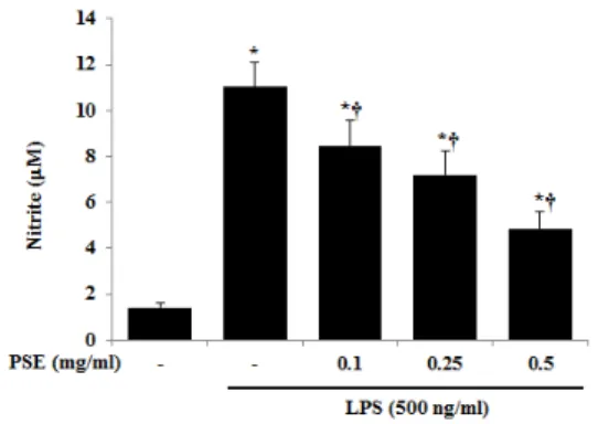
2. NO 생성에 미치는 영향
NO의 농도는 LPS로 자극하지 않은 무처치군에서 1.43±0.21 μM이었으며, LPS 로 자극한 경우 11.02±1.05 μM로 증가하 였다. PSE 0.1 mg/ml 처리군에서는 8.45
±1.10 μM, 0.25 mg/ml 처리군에서는 7.20
±1.04 μM, 0.5 mg/ml 처리군에서는 4.85
±0.77 μM으로 PSE는 NO의 생성을 농 도 의존적으로 감소시켰다(Fig. 2).
Fig. 2. The inhibitory effect of PSE on LPS-induced NO production.
The cells were pre-treated with PSE as indicated concentrations for 1 hr, and then incubated with or without LPS (500 ng/ml) for 24 hrs.
Detail methods were described in Materials and Methods.
NO release was measured by the method of Griess. Data were given as means of values
± SD from three independent experiments.
*p<0.05 in comparison with saline
†p<0.05 in comparison with LPS alone
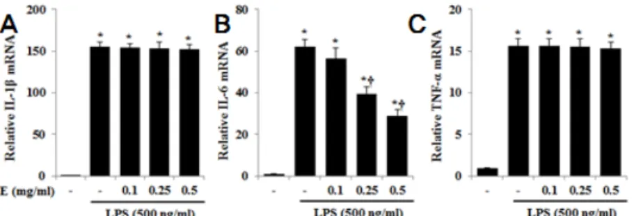
3. IL-1β, IL-6, TNF-α 생성에 미치는 영향
LPS 자극은 RAW 264.7 세포에서 IL-1β, IL-6, TNF-α의 생성을 증가시켰으며, PSE 는 IL-1β, TNF-α의 생성을 억제하지 못 하였다. IL-6의 농도는 무처치군에서는 0.26±0.06 ng/ml, LPS 자극시 67.81±2.56 ng/ml으로 증가 하였다. PSE 0.1 mg/ml 처리군에서는 56.35±5.38 ng/ml, PSE 0.25 mg/ml 처리군에서는 39.46±0.65 ng/ml, 0.5 mg/ml 처리군에서는 26.19±1.95 ng/ml 으로 IL-6의 생성을 농도 의존적으로 감소 시켰으며, PSE 0.25 mg/ml, 0.5 mg/ml 농 도에서 통계적인 유의성이 있었다(Fig. 3).
4. mRNA 수준에서 IL-1β, IL-6, TNF-α 생성에 미치는 영향
IL-1β, IL-6, TNF-α의 mRNA를 측정
한 결과 LPS 자극은 mRNA의 발현을
증가시켰으며, PSE는 IL-1β, TNF-α에
서 유의한 효과를 보이지 않았다. IL-6
에서는 무처치군의 mRNA 양을 1로 설
정하였을 때, LPS 자극시 62.17±3.51으로
증가하였으며, PSE 0.1 mg/ml 처리군에
서는 56.39±5.07, PSE 0.25 mg/ml 처리
군에서는 39.47±3.37, 0.5 mg/ml 처리군
에서는 28.96±2.96으로 IL-6의 mRNA
생성을 농도 의존적으로 감소시켰으며,
PSE 0.25 mg/ml, 0.5 mg/ml 농도에서
통계적인 유의성이 있었다(Fig. 4).
Fig. 3. Effects of PSE on the production of IL-1β, IL-6 and TNF-α in RAW 264.7 cells.
A. Effects of PSE on the production of IL-1β in RAW 264.7 cells B. Effects of PSE on the production of IL-6 in RAW 264.7 cells C. Effects of PSE on the production of TNF-α in RAW 264.7 cells
The cells were pre-treated with PSE as indicated concentrations for 1 hr, and then incubated with or without LPS (500 ng/ml) for 24 hrs. Detail methods were described in Materials and Methods. Data were given as means of values±SD from three independent experiments.
*p<0.05 in comparison with saline
†p<0.05 in comparison with LPS alone
Fig. 4. Effects of PSE on the mRNA expression of IL-1β, IL-6 and TNF-α in RAW 264.7 cells.
A. Effects of PSE on the mRNA expression of IL-1β in RAW 264.7 cells B. Effects of PSE on the mRNA expression of IL-6 in RAW 264.7 cells C. Effects of PSE on the mRNA expression of TNF-α in RAW 264.7 cells
The cells were pre-treated with PSE as indicated concentrations for 1 hr, and then incubated with or without LPS (500 ng/ml) for 24 hrs. Detail methods were described in Materials and Methods. Data were given as means of values±SD from three independent experiments.
*P<0.05 : in comparison with saline
†P<0.05 : in comparison with LPS alone
5. MAPKs 및 NF-κB 활성에 미치는 영향
MAPKs family인 ERK, JNK, p38의 활성은 각각의 인산화를 통해 이루어지며, NF-κB의 활성은 Iκ-Bα의 분해에 의존 한다. LPS 단독으로 자극한 경우와 PSE를 전 처리한 후 LPS로 자극한 경우를 각각 시간 단위로 측정한 결과 PSE는 LPS에
의한 ERK1/2, p38의 인산화는 억제하지 못하였으나 JNK의 인산화는 억제하였다.
또한 Iκ-Bα 분해를 억제하여 NF-κB의 활성을 억제하는 것으로 나타났다(Fig. 5).
앞선 실험에서 가장 유의성 높은 결과를
나타낸 PSE 0.5 mg/ml의 농도로 실험을
시행하였다.
Fig. 5. Effects of PSE on the activation of MAPKs and NF-κB in LPS-stimulated RAW 264.7 cells.
The cells were pre-treated with PSE (0.5 mg/ml) for 1 hr, and then incubated with LPS (500 ng/ml) for indicated mins. Detail methods were described in Materials and Methods. The
results shown are representative of three independent experiments.
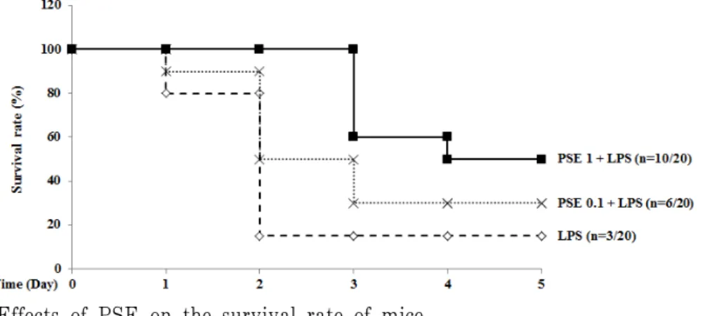
6. Mouse 모델의 생존율에 미치는 영향 LPS(37.5 mg/kg)를 복강주사 한 후, 하 루 단위로 생존율을 관찰한 결과, LPS 단 독 처리군은 2일 이후로 15%의 생존율을 보였으며, PSE 0.1 mg/kg 투여군(PSE 0.1) 에서는 3일 이후로 30%의 생존율을 보였 고, PSE 1 mg/kg 투여군(PSE 1)에서는 4일 이후로 50%의 생존율을 보여 PSE 투 여는 mouse의 생존율을 증가 시켰다(Fig. 6).
차후 실험은 더 높은 유의성을 나타낸 1 mg/kg의 농도로 실험을 진행하였다.
Fig. 6. Effects of PSE on the survival rate of mice.
The mice were pre-intraperitoneal injected with PSE for 1 h, and then intraperitoneal injected with LPS (37.5 mg/kg). After 2 days, survival rate of only-LPS-treated group was 15%. But after 3 days, the survival rate of PSE 0.1 was 30%, and after 4 days, the survival rate of PSE 1 was 50%. The number of mice of each group was 20.
7. Mouse 혈청 cytokine 생성에 미치는 영향
RAW 264.7세포에서 cytokine 생성 결 과와 동일하게 IL-1β, TNF-α 생성에는 유의한 영향을 미치지 않았으며, IL-6 생 성은 LPS 처리군의 882.27±10.26 pg/ml에 비해 PSE 1 mg/kg 투여군에서 556.05±11.52 pg/ml으로 유의하게 감소되었다(Fig. 7).
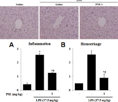
8. Mouse 간 조직 손상에 미치는 영향 간 조직의 염증 정도는 무처치군 0.43
±0.12에 비해, LPS 처리군은 2.57±0.12로
유의하게 증가하였으며, PSE 1 mg/kg
투여군에서는 1.27±0.06으로 LPS 처리군
에 비해 유의하게 감소하였다. 간 조직의
출혈 정도는 무처치군 0.50±0.00에 비해
LPS 처리군은 2.58±0.14으로 유의하게 증
가하였으며, PSE 1 mg/kg 투여군에서는 0.90±0.26으로 LPS 처리군에 비해 유의
하게 감소하였다(Fig. 8).
Fig. 7. Effects of PSE on the production of IL-1β, IL-6, and TNF-α in serum of mice.
A. Effects of PSE on the production of IL-1β in serum of mice B. Effects of PSE on the production of IL-6 in serum of mice C. Effects of PSE on the production of TNF-α in serum of mice
The serum of mice was pre-treated with PSE 1 mg/kg for 1 h, and then incubated with LPS (37.5 mg/kg) for 24 h. The level of cytokine was measured by ELISA. Data were given as means of values±SD from three independent experiments.
*P<0.05 : in comparison with saline
†P<0.05 : in comparison with LPS alone
Fig. 8. Effects of PSE on the liver tissue of mice.
A. Effects of PSE on the inflammation of liver tissue of mice B. Effects of PSE on the hemorrhage of liver tissue of mice
Representative H & E stained sections of the liver tissue in the normal mice given saline, in the control mice given LPS, and in mice given PSE 1 h before the first LPS injection. Histological sections of the liver were scored from 0 (normal) to 3 (severe) for inflammation and hemorrhage.
These figures show representative images of experiment group. The score was calculated by three pathologists who were blinded.
*P<0.05 : in comparison with saline.
†P<0.05 : in comparison with LPS alone.