LPS로 자극된 대식세포에서의 NF-κB와 MAPK 활성 조절을 통한 참까막살( Polyopes affinis) 에탄올 추출물의 항염증 효과
김민지1․김꽃봉우리1․박선희1․박소영1․최현덕2․최정수3․장미란4․임무혁5․안동현1
1부경대학교 식품공학과/식품연구소, 2부경대학교 수산과학연구소
3경남정보대학교 호텔외식조리계열, 4식품의약품안전처 건강기능식품정책과
5대구대학교 식품공학과
Anti-Inflammatory Effect of Ethanolic Extract from Polyopes affinis through Suppression of NF-κB and MAPK Activation
in LPS-Stimulated RAW 264.7 Cells
Min-Ji Kim1, Koth-Bong-Woo-Ri Kim1, Sun-Hee Park1, So-Young Park1, Hyeun-Deok Choi2, Jung-Su Choi3, Mi-Ran Jang4, Moo-Hyeog Im5, and Dong-Hyun Ahn1
1Department of Food Science and Tehcnology/Institute of Food Sciences and
2Institute of Fisheries Sciences, Pukyong National University
3Subdivision of Culinary Arts, Kyungnam College of Information and Technology
4Health Functional Food Policy Division, Ministry of Food and Drug Safety
5Department of Food Science and Biotechnology, Daegu University
ABSTRACT In this study, the anti-inflammatory effect of Polyopes affinis ethanol extract (PAEE) was investigated using LPS-stimulated RAW 264.7 cells and a croton oil-induced ICR mice model. Treatment with PAEE significantly reduced production of nitric oxide (NO) and pro-inflammatory cytokines [interleukin (IL)-6, tumor necrosis factor (TNF)-α, and IL-1β] in lipopolysaccharide (LPS)-stimulated RAW 264.7 cells. PAEE treatment also reduced expression of inducible NO synthase, cyclooxygenase-2, nuclear factor-κB, and mitogen-activated protein kinases in LPS-stimulated RAW 264.7 cells. In the croton oil-induced ear edema test, application of PAEE (10∼250 mg/kg body weight) reduced ear edema in a dose-dependent manner, and PAEE treatment at 50 mg/kg body weight showed similar inhibitory effects compared with prednisolone (10 mg/kg body weight). Histological analysis revealed reduced dermal thickness and lower number of infiltrated mast cells. These results suggest that PAEE might be used as a promising anti-in- flammatory agent for inhibition of LPS-induced inflammation and ear edema formation.
Key words: Polyopes affinis, anti-inflammatory effect, NF-κB, MAPKs, ear edema
Received 23 November 2016; Accepted 2 May 2017
Corresponding author: Dong-Hyun Ahn, Department of Food Science and Technology, Pukyong National University, Busan 48513, Korea
E-mail: [email protected], Phone: +82-51-629-5831
서 론
염증은 외부 병원체나 항원에 의한 감염 및 생체 내 자극 물질에 의한 인체 조직손상을 막는 방어기전으로 발신, 발 열, 통증과 같은 증상을 수반한다(1). 그러나 염증반응이 과 도하게 발생하거나 장기간 지속되는 경우 치매, 심혈관 질 환, 암, 대사성 증후군 등의 만성질환을 발병할 수 있는 요인 이 된다(2).
대식세포(macrophage)는 cytokines, tumor necrosis factor-α(TNF-α), lipopolysaccharide(LPS)와 같은 자극
에 의해 염증반응의 전사인자인 nuclear factor-κB(NF-κ B)와 mitogen-activated protein kinases(MAPKs)를 활 성화시키게 됨에 따라, inducible nitric oxide synthase (iNOS), cyclooxygenase-2(COX-2)를 발현시켜 nitric oxide(NO), prostaglandin E2(PGE2)를 생성하게 하고, pro-inflammatory cytokines(interleukin(IL)-6, TNF-α, IL-1β)의 분비를 증가시켜 염증반응을 유도하게 된다(3,4).
해조류는 다양한 생리활성을 지닌 2차 대사 물질을 함유 하고 있으며, 그중 항염증 활성을 지니고 있는 polyphenol 류(5), polysaccharides(6), fatty acids(7), peptides(8), chromene류(9)가 분리되었다. 홍조류인 참까막살(Polyopes affinis)은 우리나라에서 주로 남해안이나 제주도에 서식하 고 있으며, 식용이 가능하다고 알려져 있다. 참까막살에 대 한 생리활성은 OVA로 유발된 알레르기성 천식 억제(10), 랫트 비만세포 의존성 아토피 반응 억제 활성(11), 항산화
활성(12), 광보호 효과(13)에 대해 보고되어 있다. 하지만 참까막살의 대식세포에 대한 항염증 활성에 대한 보고는 되 어 있지 않다. 따라서 본 연구에서는 참까막살에 의한 LPS 로 자극된 RAW 264.7 세포에서의 염증 매개 물질의 생성 억제와 croton oil로 귀 부종을 유발한 마우스에서의 염증반 응 억제에 대해서 알아보고, 천연 항염증 소재로써 이용 가 능성을 알아보았다.
재료 및 방법
재료
본 연구에서 사용된 참까막살은 2014년도 부산 연화리에 서 채취한 것으로 담수로 깨끗하게 수세한 후 자연 건조한 다음 동결 건조하여 분말화한 후 진공 포장상태로 -20°C에 서 저장하며 사용하였다.
에탄올 추출물의 제조
참까막살 에탄올 추출물은 분말에 10배(v/w)의 95% 에 탄올을 가한 다음, 24시간 동안 실온에서 shaker(H-0820, Dongwon Science Co., Busan, Korea)를 이용하여 추출하 였다. 그런 다음 원심분리기(UNION 32R, Hanil Co., Kim- po, Korea)를 이용하여 1,977×g에서 10분간 원심분리 한 후 상층액을 취하고, 남은 잔사를 이와 같은 방법으로 2회 반복 추출하였다. 상층액은 37°C에서 감압농축기(RE 200, Yamoto Co., Tokyo, Japan)로 농축하였으며, 농축하여 건 조된 시료는 -20°C에서 보관하며 실험에 이용하였다.
세포배양
한국세포주은행(Seoul, Korea)에서 분양받은 murine macrophage cell line RAW 264.7 세포(KCLB 40071)는 Dulbecco’s modified Eagle’s medium(DMEM, Gibco, Grand Island, NY, USA)에 10% inactivated fetal bovine serum(HyClone, Victoria, Austria)과 1% penicillin-strep- tomycin(HyClone)을 포함한 배지를 사용하여 37°C, 5%
CO2 incubator(MCO-15AC, Sanyo, Tokyo, Japan)에서 배양하였다.
세포독성 측정
참까막살 에탄올 추출물(PAEE)의 세포독성은 MTT as- say를 실시하였다. RAW 264.7 세포를 1×10⁶ cells/well 농도로 분주하고 20시간 배양하였다. 배양 후 PAEE를 농도 별(0.1, 1, 10, 50, 100 μg/mL)로 첨가하여 37°C, 5% CO2 incubator(MCO-15AC)에서 22시간 배양하였다. 배양 후 5 mg/mL 농도인 thiazolyl blue tetrazolium bromide (MTT, Sigma-Aldrich Co., St. Louis, MO, USA) 시약을 첨가하고 2시간 동안 CO2 배양기에서 반응시킨 후 이를 4°C, 879×g에서 10분간 원심분리 하여 상층액을 제거하였 다. 그 후 각 well에 dimethyl sulfoxide(Sigma-Aldrich
Co.)를 첨가하고 이를 microplate reader(Model 550, Bio- Rad, Hercules, CA, USA)를 이용하여 540 nm에서 흡광도 를 측정하였다.
동물실험
귀 부종 실험에 사용한 마우스는 생후 8주령의 수컷, ICR 종으로 오리엔트바이오(Seongnam, Korea)로부터 구입하 였다. 마우스는 온도 20±2°C, 습도 50±10%, 12시간 명암 주기가 유지되는 동물실에서 1주일간 예비 사육한 후 실험 에 사용하였다. 본 실험은 부경대학교 동물윤리실험윤리위 원회로부터 동물실험 승인을 받아 수행하였다(승인번호 2015-04).
Croton oil에 의한 in vivo 귀 부종 실험
PAEE의 항염증 효과를 in vivo 상에서 알아보기 위하여 Kim 등(14)과 Saraiva 등(15)의 방법으로 귀 부종 실험을 실시하였다. ICR 마우스에 PAEE를 10, 50 및 250 mg/kg・
body weight 농도가 되도록 200 μL씩 경구 투여하고 한 시간 후, 오른쪽 귀에 2.5% croton oil을 20 μL/ear 농도로 도포하였다. 도포 5시간 후 귀 두께를 측정하였고 croton oil(TCI Chemical, Portland, OR, USA)의 처리로 귀 두께 가 증가한 것을 부종의 형성으로 간주하였다. 귀 조직 관찰 은 ICR 마우스의 오른쪽 귀에 PAEE를 100 mg/mL 농도로 20 μL씩 도포하고 15분 후 5% croton oil을 20 μL씩 도포하 였다. 6시간 후 diethyl ether로 마취사 시키고, 귀 조직을 절제하여 10% formaldehyde에 72시간 고정하였다. 고정 후 파라핀 블록을 만들어 박편을 제조하고 hematoxylin- eosin과 toluidine-blue 염색을 하여 조직을 관찰하였다. 부 종 생성률은 다음과 같은 식에 의해 계산하였다.
Edema formation (% of control)=(Ear thickness of sample/ Ear thickness of control)×100
NO 및 pro-inflammatory cytokine 생성량 측정 NO의 농도는 배양액 내의 nitrite 농도를 Griess 반응을 이용하여 측정하였다(16). RAW 264.7 세포는 DMEM 배지 를 이용하여 24-well plate에 2.5×10⁵ cells/mL로 분주하 고 5% CO2 배양기에서 20시간 배양하였다. 배양 후 LPS(1 μg/mL)와 PAEE를 농도별(0.1, 1, 10, 50, 100 μg/mL)로 처리하여 24시간 배양하였다. 배양액의 상층액을 얻은 후 동량의 Griess 시약(1% sulfanilamide+0.1% naphthalene diamine dihydrochloride, 1:1)을 첨가하여 실온에서 10분 간 반응시키고, microplate reader를 이용하여 540 nm에 서 흡광도를 측정하였다. 세포 배양액 내 NO의 농도는 so- dium nitrite(NaNO2)의 농도별 표준곡선과 비교하여 산출 하였다. Pro-inflammatory cytokine의 분비량 측정의 경 우 2.5×10⁵ cells/mL로 24-well plate에 분주하고 5% CO2
배양기에서 18시간 배양한 후, LPS(1 μg/mL)와 PAEE(0.1, 1, 10, 50, 100 μg/mL)를 처리하여 12시간 배양하였다. 세
0 20 40 60 80 100 120
PBS 0.1 1 10 50 100
Proliferation index (% of conrol) .
(μg/mL) ND
Fig. 1. Effect of ethanolic extract from Polyopes affinis on pro- liferation of RAW 264.7 cells. Proliferation index (% of con- trol)=[sample absorbance/ control absorbance (PBS)]×100. ND:
not significantly different.
포배양액 내의 TNF-α, IL-6 및 IL-1β 사이토카인의 분비 량을 ELISA kit(Mouse ELISA set, BD Biosciences, San Diego, CA, USA)을 이용하여 정량하였다.
Western blot에 의한 단백질 발현량 측정
iNOS와 COX-2의 발현량을 관찰하기 위해서는 세포에 LPS(1 μg/mL)와 PAEE(0.1, 1, 10, 50, 100 μg/mL)를 처리 한 후 18시간 동안 배양하였으며, p-NF-κB p65, p-IκBα와 인산화된 MAPKs(p-ERK, p-JNK, p-p38)의 발현량은 세 포에 LPS와 PAEE를 처리하고 30분 동안 배양하였다.
p-NF-κB p65, p-IκBα, iNOS, COX-2 및 p-MAPKs의 경 우 cytosol lysis buffer[50 mM HEPES(pH 7.4), 150 mM NaCl, 5 mM EDTA, 1% deoxycholate, 5 mM phenyl- methylsulfonyl fluoride, 1 μg/mL aprotinin, 1% Triton X-100, 0.1% NP-40]를 이용하였으며, NF-κB p65의 경 우 nucleus lysis buffer(10 mM HEPES, 100 mM NaCl, 1.5 mM MgCl2, 0.1 mM EDTA, 0.1 mM dithiothreitol)를 첨가하여 30분간 4°C에서 lysis 시킨 후 15,520×g에서 20 분간 원심분리 하여 세포막 성분 등을 제거하였다. BCA protein assay kit(Pierce, Rockford, IL, USA)을 사용하여 단백질을 정량하였으며 30 μL의 lysate를 Laemmli(17)의 방법을 사용하여 10% SDS-PAGE로 분리하였다. 전기영동 한 겔은 Towbin 등(18)의 방법을 참고하여 polyvinylidene difluoride membrane(Bio-Rad)에 1시간 동안 전사시켜 5% skim milk가 함유된 tris-buffered saline(TBS, pH 7.5) 용액으로 상온에서 2시간 동안 blocking 하였다. iNOS, COX-2, NF-κB p65, p-NF-κB p65, p-IκBα의 발현량을 검토하기 위한 항체로는 anti-mouse iNOS, COX-2, NF- κB p65, p-NF-κB p65 및 p-IκBα(Santa Cruz Biotech- nology, Dallas, TX, USA)를 사용하여 1:500으로 희석하 여 사용하였다. 인산화된 c-Jun N-terminal kinase(JNK), extracellular signal-regulated kinase(ERK), p38 pro- tein kinase(p38) 및 JNK, ERK, p38의 발현량을 검토하기 위하여 anti-mouse p-JNK, JNK, p-ERK, ERK, p-p38 및 p38(Cell Signaling Technology Inc., Danvers, MA, USA) 항체를 1:500으로 희석하여 상온에서 2시간 반응시킨 후 TBS로 3회 세정하였다. 2차 항체로 horseradish perox- idase가 결합된 anti-mouse IgG 및 anti-rabbit IgG를 1:2,000으로 희석하여 상온에서 1시간 반응시킨 다음 TBS 로 3회 세정하여 ECL 기질과 1~3분간 반응 후 각각의 단백 질 밴드는 Gene tool(GeneGnome5, Syngene, Cambridge, UK)을 이용하여 가시화하였다.
통계처리
실험 결과의 통계처리는 SAS program(Statistical Ana- lytical System V8.2, SAS Institute Inc., Cary, NC, USA) 을 이용하여 평균값을 분산분석한 후, Duncan의 다중검정법 으로 P<0.05 수준에서 항목 간의 유의적 차이를 검정하였다.
결과 및 고찰
세포독성
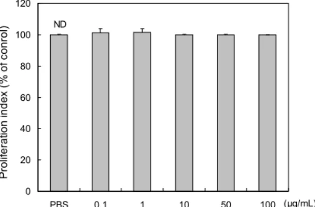
PAEE의 RAW 264.7 세포에 대한 독성을 MTT법으로 측정한 결과(Fig. 1), PAEE는 0.1~100 μg/mL 농도 범위에 서 세포독성을 나타내지 않았다. 따라서 in vitro 실험은 100 μg/mL 이하에서 진행하였다.
NO 분비량과 iNOS와 COX-2의 발현량
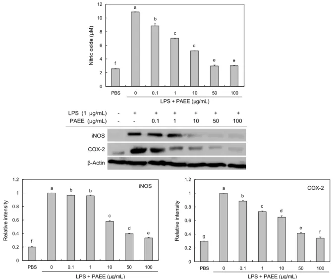
일반적인 iNOS에 의한 NO의 생성은 세균이나 종양을 제 거하는 역할을 하지만(19), 과도한 생성은 염증을 유발하여 조직의 손상, 유전자 변이, 신경손상 등을 일으키는 것으로 알려져 있다(20). 이에 LPS로 자극된 RAW 264.7 세포에서 PAEE 처리가 NO와 NO 생성과 관련된 효소인 iNOS의 발 현을 억제하는지 알아보았다. 그 결과 LPS에 의해 증가한 NO의 생성이 PAEE를 0.1~100 μg/mL 농도로 처리 시 농 도 의존적으로 유의하게 억제되었다(Fig. 2). 또한, LPS에 의해 NO를 생합성하는 효소인 iNOS의 세포 내 발현량을 측정한 결과, PAEE(0.1~100 μg/mL) 처리에 의해 LPS에 의해 증가한 iNOS의 발현이 농도가 증가함에 따라 유의하 게 억제됨을 확인하였다(Fig. 2). PAEE가 염증 매개물질인 PGE2 생성과 관련된 주요 효소인 COX-2 발현에 미치는 영향을 조사하였다. COX-2는 염증성 자극, 호르몬, 성장 인자에 의해 단시간에 유도되어 발현되며, 염증과 관련된 prostanoid 생성에 중요한 역할을 한다(21). 또한, COX-2 의 지속적인 과다 발생은 NO의 분비량 증가와도 관련성이 있다고 알려져 있다(22). Fig. 2에서 COX-2의 발현은 LPS 에 의해 증가하였으며, 농도 의존적으로 억제되었다. 따라서 PAEE는 RAW 264.7 세포에서 iNOS의 발현이 감소하여 NO 생성을 감소시키는 것으로 생각되며, COX-2의 발현 억제에도 감소 효과를 나타내어 항염증 효과를 나타내는 것 으로 확인되었다.
0 2 4 6 8 10 12
PBS 0 0.1 1 10 50 100
LPS + PAEE (μg/mL)
Nitric oxide (μM) .
a b
c
d
e e
f
iNOS COX-2 β-Actin
LPS (1 μg/mL) - + + + + + + PAEE (μg/mL) - - 0.1 1 10 50 100
0 0.2 0.4 0.6 0.8 1 1.2
PBS 0 0.1 1 10 50 100
LPS + PAEE (μg/mL)
Relative intensity .
e
a b b
c
d
f
iNOS
0
0.2 0.4 0.6 0.8 1 1.2
PBS 0 0.1 1 10 50 100
LPS + PAEE (μg/mL)
Relative intensity .
f a
b c
d
e g
COX-2
Fig. 2. Inhibitory effects of ethanolic extract from Polyopes affinis (PAEE) on NO production and the expression of iNOS and COX-2 in LPS-induced RAW 264.7 cells. The levels of iNOS and COX-2 in the cytosolic protein were determined by western blot analysis. RAW 264.7 cells were treated with the indicated concentrations of PAEE (0.1, 1, 10, 50, and 100 μg/mL) and LPS (1 μg/mL) for 30 min and the proteins were detected using specific antibodies. For quantification, the expression data were normalized to the β-actin signal. Data are obtained from three independent experiments and expressed as mean±SD. Means with different letters (a-g) above the bars are significantly different (P<0.05).
Pro-inflammatory cytokines 분비 억제 효과
활성화된 대식세포인 RAW 264.7 세포에서는 TNF-α, IL-6, IL-1β와 같은 pro-inflammatory cytokine들을 증가 시키는 것으로 알려져 있다(23). TNF-α는 대식세포나 단핵 세포에서 세포의 분화와 성장 등의 기능에 관여하고 염증이 발생하게 되면 그 부위에 백혈구를 모이게 한다(24). IL-1β 는 단핵구, 대식세포, B 세포 등에서 분비되어 여러 면역 반응들과 연관되어 있는데, 염증반응의 초기와 발달에 있어 중요한 역할을 한다(25). IL-6는 단핵구를 포함한 여러 세 포에서 분비가 되고 항염증과 전염증성의 특징을 지니고 있 는 면역반응에 중요한 기능을 담당하고 있지만, 류마티즘 관절염과 같은 염증 질환에서 분비가 증가한 것을 볼 수 있 다(26). 본 연구에서 PAEE 처리가 pro-inflammatory cy- tokine의 분비를 농도 의존적으로 IL-6, TNF-α 및 IL-1β 의 분비량을 억제하였다(Fig. 3). 특히 IL-6의 경우 50 μg/
mL 이상에서 약 70% 이상의 억제율을 나타내었으며, TNF- α와 IL-1β의 경우 100 μg/mL 농도에서 약 50% 및 40%
정도의 억제율을 나타내었다. 따라서 PAEE는 IL-6, TNF- α 및 IL-1β와 같은 pro-inflammatory cytokine의 생성을 억제하여 항염증 활성을 나타내는 것으로 생각된다.
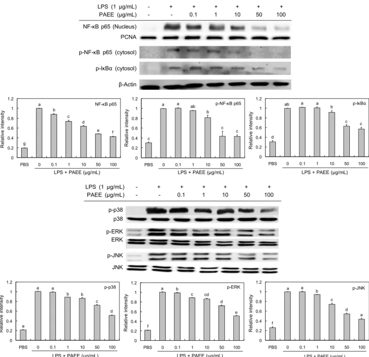
NF-κB 및 MAPKs(JNK, ERK, p38)의 발현 억제 효과 NF-κB는 세포질에서 IκB와 결합되어 있는 경우 전사인 자로서 역할을 못하지만, IκB가 LPS와 같은 자극원에 의해 인산화가 되면 분해되어 NF-κB가 핵 내로 이동하여 다양한 염증성 매개물질의 합성을 촉진하게 된다(27). PAEE의 항 염증 활성이 NF-κB 발현에 미치는 영향을 확인한 결과 (Fig. 4), 핵 내에서 LPS에 의하여 증가한 발현이 PAEE 처 리에 의해 농도 의존적으로 감소함을 확인하였다. 세포질에 서 인산화된 NF-κB p65와 인산화된 IκBα의 발현량을 측정
0 100 200 300 400 500 600 700 800
PBS 0 0.1 1 10 50 100
LPS + PAEE (μg/mL)
IL-6 (pg/mL) .
f
e e d c b a
0 500 1000 1500 2000 2500
PBS 0 0.1 1 10 50 100
LPS + PAEE (μg/mL)
TNF-α (pg/mL) .
a b
c c
d d
e
0 10 20 30 40 50 60 70
PBS 0 0.1 1 10 50 100
LPS + PAEE (μg/mL)
IL-1β (pg/mL) .
a
ab b bc
c d
e Fig. 3. Inhibitory effect of ethanolic extract from Polyopes affi-
nis (PAEE) on production of IL-6, TNF-α, and IL-1β in LPS-in- duced RAW 246.7 cells. Means with different letters (a-f) above the bars are significantly different (P<0.05).
한 결과(Fig. 4) LPS에 의해 증가하였으며, PAEE 처리에 의해 농도가 증가할수록 감소한 것으로 나타났다. 따라서 IκB의 인산화가 억제됨으로써 LPS에 의한 NF-κB p65의 핵 내로의 이동이 억제된 것으로 생각된다. 한편 MAPK는 세포의 성장, 분열, 스트레스나 사이토카인에 의한 세포반응 의 조절 등에 중요한 역할을 하며(28), 외부자극이 가해지면 ERK, JNK, p38에 의한 신호 전달 경로에 의해 세포 내에서 활성화되어 세포의 형태 변화와 사이토카인 전사 등을 초래 한다(4). 본 연구에서 PAEE에 의한 항염증 활성이 MAPK 발현 조절에 영향을 미치는지 조사하였다(Fig. 4). 그 결과 LPS 자극에 의해 증가한 인산화된 p38, ERK, JNK의 발현 은 PAEE 처리에 의해 농도 의존적으로 억제됨을 확인하였 다. 이상으로 PAEE는 LPS 자극에 의해 활성화된 RAW 264.7 세포에서 염증매개물질인 iNOS와 COX-2의 발현 및 NO와 pro-inflammatory cytokine의 생성이 NF-κB와 MAPKs의 활성 억제를 통하여 항염증 활성을 나타내는 것 으로 나타났다.
귀 부종 억제 효과
Croton oil을 이용한 귀 부종 실험은 급성 염증에 대한 항 염증 효과 평가법으로 많이 이용되고 있는 방법이다. Cro- ton oil에 의한 염증반응은 phospholipase A2 활성에 의해 유도되며, 이는 세포막에서 arachidonic acid를 prosta- glandins과 leukotrienes으로 전환됨에 따라 발생하게 된다 (29). 따라서 croton oil로 염증을 유발하여 PAEE(10, 50 및 250 mg/kg)의 경구투여에 의한 귀 두께 변화를 측정하였
다(Fig. 5). 그 결과 croton oil에 의해 귀 부종이 형성됨을 확인하였고, PAEE 처리 시 모든 농도에서 귀 두께가 유의하 게 억제됨을 확인하였다. 특히 PAEE를 50 mg/kg 농도로 경구 투여하였을 때 시판 항염증제인 prednisolone 10 mg/
kg 처리구와 유사한 귀 부종 억제 효과를 나타내었다. 조직 관찰 결과(Fig. 5A)에서는 croton oil만을 처리한 경우에 경 피와 진피의 두께가 증가하였고, PAEE를 100 mg/mL 농도 로 처리한 경우 경피 및 진피 두께가 얇아진 것을 확인하였 다. Toluidine-blue 염색을 통해 진피로 비만세포의 침윤된 정도를 확인한 결과(Fig. 5B), PAEE 처리구는 predniso- lone 처리구와 유사한 정도로 mast cell 수가 감소함을 확인 하였다. 따라서 PAEE는 croton oil로 유도된 귀 부종 억제 에 효과가 있음을 확인하여 염증반응의 하나인 부종 억제를 위한 예방 또는 치료제로써 이용 가능성이 높은 것으로 확인 되었다.
요 약
본 연구에서는 참까막살 에탄올 추출물(PAEE)의 항염증 활 성을 확인하기 위하여 LPS로 자극된 RAW 264.7 세포에서 의 pro-inflammatory cytokine 및 NO의 분비 생성량과 western blot으로 단백질 발현량을 측정하였다. 또한, cro- ton oil로 유도된 귀 부종 모델을 이용하여 알아보았다. RAW 264.7 세포에서 PAEE를 0.1~100 μg/mL 농도로 처리 시 세포독성을 나타내지 않음을 확인하였다. 그 결과 PAEE는 pro-inflammatory cytokine(IL-6, TNF-α, IL-1β) 및 NO
LPS (1 μg/mL) - + + + + + + PAEE (μg/mL) - - 0.1 1 10 50 100 NF-κB p65 (Nucleus)
PCNA p-NF-κB p65 (cytosol)
p-IκBα (cytosol)
β-Actin
0 0.2 0.4 0.6 0.8 1 1.2
PBS 0 0.1 1 10 50 100
LPS + PAEE (μg/mL)
Relative intensity . NF-κB p65
g
e f d c b a
0 0.2 0.4 0.6 0.8 1 1.2
PBS 0 0.1 1 10 50 100
LPS + PAEE (μg/mL)
Relative intensity . p-NF-κB p65a a ab
b
c c
c
0 0.2 0.4 0.6 0.8 1 1.2
PBS 0 0.1 1 10 50 100
LPS + PAEE (μg/mL)
Relative intensity . p-IκBαab a a b
c c
d
LPS (1 μg/mL) - + + + + + + PAEE (μg/mL) - - 0.1 1 10 50 100
p-p38 p38 p-ERK ERK
p-JNK JNK
0 0.2 0.4 0.6 0.8 1 1.2
PBS 0 0.1 1 10 50 100
LPS + PAEE (μg/mL)
Relative intensity . p-p38
e
d c b b a a
0 0.2 0.4 0.6 0.8 1 1.2
PBS 0 0.1 1 10 50 100
LPS + PAEE (μg/mL)
Relative intensity . p-ERK
f
e d c cd
a b
0 0.2 0.4 0.6 0.8 1 1.2
PBS 0 0.1 1 10 50 100
LPS + PAEE (μg/mL)
Relative intensity . p-JNK
f
e d c a b
a
Fig. 4. Inhibitory effects of ethanolic extract from Polyopes affinis (PAEE) on the protein expression of NF-κB p65, p-IκBα, p-p38, p-ERK, and p-JNK in LPS-induced RAW 264.7 cells. The levels of p-IκBα, p-NF-κB p65, and p-MPAKs (p-p38, p-ERK, and p-JNK) in the cytosolic protein and the NF-κB p65 in nuclear protein were determined by western blot analysis. RAW 264.7 cells were treated with the indicated concentrations of PAEE (0.1, 1, 10, 50, and 100 μg/mL) and LPS (1 μg/mL) for 30 min, and the proteins were detected using specific antibodies. For quantification, the expression data were normalized to PCNA or β-actin or the total MAPKs signals. Data are obtained from three independent experiments and expressed as mean±SD. Means with different letters (a-g) above the bars are significantly different (P<0.05).
의 분비량을 농도 의존적으로 억제시켰으며, iNOS와 COX- 2의 발현량도 감소시킴을 확인하였다. 이러한 항염증 활성 결과는 NF-κB와 MAPKs 전사인자의 활성 억제에 의한 것 으로 확인되었다. 또한, croton oil로 유도된 귀 부종 모델에 서 PAEE를 50 mg/kg body weight 처리 시 귀 부종이 pre- dnisolone(10 mg/kg body weight)과 유사한 정도로 억제
됨을 확인하였다. 귀 조직 관찰에서도 PAEE는 croton oil에 의해 증가한 진피와 경피의 두께를 감소시켰으며, 진피로 침윤된 mast cell의 수도 감소시켰다. 이 결과를 종합해 보 면 참까막살 에탄올 추출물은 NF-κB와 MAPKs의 활성화 억제를 통해 염증 매개 물질의 생성을 억제시켜 항염증 활성 을 나타내는 것으로 확인되었다.
0 20 40 60 80 100 120
Control 10 50 10 50 250
Edema formation (% of control) .
Prednisolone PAEE
(mg/kg) a
bc d
b b
c
1 2
a b
c d
a b
c d
A B
Fig. 5. Inhibition of ethanolic extract from Polyopes affinis (PAEE) against croton oil-induced mouse ear edema (n=5) and photomicro- graph of transverse sections of mice ears sensitized with topical application of 5% croton oil (v/v) in acetone (a-c) or vehicle acetone (d), stained with (A) hematoxylin-eosin and (B) toluidine-blue examined under light microscopy (magnification: 200×).
Treatments: vehicle 2% Tween 80 (a), prednisolone 0.08 mg/ear (b) and PAEE 20 μL/ear (c). The numbers 1 and 2 indicate dermis and epidermis, respectively and the arrow in (B) means infiltration of mast cells. Means with different letters (a-d) above the bars are significantly different (P<0.05).
감사의 글
이 논문은 2016년도 정부(교육부)의 재원으로 한국연구재 단의 지원을 받아 수행된 기초연구사업입니다(No. 2012R1 A6A1028677).
REFERENCES
1. Nathan C. 2002. Points of control on inflammation. Nature 420: 846-852.
2. Medzhitov R. 2008. Origin and physiological roles of in- flammation. Nature 454: 428-435.
3. Jeong DH, Kim KB, Kim MJ, Kang BK, Ahn DH. 2013.
Anti-inflammatory activity of ethanolic extract of Sargassum micracanthum. J Microbiol Biotechnol 23: 1691-1698.
4. Waetzig V, Czeloth K, Hidding U, Mielke K, Kanzow M, Brecht S, Goetz M, Lucius R, Herdegen T, Hanisch UK.
2005. c-Jun N-terminal kinases (JNKs) mediate pro-inflam- matory actions of microglia. Glia 50: 235-246.
5. Jung WK, Heo SJ, Jeon YJ, Lee CM, Park YM, Byun HG, Choi YH, Park SG, Choi IW. 2009. Inhibitory effects and molecular mechanism of dieckol isolated from marine brown alga on COX-2 and iNOS in microglial cells. J Agric Food
Chem 57: 4439-4446.
6. Kim MJ, Bae NY, Bark SW, Kim KBWR, Park JH, Park SH, Ahn DH. 2015. Anti-inflammatory effect of alginate oligosaccharides produced by an alginate-degrading enzyme from Shewanella oneidensis PKA1008 on LPS-induced RAW 264.7 cells. Korean J Fish Auqat Sci 48: 888-897.
7. Dang HT, Lee HJ, Yoo ES, Shinde PB, Lee YM, Hong J, Kim DK, Jung JH. 2008. Anti-inflammatory constituents of the red alga Gracilaria verrucosa and their synthetic ana- logues. J Nat Prod 71: 232-240.
8. Kim SK, Wijesekara I. 2010. Development and biological activities of marine-derived bioactive peptides: A review.
J Funct Foods 2: 1-9.
9. Jeong DH, Kim KB, Kim MJ, Kang BK, Ahn DH. 2014.
Anti-inflammatory activity of methanol extract and n-hex- ane fraction mojabanchromanol b from Myagropsis mya- groides. Life Sci 114: 12-19.
10. Lee DS, Park WS, Heo SJ, Cha SH, Kim D, Jeon YJ, Park SG, Seo SK, Choi JS, Park SJ, Shim EB, Choi IW, Jung WK. 2011. Polyopes affinis alleviates airway inflammation in a murine model of allergic asthma. J Biosci 36: 869-877.
11. Na HJ, Moon PD, Lee HJ, Kim HR, Chae HJ, Shin T, Seo Y, Hong SH, Kim HM. 2005. Regulatory effect of atopic allergic reaction by Carpopeltis affinis. J Ethnopharmacol 101: 43-48.
12. Heo SJ, Cha SH, Lee KW, Jeon YJ. 2006. Antioxidant ac- tivities of red algae from Jeju Island. Algae 21: 149-156.
13. Hyun YJ, Piao MJ, Kim KC, Zheng J, Yao CW, Cha JW, Kang HK, Yoo ES, Koh YS, Lee NH, Ko MH, Hyun JW.
2014. Photoprotective effect of a Polyopes affinis (Harvey) Kawaguchi and Wang (Halymeniaceae)-derived ethanol ex- tract on human keratinocytes. Trop J Pharm Res 13: 863- 871.
14. Kim DW, Chi YS, Son KH, Chang HW, Kim JS, Kang SS, Kim HP. 2002. Effects of sophoraflavanone G, a prenylated flavonoid from Sophora flavescens, on cyclooxygenase-2 and in vivo inflammatory response. Arch Pharm Res 25:
329-335.
15. Saraiva RA, Araruna MK, Oliveira RC, Menezes KD, Leite GO, Kerntopf MR, Costa JG, Rocha JB, Tomé AR, Campos AR, Menezes IR. 2011. Topical anti-inflammatory effect of Caryocar coriaceum Wittm. (Caryocaraceae) fruit pulp fixed oil on mice ear edema induced by different irritant agents.
J Ethnopharmacol 136: 504-510.
16. Lee ST, Jeong YR, Ha MH, Kim SH, Byun MW, Jo SK.
2000. Induction of nitric oxide and TNF-α by herbal plant extracts in mouse macrophages. J Korean Soc Food Sci Nutr 29: 342-348.
17. Laemmli UK. 1970. Cleavage of structural proteins during the assembly of the head of bacteriophage T4. Nature 227:
680-685.
18. Towbin H, Staehelin T, Gordon J. 1979. Electrophoretic transfer of proteins from polyacrylamide gels to nitrocel- lulose sheets: procedure and some applications. Proc Natl Acad Sci U S A 76: 4350-4354.
19. Weisz A, Cicatiello L, Esumi H. 1996. Regulation of the mouse inducible-type nitric oxide synthase gene promoter by interferon-gamma, bacterial lipopolysaccharide and NG-
monomethyl-L-arginine. Biochem J 316: 209-215.
20. Mu MM, Chakravortty D, Sugiyama T, Koide N, Takahashi K, Mori I, Yoshida T, Yokochi T. 2001. The inhibitory ac- tion of quercetin on lipopolysaccharide-induced nitric oxide production in RAW 264.7 macrophage cells. J Endotoxin Res 7: 431-438.
21. Dubois RN, Abramson SB, Crofford L, Gupta RA, Simon LS, Van De Putte LB, Lipsky PE. 1998. Cyclooxygenase in biology and disease. FASEB J 12: 1063-1073.
22. Perkins DJ, Kniss DA. 1999. Blockade of nitric oxide for- mation down-regulates cyclooxygenase-2 and decreases PGE2 biosynthesis in macrophages. J Leukoc Biol 65: 792-799.
23. Zhang JM, An J. 2009. Cytokines, inflammation and pain.
Int Anesthesiol Clin 5: 27-37.
24. Aggarwal BB, Natarajan K. 1996. Tumor necrosis factors:
developments during the last decade. Eur Cytokine Netw 7: 93-124.
25. Kim EY, Moudgil KD. 2008. Regulation of autoimmune inflammation by pro-inflammatory cytokines. Immunol Lett 120: 1-5.
26. Van Q, Nayak BN, Reimer M, Jones PJ, Fulcher RG, Rempel CB. 2009. Anti-inflammatory effect of Inonotus obliquus, Polygala senega L., and Viburnum trilobum in a cell screen- ing assay. J Ethnopharmacol 125: 487-493.
27. Nam NH. 2006. Naturally occurring NF-κB inhibitors. Mini Rev Med Chem 6: 945-951.
28. Johnson GL, Lapadat R. 2002. Mitogen-activated protein kinase pathways mediated by ERK, JNK, and p38 protein kinases. Science 298: 1911-1912.
29. Satyam SM, Bairy KL, Musharraf S, Fernandes DL. 2014.
Inhibition of croton oil-induced oedema in rat ear skin by topical nicotinamide gel. Pharmacologyonline 3: 22-25.