• 검색 결과가 없습니다.

Articular loose body, Synovial Chondromatosis of the Temporomandibular Joint : a Case Report

N/A
N/A
Protected

Academic year: 2021

Share "Articular loose body, Synovial Chondromatosis of the Temporomandibular Joint : a Case Report"

Copied!
2
0
0

로드 중.... (전체 텍스트 보기)

전체 글

(1)

대구외지 2009;35:310-311

310

Abstract (J. Kor. Oral Maxillofac. Surg. 2009;35:310-311)

Introduction

Radiopacities are sometimes observed inside or outside of the temporomandibular joint space. These loose bodies may be present in degenerative joint diseases

1)

. In rare cases, synovial chondromatosis or chondrosarcoma may also mimic the appearance of loose bodies
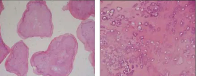
2)

. Synovial chondromatosis is a benign pathologic condition characterized by the development of nodules of cartilage within the synovial connective tissues of articulating joints and formation of multiple small foci of hyaline cartilage or hyperplastic synovium in the joints

3)

. The etiology of the disease has not been completely clarified, but may involve secondary reactive metaplasia after trauma or chronic abnormal loading leading to detachment of chondro- cytes

4)

. Cartilaginous nodules develop which may remain attached to the synovial membrane or detach, calcify and form loose bodies inside the articular space

5)

.

Case Report

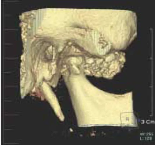
A 46 years-old female was referred from the Department of Periodontics for the evaluation of right Condyle head(Fig. 1).

She had not any discomfort symptoms but many radiopaque calcification were seen her right Temporomandibular joint area

in panoramic view and cone beam CT(Fig. 2).

Mouth opening limitation, tenderness and swelling around on right TMJ were not observed. Slightly flattening was existed on condyle head but it did not cause any other problems.

In fact, she was a patient who had a squamous cell carcinoma on her Mandible. So, we had to operate on marginal mandiblectomy under general anesthesia. After marginal

이 백 수

130-701

서울특별시 동대문구 회기동

1

경희대학교 치과대학 구강악안면외과학교실 Baek-Soo Lee

Department of Oral & Maxillofacial Surgery Kyung Hee University Dental School 1 Hoegi-dong Dongdaeman-gu, Seoul 130-701, Republic of Korea

Tel: 82-2-958-9440 Fax: 82-2-966-4572 E-mail: leebs@khu.ac.kr

Articular loose body, Synovial Chondromatosis of the Temporomandibular Joint : a Case Report

Byung-Joon Choi, Baek-Soo Lee, Yeo-Gab Kim, Yong-Dae Kwon, Young-Ran Kim Department of Oral & Maxillofacial Surgery, Kyung Hee University Dental School, Seoul, Korea

Synovial chondromatosis is an uncommon disease of cartilage transformation of synovial membrane with formation of loose bodies within the joint space. The involvement of temporomandibular joint is very rare. Symtoms include swelling, pain, stiffness of the jaw, and inability to close the jaw. A case involving the temporomandibular joint(TMJ) and non-symptoms is presented.

Key words: Synovial chondromatosis, TMJ, loose body

[원고접수일 2009. 8. 21 / 1차수정일 2009. 8. 28 / 2차수정일 2009. 9. 7 / 게재확정일 2009. 9. 22]

Fig. 1. multiple radiopaque mass at Rt. Condyle head.

Fig. 2. multiple calcification in cone beam CT.

(2)

Articular loose body, synovial chondromatosis of the temporomandibular joint : A case report

311 mandiblectomy, we removed multiple calcified masses in the

superior joint space(Fig. 3). About 30 calcifications were vari- ous size and chondrocytes with hyaline cartilage were observed in microscopic view(Fig. 4). The final diagnosis of synovial chondromatosis was confirmed. Also any other symp- toms were not appeared after operation. At present, there has been no evidence of recurrence during the 1 year of follow-up period.

Discussion

Synovial chondromatosis is an uncommon articular disorder of unknown etiology. When the TMJ is involved, the most fre- quent clinical symptoms are pre-auricular swelling together with joint pain and limitation of mandible movement, and clicking or crepitus on joint motion. But in this case patient did not have any symptom around TMJ. Multiple calcifications were observed around right Condyle head in CT scan. MRI can better define the anatomy of the articular disc and joint cap-

sule, and visualize the boundaries of the lesion with the neigh- boring anatomical structures, especially extensions out of the joint. If the patient had not been a squamous cell carcinoma patient, surgical excision of the radiopaque mass might have been unnecessary because there were no pre-auricular swelling and no limitation of mouth opening. Under general anesthesia marginal mandiblectomy and multiple calcified masses removal were done in order. Various treatments have been used for synovial chondromatosis of the TMJ. When radi- ographic findings confirm its location in the upper space, and loose bodies are smaller than the diameter of the arthroscope, it can be treated by arthroscopic surgery. If we did arthroscopic surgery, it was lesser invasive than TMJ open surgery.

Therefore further investigations and differential diagnosis are essential before deciding on TMJ open surgery.

Reference

1. Kim IK, Choi J, Oh SS, Oh NS, Kim HD, Lee SH, Yang DH.

SYNOVIAL CHONDROMATOSIS OF THE TEMPORO- MANDIBULAR JOINT: A CASE REPORT: J. Kor. Oral Maxillofac. Surg. 2000; 26: 109-113.

2. White SC, Pharoah MJ. Oral Radiology Principles and Interpretation, 4

th

ed. St. Louis, Mosby, 2000, p.520-521.

3. Xu WH, Ma XC, Guo CB, Yi B, Bao SD. Synovial chondro- matosis of the temporomandibular joint withmiddle cranial fossa extension: Int. J. Oral Maxillofac. Surg. 2007; 36: 652-655.

4. Yokota N, Inenaga C, Tokuyama T, Nishizawa S, Miura K, Namba H. Synovial Chondromatosis of the Temporomandibular Joint With Intracranial Extension: Neurol Med Chir(Tokyo) 2008; 48: 266-270.

5. Adachi PL, Kaba SP, Martins MT, Hueb CH, Shinohara EH.

Arthrocentesis in the treatment of loose bodies of the temporo- mandibular joint associated with synovial chondromatosis: Br J Oral Maxillofac Surg 2008; 46: 320-321.

6. Sembronio S, Albiero AM, Toro C, Robiony M, Politi M.

Arthroscopy with open surgery for treatment of synovialchondro- matosis of the temporomandibular joint: Br J Oral Maxillofac Surg 2008; 46: 582-584.

Fig. 3. removed calcified mass.

Fig. 4. hyaline cartilage and calcification in microscopic view (H-E stain. ×20 , ×200 original magnification).

수치

Fig. 2. multiple calcification in cone beam CT.
Fig. 4. hyaline cartilage and calcification in microscopic view (H-E stain. ×20 , ×200 original magnification)

참조

관련 문서

Should a module be used with any of the absolute maximum ratings exceeded, the characteristics of the module may not be recovered, or in an extreme case, the module

1 John Owen, Justification by Faith Alone, in The Works of John Owen, ed. John Bolt, trans. Scott Clark, "Do This and Live: Christ's Active Obedience as the

•  Each observed shape is now a point (vector) x in 2*K dimensional space. •  The “mean shape” is the center of mass of

Grade 3: definite osteophytes, moderate diminu- tion of joint space narrowing.. Grade 4: large

Underdal, Sharing the Burden of Greenhouse Gas Mitigation: Final report of the joint CICERO-ECN project on the global differentiation of emission

In contrast, Chiropractic has shown better result than sports massage in the range of motion of the shoulder joint, Thoracic Scapular rhythm test, and the

ƒ Monosaccharide may be present in the form of Monosaccharide may be present in the form of a linear or ring structure. ƒ In solution it is in the form of

- The best value of a parameter of a probability distribution should be that value which maximizes the likelihood or joint probability of occurrence of