Ⅰ. 서 론
신혈관 생성은 혈관 발육과 태아에서의 분화, 창상치유, 기 관재생 등의 다양한 생물학적 과정에 매우 중요한 요소이다1)
.
이런 신혈관 생성은 정교하게 조절되고 계획된, 자체적으로 조절 기전을 가진 과정이며 이러한 조절기전은 혈관 생성 자 극 요소와 억제 요소간의 균형활동의 결과이다1).
실제적으로 는 존재하지 않는 것처럼 보이며, 전체 기관 증식시에 나타나는 모든 혈관 endothelial cells의 0.1 % 미만 밖에는 되지 않는다1-4)
.
그러나 류마티스성 관절염, 건선, retrolental fibroplasias, 당뇨병 성 망막병변, 혈관종, 장기 이식의 거부반응, 종양 성장 및 전이 과정 등의 많은 병적 상태는 지속적인 혈관 생성에 의해 유도 되며, 10% 이상의 vascular endothelial cell들이 활발하게 증식 한다1-4).
성장, 회복, 그리고 재형성 등의 골과 관련된 전체 반응들은 새로운 혈관의 형성을 필요로 한다. 새로운 골이 형성되는 부 위에서는 조골세포 및 골세포 전구체 등이 혈관의 endothelial
cell 주변에 위치하게 되며 이러한 사실로 혈관 재생과 골형성
은 서로 연관을 가짐을 유추할 수 있다2-5). 연골내에서 골이 형
성되는 것을 관찰하였을 때, 성장판의 골단 종말부(metaphysealend)로 모세혈관이 자라들어가는 현상은 연골이 광화된 골로
대치되는데 매우 중요하다6,7).
골절의 치유과정에서 조골세포 에 의한 골화과정은 가골내로의 모세혈관 투과와 공간적으로 정 필 훈110-744
서울 종로구 연건동28
서울대학교 치과대학 구강악안면외과학교실 Pill-Hoon Choung
Department of OMFS, College of Dentistry, Seoul National University, Yeon-gun dong 28, Jongno-Gu, Seoul, 110-744, Korea
Tel: 82-2-2072-3477 Fax: 82-2-745-3477 E-mail: [email protected]
MG-63 세포주에서 Vascular Endothelial Growth Factor (VEGF) mRNA 발현에 대한 Insulin-like Growth Factor I (IGF-I)의 효과에 대한 연구
서제덕∙명 훈*∙강나라**∙정필훈*
서울대학교 보라매병원 치과 구강악안면외과, *서울대학교 치과대학 구강악안면외과학교실,
**이화여자대학교 의과대학 구강악안면외과
Abstract (J. Kor. Oral Maxillofac. Surg. 2005;31:363-369)
THE EFFECTS OF INSULIN-LIKE GROWTH FACTOR I (IGF-I) ON EXPRESSION OF VASCULAR ENDOTHELIAL GROWTH FACTOR (VEGF) MRNA IN MG-63 OSTEOBLASTLIKE CELLS
Je-Duck Suh, Hoon Myung
*, Nara Kang
**, Pill-Hoon Choung
*Department of Oral and Maxillofacial Surgery, Seoul National University Boramae Hospital
*Department of Oral and Maxillofacial Surgery, Seoul National University, College of Dentistry
**Department of Oral and Maxillofacial Surgery, Ewha Womans University, College of Medicine
Purpose: To determine the role of Insulin-like Growth Factor-I (IGF-I) in the regulation of Vascular Endothelial Growth Factor (VEGF) expression in MG-63 cells and then to find the mechanism b which this regulation occurs.
Materials and methods: MG-63 cells were grown to confluence in 60-mm dishes. To determine the effects of IGF-I on expression of VEGF mRNA according to time and concentration, the cells were treated with 10 nM IGF-I, following isolation of total RNA and Northern blot analysis after 1, 2, 4, 8, 12, 24 hours and after 2 hours of treatment with 0.5, 2, 10, 25, 50 nM IGF-I respectively, isolation of total RNA and Northern blot analysis were followed. To determine the mechanism of action of IGF-I, inhibitors such as hydroxyurea (76.1 ㎍/㎖), actinomycin D (2.5 ㎍/㎖), cyclohex- imide (10 ㎍/㎖) were added 1 hour after treatment of 10 nM IGF-I.
Results: 1. the expression of VEGF mRNA was increased with treatment of IGF-I.
2. The expression of VEGF mRNA was increased according to time- and concentration dependent manner of IGF-I.
3. The effect of IGF-I was decreased by hydroxyuera, actinomycin D, but not by cycloheximide.
Conclusion: IGF-I regulate the expression of VEGF mRNA in the level of DNA synthesis and transcription. These results could suggest that IGF-I plays an important role in angiogenesis in the process of new bone formation and remodeling.
Key words: Osteogenesis, Angiogenesis, Vascular endothelial growth factor, Insulin-like growth factor I, MG-63 cell line
서로 연관된다6,8)
.
다양한 성장인자들은 혈관 재생과 관련이 있으며 이러한 성 장인자들로는 tumor necrosis factor (TNF), transforming growth fac-
tor-β(TGF-beta), basic fibroblast growth factor (bFGF) 등이 거론된
다1)
. 기존에 존재하던 모세혈관들로부터 새로운 혈관이 생성
되는 과정은 angiogenic factor, 세포외 기질, 그리고 protease 등의 서로 복합적으로 작용하여 이루어진다. 혈관 재생과 관련한 요소들 중에서 가장 중요한 매개체 중의 하나가 Vascular
Endothelial Growth Factor(VEGF) 이며 이는 Vascular Permeability Factor(VPF) 또는 Vasculotropin이라고도 불린다. 대표적인 기능
으로는 혈관의 투과성을 증가시키며, endothelial cell 증식을 유 도한다. VEGF는 Platelet-derived Growth Factor(PDGF)와 동질성 을 공유하는 glycoprotein이며 생체내에서 모세혈관 형성을 자 극하는 가장 중요한 혈관형성 요소로 알려져 있다2,6). 또한 endothelial cell에 대해 제한적으로 유사분열(mitogenic)을
촉진하며 화학 주성을 가진다9).
최소한 4가지의 VEGF-VEGF
121,165,189,206-가 존재하지만 단일 유전자로부터 형성되어
선택적인 절편화(alternative splicing)에 의해 서로 다른 형태가
형성된다1,10)
. 그러나 이중 VEGF 165가 가장 중요한 생물학적
활동력을 가지며 생체내에서 가장 많은 부분을 차지한다.
VEFG 121도 정상 또는 병적 상황에서 발현되지만 VEGF 165에
비해 10~100배정도 약한 생물학적 특성을 갖는다1). VEGF는 2
개의 tyrosine kinase receptors-Flk-1(KDR), Flt-1-와 결합하여 작용한다11-13)
. 이러한 수용체들은 주로 endothelial cell에 제한적으로
발현한다5)
. 그러나, VEGF는 많은 세포로부터 분비되며 다양한
성장인자들과 cytokine에 의하여 분비가 조절된다. 또한 VEGF 는 조골세포에서의 alkaline phosphatase활동을 유도하며 parathy-
roid hormone에 대한 조골세포의 반응성을 강화시키는 것으로
보고되었다5). 조골세포는 VEGF 수용체에 대해 가장 높은 친화
성을 보이며 이는 조골세포내에 기능적인 VEGF 수용체들이 다수 존재함을 알려준다5). 골반응에 대한 모세혈관의 본질적
인 역할과 VEGF가 골세포에서 매우 조절되는 양태로 생산된 다는 점을 고려하였을 때 VEGF와 같은 혈관 생성 인자들이 골 의 성장과 수복에 매우 중요한 역할을 하는 것으로 생각된다6).
예를 들어 endogeneous VEGF의 기능을 차단하면 골 생성과 흡 수가 차단된다14).
Insulin-like Growth Factor-I (IGF-I)은 insulin, insulin-like growth factor-II, relaxin 등과 동질성을 공유하며 인슐린 자체에 대한 세
포적인 반응 등을 공유한다1,15). IGF-I은 대다수의 조직에서 생
산되나 growth hormone에 의해 합성이 유도되어 주로 간세포에 서 분비가 이루어진다1-3,6,7). IGF-I은 간엽조직의 성장에 중요한
매개체이며 혈장과 조직액에 insulin-like growth factor bindingprotein(IGFBP) 등과 함께 nanomolar 농도로 존재한다
15). 이러한
결합 단백질은 몇 가지의 복잡한 기전을 통하여 IGF 의 작용을 조절하게 된다. Insulin 수용체와 유사한 membrane bound tyrosinekinase인 제1형 IGF 수용체와 IGF의 결합으로 유사분열이 시작
된다15)
. 또한 많은 형태의 세포에서 VEGF를 포함한 서로 다른
유전자의 발현을 유도한다1)
. IGF 는 순환하는 혈액내에서와 세
포외 공간에서 IGFBP-I 에서 - VI까지의 IGF 결합 단백질과 결 합하는 것으로 알려져 있다16)
. IGF는 골에서 가장 풍부한 성장
인자들 중 하나이며 IGF 조절기전은 골 형성과 재형성에 중요 하다. 또한 human osteoblast-like cell과 조골 세포 등에서 DNA,alkaline phosphatase 그리고 type I collagen 등의 합성을 유도하는
것으로 알려져 있다15). IGF의 발현 및 조절기전은 세포의 생리
학적인 상태에 따라 매우 다를 수 있으며 또한 골세포의 성장 발육 조절에 매우 중요한 작용을 한다15).
골에 대한 IGF의 뚜렷한 이화작용과 골 성장, 수복, 재형성에 있어서 모세혈관 형성 등을 통하여 본 저자 등은 IGF-I이 조골 세포에서의 VEGF 발현을 증가시킬 수 있을 것이라고 유추할 수 있었다. 그리하여 저자 등은 조골세포와 유사한 기능을 갖 는 것으로 알려진 MG-63 세포주를 이용하여 VEGF 발현에 대 한 IGF-I의 역할 등을 알아보고자 하였으며 어떠한 기전을 통 하여 작용하는 지에 대하여 알아보고자 한다.
Ⅱ. 재료 및 방법 1. 세포배양
MG-63세포주를 95% 의 공기와 5%의 이산화탄소로 이루어
진 습기화된 대기 중에서 37℃에서 10%의 우태아혈청(fetalbovine serum) (Gibco, grand island, NY, USA)과 1% 항생제(100
unit/ml
페니실린, 스트렙토마이신)을 혼합한 Dulbecco’sModified Eagle’ s Medium(Gibco, grand island, NY, USA)에서 배양
하였고 60-mm 조직배양 접시에서 배양하였다.2. RNA 분리 및 northern blot analysis
제조사의 설명에 따라 TRI-REAGENT�
(Molecular Research Center, Cincinnati, OH, USA)를 이용하여 60-mm 조직 배양 접시
에서 자라 밀생에 도달한 세포들로부터 total cellular RNA를 추 출하였다.세포들을 모은 후 1.0 ml의 TRI-REAGENT에 반복적인 피펫 팅으로 분해시키고, 세포의 핵내 단백질 복합체(nucleoprotein
complex)의 완전한 분해를 위해 실온에서 약 5분간 보관하였
다. 그리고, 1.0 ml의 TRI-REAGENT당 0.2 ml의 chloroform과 15 초간 강하게 흔들어 혼합하였다. 이후 실온에서 약 10분간 보 관하고 4℃의 온도, 12,000 g의 조건으로 25분간 원심분리하였 다. 이러한 원심분리를 거치면 3층으로 구별된다. 이중 RNA는 최상층인 aqueous phase에 존재하므로 이 층을 fresh tube로 옮긴 후 0.5 ml의 isopropanol로 처리하면 침전물이 형성된다. 얼음에 서 약 10분간 보관하고 12,000 g로 8분 동안 원심분리하였다. 원 심분리후 상청액은 제거하고, 남은 RNA pellet은 1.0 ml 의 75%ethanol로 세척한 후 다시 5분간 원심 분리하였고 다시 에탄올
로 세척하였다. 이러한 일련의 세척 과정을 거친 RNA를DEPC(diethylpyrocarconate) - treated H
2O에 용해시켰다. 발생
가능한 DNA 오염을 제거하기 위하여 RNase-free DNase I(Amersham Pharmacia Biotech, 도시, IL, USA)으로 total RNA를 처
리하였다. 정량화를 위해 260 nm에서 Ultraspec 2000 UV/VisibleSpectrometer (Pharmacia Biotech, Piscataway, NJ, USA)를 이용하였
다. 20μg 의 RNA 표본을 MOPS 완충액하에서 formaldehydeagarose gel electrophoresis 로 용해시켰고, intactness를 검증하였
다. 전기영동 이후에 12시간동안 capillary transfer를 이용하여gel로부터 나일론 막 (Nytran
�, Keene, NH, USA)으로 흡착하였
고 UV irradiation (200 mJ/cm2)으로 cross-link시켰다. 이 Blots들은 random priming method에 의해 인지되는 적절한 probe에 12시간
동안 55℃에서 하이브리드화 하였고 cDNA는 DIG-dUTP를 이 용, labelling하였다. Label된 cDNA probe는 DIG-detectionkit(Boehringer Mannheim, Mannheim, Germany)를 이용하여 관찰
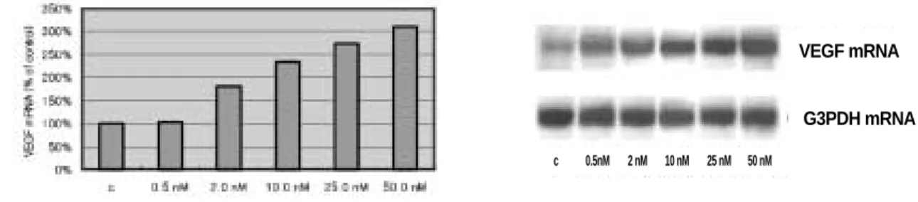
하였다. Northern blot은 이후 각 시편당 유사한 양의 RNA가 적 절히 loading 및 transfer되었는지를 확인하기 위하여 G3PDH를 이용, reprobing을 시행하였다.3. IGF-I 처리의 시간과 농도에 따른 VEGF mRNA 발현에 대한 연구
우선 MG-63 세포주에서 IGF-I이 VEGF mRNA발현을 변화시 키는지에 대한 실험을 시행하였다. 우선 대조군으로 설정된 밀생을 이룬 해당 세포주에서 안정 수준의 VEGF mRNA 를
RT-PCR을 이용, 측정하였다. 196 bp의 크기를 갖는 PCR 생산
물이 urea-containing polyacrylamide gel상에 확인되었다. (Table 1,Fig. 1)
해당 세포주를 10 nM의 IGF-I으로 미리 설정된 시간-
1,2,4,8,12,24 시간-동안 처리한 후 전체 RNA를 분리, 그 발현을 northern blot analysis를 통해 분석하였다.
IGF-I 농도에 따른 VEGF mRNA 발현 변화를 확인하기 위하
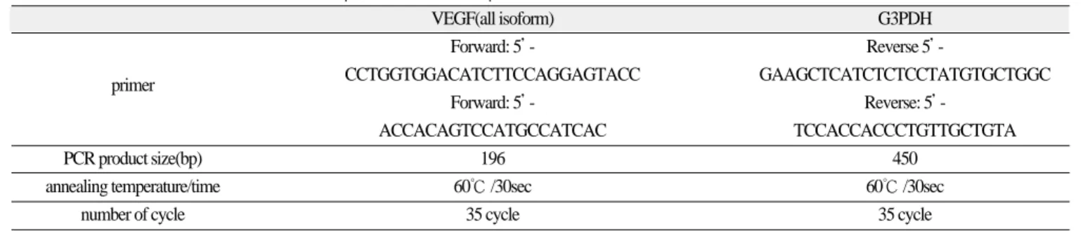
여 2시간동안 서로 다른 농도-0.5, 2.0, 10.0, 25.0, 50.0 nm-로 처리 한 후 분석을 시행하였다.4. VEGF 발현에 대한 IGF-I 기전에 대한 연구
세포 분열 단계, RNA 합성 단계, 새로운 단백질 합성 등의 여 러 단계에 대한 차단을 통하여 그 관여하는 기전을 알아보기 위하여 hydroxyurea (76.1 ㎍/㎖), actinomycin D (2.5 ㎍/㎖), 그리고
cycloheximide (10.0 ㎍/㎖) 등을 IGF-I으로 처리, 1시간 후에 밀생
을 이루고 있는 세포주에 가하였다. 그리고 전체 RNA를 24시 간 후에 분리하여 분석을 시행하였다.Ⅲ. 결 과
1. IGF-I 처리의 시간과 농도에 따른 VEGF mRNA 발현에 대한 연구
VEGF mRNA의 발현 정도는 처리된 mRNA의 농도와 시간에
따라 변화하는 양상을 보였다. 즉, IGF-I으로 처리된 시간 및 농 도를 증가시킬수록 VEGF mRNA의 발현정도는 증가하는 것으 로 나타났다. IGF-I으로 처리한 군은 그렇지 않은 군에 비해 약3.5배 정도 많은 VEGF mRNA 발현을 보였다. 그리고 VEGF mRNA의 발현은 IGF-I으로 24시간 동안 처리한 군에서 최대 발
현을 나타냈다. IGF-I에 의한 VEGF mRNA발현은 처리 시간에 따라 증가하는 것으로 나타났다. (Fig. 2)VEGF mRNA의 발현은 처리된 IGF-I 의 농도에 따라 달라지
는데 실험된 세포주에서는 VEGF mRNA의 최대 발현은 사용 된 최대 농도로 처리하였을 때 나타나는 것으로 나타났다.(50nM) (Fig. 3) Table 1.The condition of PCR and sequences of each primer
VEGF(all isoform) G3PDH
Forward: 5’ - Reverse 5’ -
primer CCTGGTGGACATCTTCCAGGAGTACC GAAGCTCATCTCTCCTATGTGCTGGC
Forward: 5’ - Reverse: 5’ -
ACCACAGTCCATGCCATCAC TCCACCACCCTGTTGCTGTA
PCR product size(bp) 196 450
annealing temperature/time 60℃ /30sec 60℃ /30sec
number of cycle 35 cycle 35 cycle
Fig. 1. mRNA expression of VEGF in RT-PCR.
mRNA expressions of VEGF are shown at the expect- ed location(196 bp) in the gel. (M: size marker)
M
281 bp
234 bp
190 bp
2. VEGF 발현에 대한 IGF-I 기전에 대한 연구
해당 세포주에서 IGF-I에 의해 유도된 VEGF mRNA의 발현 에 관련된 기전을 알아보기 위하여 서로 다른 단계에 작용하 는 억제제(inhibitor)의 효과를 확인하였다.
DNA 합성 과정을 차단하는 hydroxyurea로 세포주를 처리하
였을 때 VEGF mRNA발현에 대한 IGF-I의 발현 자극 효과가 차 단되었다. 또한 actinomycin D에 의한 transcription 차단은 아무것도 처리하지 않은 세포주에서 VEGF transcription을 감소시켰 으며 IGF-I에 의한 VEGF mRNA의 발현이 거의 완전히 차단되 는 결과를 나타내었다. IGF-I의 VEGF mRNA에 대한 효과가 새 로이 합성된 단백질에 의한 것인지를 확인하기 위하여 cyclo-
heximide로 처리하였다. 이때 cycloheximide는 아무것도 처리하
지 않은 대조군 세포주에서는 VEGF mRNA발현을 증가시켰으 나IGF-I의 효과에 대해서는 거의 영향을 미치지 않았던 것으로 나타났다. (Fig. 4)Fig. 2. Time course of IGF-I action on the abundance of VEGF mRNA in MG-63 osteoblast-like cells.
The cells were treated for the indicated times - 1, 2, 4, 8, 12 and 24 hours with 10 nM IGF-I, the total RNA were isolated, an expression of VEGF was determined by Northern blot analysis.
Fig. 3. Concentration dependence of IGF-I on the abundance of VEGF mRNA in MG-63 osteoblast-like cells.
The cells were treated with different concentration of IGF-I, which was 0.5, 2.0, 10.0, 25.0 and 50.0 nM, for 2 hours.
Fig. 4. The effects of different inhibitors on expression of VEGF mRNA.
1: none, 2: IGF-I only, 3: IGF-I � Hydroxyurea, 4: IGF-I � Actinomycin D, 5: IGF-I � Cycloheximide VEGF mRNA
G3PDH mRNA
VEGF mRNA
G3PDH mRNA
VEGF mRNA
G3PDH mRNA
1 2 3 4 5
c 1 hr 2 hrs 4 hrs 8 hrs 12 hrs 24 hrs
c 0.5nM 2 nM 10 nM 25 nM 50 nM
Ⅳ. 고 찰
아직까지 그 기전이 어떠한 방식으로 서로 연결되어 있는 가 에 대해서는 정확히 알려지지 않았으나 골에서 유래된 국소인 자(local factors)들은 혈관형성을 자극할 수 있을 것이며 그 반대 로 혈관내피세포에서 분비된 인자들이 골형성을 자극할 수 있 을 것이다6)
. 인간의 조골세포와 탯줄 정맥 혈관세포를 같이 배
양하여 실행한 연구에 의하면 조골세포에서 유래된 VEGF는 주변 endothelial cell들의 증식을 촉진하였다18).
Endothelial cell들은 골의 분화와 성장에 영향을 미칠 수 있는
cytokine과 성장인자들을 발현한다
17).
조골세포 및 파골세포와매우 밀접한 관계에 있는 endothelial cell들은 매우 다양한
cytokine이나 성장인자들-fibroblast growth factor, interleukin-I, -6, colony-stimulating factors, prostaglandin, endothelin-1, nitric oxide, oxygen radicals 등
6,19,20)과 같은 성장인자들을 생산한다.골 내에 존재하는 혈관들은 오랫동안 골의 재형성과 성장에 중요한 본질적 요소로 인식되어 왔다. 그러나 혈관 세포와 골 간의 정확한 분자적인 관계는 여전히 알려져 있지 않다6)
.
다양한 성장인자들-tumor necrosis factor, transforming growth
factor-β , basic fibroblast growth factor 등-이 혈관 형성과 관련이 있
음이 알려져 있다1). 그러나, 이러한 인자들은 직접적으로 혈관
형성에 관여하는 것이 아니라 간접적으로 작용하는 것으로 추 측된다. 그러므로 비록 다양한 요소들이 잠재적으로 혈관 형 성을 유도하는 것으로 알려져 있을 지라도 이 중 VEGF 가 가장 분열을 촉진할 수 있고 운동성을 유도하는 분자이다7). VEGF는
특별히 endothelial cell 에만 선택적으로 그 기능을 수행하며,VEGF형질이 상실된 경우 embryo의 생존에 치명적이라는 결
과로 인해 인간 태생 발육에 있어서 매우 중요한 역할을 함을 보고하는 연구 결과가 점점 늘어나고 있다17,21,22). VEGF는 순환
하는 거대분자들(circulating macromolecule)에 대해 투과도가 증 가하도록 하는 venule들을 만들어 내는 tumor에 의해 분비되는 단백질로 처음 알려졌고23,24)endothelial mitogen으로써 pituitary follicular cell로부터 분리되었다
25). VEGF는 다양한 혈관 생성 환
경하에서 매우 중요한 역할을 수행한다. 즉, 순환기계(circulat-ing system), embryonic development시의 기관들의 형성과 창상
치유, 당뇨성 신혈관화 등의 다양한 과정에 매우 중요한 역할 을 수행하며, 신혈관 형성이라는 과정은 또한 종적 골 성장에 매우 중요한 성장판에서의 endochondral ossification26), tumor의
성장과 파생9)등의 과정에 중요한 역할을 수행한다.다양한 자극들-저산소성 상태28)
, phorbol esters
29), cAMP ana- logue
29), 중금속 이온들(카드뮴, 코발트, 니켈 등)
30), interleukin -1
31), -6
32), transforming growth factor-β
33), platelet-derived growth factor-β
34), IGF
35), H
2O
2, Ultraviolet B light irradiation
36)-로 세포들을
처리함으로써 VEGF mRNA는 발현된다.본 연구에서 실험 대상으로 이용된 MG-63 세포주에서 세포 들을 IGF-I으로 처리시 VEGF mRNA가 약 3.5배정도 증가되어 발현되었고, 이는 처리된 시간과 처리한 농도에 따라 발현이 조절될 수 있었다. 생리적 농도의 IGF-I은 인간 SaOS-2
osteoblast-like cell, murine osteoblast-like cell, non-malignant untrans- formed osteoblast-like cells-mouse preosteoblast-like cell line(KS483)
등에서 VEGF mRNA과 VEGF protein의 발현을 증가시키는 것 으로 보고되었다6). 또한, VEGF 및 그 수용체의 발현과 생산은
해당 세포의 분화 상태에 따라 다르다. VEGF-A의 발현은 분화 과정의 진행에 따라서 증가되며 이러한 발현, 생산과정은 세 포의 분화 단계를 조절하는 인자들에 의해 조절될 수 있으며 외부에서 가해진 exogeneous VEGF-A를 가하는 경우 조골세포 의 분화도가 증가하게 된다.인슐린과 IGF-I에 의한 VEGF 발현 유도는 다양한 신호전달 과정에 의해 일어난다. 인슐린의 신호전달과정은 phosphatidyli-
nositol 3-kinase/protein kinase B 를 포함하며, IGF-I의 신호전달과
정은 MEK/mitogen-activated protein kinase를 포함한다1). VEGF 발
현을 유도하는 자극의 기전은 매우 다른 요인에 의해 매우 다 른 기전을 통한다.VEGF mRNA 발현의 증가는 세포 복제와 new transcription에 따
라 달라질 수 있음이 추측되고 있다. 그러나 새롭게 합성된 단 백질은 VEGF mRNA의 증가에 큰 영향을 미치지 않는 듯하다.IGF-I의 역할은 actinomycin D가 VEGF mRNA에 대한 IGF-I의
자극을 차단하는 것으로 보이기 때문에 VEGF 유전자, 또는VEGF 전사과정을 조절하는 다른 유전자 등이 전사되는 과정
에서 조절된다. 그러나 이러한 인자들이 VEGF promoter 또는 공통된 조절 전사 요인들에 대한 공통된 활성 요소들을 어떠 한 방식으로 공유하는지에 대해서는 아직 알려져 있지 않다.새로운 단백질의 합성은 fetal rat calvarial cells에 대한 IGF-I의
VEGF mRNA발현 자극에 반드시 필수적인 요소는 아닌 것으
로 나타났다. 그러나 cycloheximide는 prostaglandin E2에 의한VEGF mRNA의 증가를 강화한다
35).
골에서 IGF-I은 autocrine 또는 paracrine의 방식으로 pre-
osteoblast의 증식을 촉진하며 분열과정과는 무관하게 분화된
조골세포에 의한 골 기질의 형성을 증진한다3).
본 연구의 결과들로 MG-63 osteoblast-like cell 등은 VEGF를 발 현한다는 것과 이러한 VEGF의 발현은 IGF-I에 의해 증가된다 는 점을 알 수 있었다. 이러한 결과를 통해 재형성 및 재생 과정 에 있어서 혈관들과 조골세포간의 상호 관계에 대한 한 모델 을 추론할 수 있었다. 흡수과정중의 골 기질로부터 분비되거 나 부갑상선 호르몬 또는 성장 인자 등의 systemic hormone에 대 한 반응으로 조골 세포로부터 분비되는 IGF-I은 autocrine 또는
paracrine의 방식으로 조골세포를 자극하여 VEGF 발현을 증진
시킨다. 이렇게 증가된 VEGF의 발현을 통하여 모세혈관들의 형성이 유도되고 이로 인해 골의 성장, 재생, 재형성 등이 진행 된다.이러한 결과들을 통하여 골 형성을 증가시키는 인자들은 부 분적으로 조골세포에 의한 VEGF 생산을 증가시키며 그로 인 해 혈관 형성을 증가시킨다. 본 저자 등은 골 형성은 생체 내에 서 부분적으로 VEGF와 같은 endothelial growth factor 등을 통해 혈관 형성을 조절함으로써 골 형성 등도 조절할 수 있을 것이 라는 가능성을 확인하였다.
Ⅴ. 결 론
본 연구를 통하여 MG-63 조골세포유사세포주에서 VEGF
mRNA의 발현은 IGF-I의 처리에 의하여 증가되며 IGF-I에 의한
조절 양상은 시간과 사용된 농도에 따라 그 발현이 조절된다.IGF-I에 의한 VEGF mRNA 발현 양상은 hydroxyurea와 actino- mycin D의 추가 처치에 의하여 발현이 감소하지만 cyclohex- imide에 의하여는 발현 양상이 영향을 받지 않으므로 IGF-I에
의한 VEGF mRNA의 발현은 DNA 합성과 전사 단계에 작용하 여 그 영향을 나타낸다는 결론을 도출해 낼 수 있었으며 이로 인하여 IGF-I은 신생골의 형성 및 재형성 과정에 중요한 신혈 관생성과정에 있어서 매우 중요한 역할을 함을 알 수 있었다.참고문헌