Long-Term Effects of Bone Marrow-Derived Mesenchymal Stem Cells in
Dextran Sulfate Sodium-Induced Murine Chronic Colitis
Hyun Jung Lee, Sun-Hee Oh, Hui Won Jang, Ji-Hee Kwon, Kyoung Jin Lee, Chung Hee Kim, Soo Jung Park, Sung Pil Hong, Jae Hee Cheon, Tae Il Kim, and Won Ho Kim
Department of Internal Medicine and Institute of Gastroenterology, Yonsei University College of Medicine, Seoul, Korea
Background/Aims: Bone marrow-derived mesenchymal stem cells (BM-MSCs) have shown beneficial effects in ex-perimental colitis models, but the underlying mechanisms are not fully understood. We investigated the long-term ef-fects of BM-MSCs, particularly in mice with chronic colitis. Methods: Chronic colitis was induced by administering 3% dextran sulfate sodium (DSS) in a series of three cycles. BM-MSCs were injected intravenously into DSS-treated mice three times during the first cycle. On day 33, the therapeutic effects were evaluated with clinicopathologic profiles and histological scoring. Inflammatory mediators were measured with real-time polymerase chain reaction. Results: Systemic infusion of BM-MSCs ameliorated the severity of colitis, and body weight restoration was significantly promoted in the BM-MSC-treated mice. In addition, BM-MSC treatment showed a sustained beneficial effect throughout the three cycles. Microscopic examination revealed that the mice treated with BM-MSCs had fewer inflammatory infiltrates, a lesser extent of inflammation, and less crypt structure damage compared with mice with DSS-induced colitis. Anti-inflammatory cyto-kine levels of interleukin-10 were significantly increased in the inflamed colons of BM-MSC-treated mice compared with DSS-induced colitis mice. Conclusions: Systemic infusion of BM-MSCs at the onset of disease exerted preventive and rapid recovery effects, with long-term immunosuppressive action in mice with repeated DSS-induced chronic colitis. (Gut Liver 2016;10:412-419)
Key Words: Bone marrow; Mesenchymal stem cell; Dextran sulfate sodium; Chronic colitis; Inflammatory bowel disease
INTRODUCTION
Inflammatory bowel disease (IBD), ulcerative colitis (UC) and Crohn’s disease (CD), are chronic relapsing inflammatory dis-orders of the gastrointestinal tract. Although the pathogenesis of UC and CD is still unclear, growing evidence has shown that epithelial barrier defects and dysregulated immune system reactions in genetically susceptible individuals contribute to sustained mucosal inflammation.1,2 An inappropriate adaptive
immune response activated by commensal microbes is known to directly cause tissue damage, and recent studies report that clearance of over-reactive or autoreactive T-cells and balance of regulatory and effector T cells (Th1 and Th2) are disturbed in patients with IBD.2,3 As for the treatment of IBD, because the
need for bowel resection due to failure of medical treatments is remained yet, there is an unmet demand for new therapeutic approaches targeting uncontrolled inflammatory activity to complement the limited efficacy of current immunosuppressive and biologic agents.4-6
Candidates for new alternative treatments include cell ther-apy. The first use of hematopoietic stem cells (HSC) as a treat-ment for IBD was reported in patients with CD, who remained in remission after an HSC transplant for non-Hodgkin’s lym-phoma.7 With the development of autologous bone marrow (BM)
stem cell transplantation for CD,8 mesenchymal stem cells (MSCs)
have been also reported as an effective source of cell therapy. MSCs, derived from various tissues like BM and adipose tis-sue, are a heterogeneous population of stromal stem cells that can self-renew and differentiate into different cell types, such as bone, cartilage, and fat cells.9 In addition to their ability to
promote tissue regeneration from damaged tissue progenitors,9,10
MSCs have been shown to regulate both innate and adaptive
This is an Open Access article distributed under the terms of the Creative Commons Attribution Non-Commercial License (http://creativecommons.org/licenses/by-nc/4.0) which permits unrestricted non-commercial use, distribution, and reproduction in any medium, provided the original work is properly cited.
Correspondence to: Tae Il Kim
Division of Gastroenterology, Department of Internal Medicine, Yonsei University College of Medicine, 50 Yonseiro, Seodaemun-gu, Seoul 03722, Korea
Tel: +82-2-2228-1965, Fax: +82-2-393-6884, E-mail: [email protected] Received on May 20, 2015. Revised on July 16, 2015. Accepted on July 16, 2015. pISSN 1976-2283 eISSN 2005-1212 http://dx.doi.org/10.5009/gnl15229
immune responses by inhibiting T cell proliferation, B cell func-tion, and dendritic cell maturation.10-12 Therefore, MSCs are
emerging as a candidate for treatment of immune-mediated disease, including IBD. Indeed, an in vivo immunosuppressive effect was reported in 94% of patients with acute, severe graft-versus-host disease who were treated with intravenous infusions of MSCs.13,14 In addition, with beneficial effect of MSCs in
ex-perimental colitis models,15-17 some clinical trials demonstrated
positive results of MSCs therapy in Crohn’s perianal fistula and refractory luminal CD.18,19 However, most experiments have
fo-cused on the prevention and improvement of inflammation in a relatively short period after injection of MSCs and the underly-ing mechanisms for the beneficial effects of MSCs are not yet fully understood. Therefore, we investigated the long-term effect of BM-MSCs in mice with chronic colitis induced by repeated administration of dextran-sulfate sodium (DSS).
MATERIALS AND METHODS
1. Animals
Female C57BL/6 mice (aged 8 weeks, weighing 17 to 21 g) were purchased from The Jackson Laboratory (Bar Harbor, ME, USA; http://www.jax.org). Mice were housed under specific pathogen-free conditions in a controlled temperature (24oC) and
12–12 hour light–dark cycle and provided standard diet and water ad libitum. All animal experiments were approved by the Yonsei University Institutional Animal Care and Use Committee (approval number: 09-093-3) and performed according to the Guide for the Care and Use of Laboratory Animal (National Re-search Council, USA, 2010).
2. BM-MSCs isolation and culture
Green fluorescent protein-transgenic (GFP-Tg) C57BL/6 mouse BM-MSCs were isolated and cultured as described previ-ously.20 The BM cells were obtained by flushing the femurs,
tib-ias, and iliac crests from 6-week-old mice with phosphate buff-ered saline (PBS) supplemented with 2% fetal bovine serum (FBS; HyClone Laboratories Inc., Logan, UT, USA). The collected cells were cultured and maintained in a 75-cm2 culture flask
contain-ing Dulbecco’s modified Eagle medium (HyClone Laboratories Inc.) supplemented with 10% FBS, 100 U/mL penicillin (HyClone Laboratories Inc.), and 100 g/mL streptomycin (HyClone Labo-ratories Inc.). Nonadherent cells were removed after 3 days, and only adherent cells were maintained in culture for 2 to 3 weeks until almost confluent. Then, adherent cells were detached using 0.25% trypsin-ethylenediaminetetraacetic acid (HyClone Labo-ratories Inc.). Subsequent passaging and seeding of the cells was performed at a density of 1×10⁷ cells.
3. Colitis induction and experimental design
Chronic colitis was induced by administering 3% (w/v) DSS (molecular weight: 36,000 to 50,000 Da; MP Biochemicals,
Santa Ana, CA, USA) via drinking water in a cyclic manner, which consisted of three cycles (4 days/cycle) of DSS and 6 days
of normal drinking water ad libitum.21 GFP-Tg mice BM-MSCs
(1×10⁷ cells/200 µL) were injected intravenously into the
DSS-treated mice (n=5) on days 1, 3, and 5 during the first cycle (BM-MSC-treated group). Dosage and frequency of BM-MSCs were determined on the basis of our preliminary data and previ-ous studies.16,17 In the control group, DSS-treated mice received
PBS (n=5). All mice were sacrificed at day 33 after being fed DSS water, and the colon was processed for histological analysis or frozen in liquid nitrogen for RNA extraction. The therapeutic effect of BM-MSC treatment was evaluated by clinicopathologic profiles of body weight, colon length, and histological scoring.
4. Histological analysis
The entire colon was removed from the cecum to the anus, fixed in 4% paraformaldehyde, embedded in paraffin, and sliced into 4-µm sections. After hematoxylin and eosin (H&E) stain-ing, histological analysis was performed in a blind manner. The following three parameters were studied as described previously:22
inflammation severity (0=none, 1=mild, 2=moderate, and 3=se-vere), inflammation extent (0=none, 1=mucosa, 2=mucosal and submucosa, and 3=transmural), and crypt damage (0=none, 1=basal 1/3 damaged, 2=basal 2/3 damaged, 3=crypts loss, but surface epithelium present, 4=both crypts and surface epithe-lium loss). Total histological score was defined as the sum of the three parameters.
Immunohistochemical (IHC) staining for GFP, using a Ve-castain ABC kit (Vector Laboratories, Burlingame, CA, USA), was performed to identify the in vivo localization of trans-planted BM-MSCs in the inflamed colon. Colonic tissue samples were incubated first with the primary anti-GFP antibody (Abcam, Cambridge, MA, USA) overnight at 4oC, then with a biotinylated
secondary linking antibody, and finally with a streptavidin-peroxidase complex for 1 hour. The final color product was de-veloped using aminoethylcarbazole (Dako, Glostrup, Denmark). Sections were counterstained with hematoxylin, and tissues were photographed using an Olympus photomicroscope (Olym-pus Corp., Tokyo, Japan).
5. Cytokine measurement
mRNA expression of inflammatory mediators, such as IL-10, transforming growth factor β (TGF-β), and tumor necrosis factor α (TNF-α), in the inflamed colon was measured by real-time polymerase chain reaction (PCR). Isolated colons were frozen in liquid nitrogen and stored at -70oC until use. Tissue
samples were homogenized using a Polytron®
System PT1200E (Kinematica AG, Luzern, Switzerland). Total RNA was extracted from the colonic tissues using a miRNeasy®
Mini Kit (QIAGEN, Hilden, Germany) according to the manufacturer’s instructions and then reverse transcribed using the PrimerScriptTM RT Master
by PCR using the Applied Biosystems (Foster City, CA, USA) StepOnePlusTM real-time PCR system and the following primers:
GGT TGC CAA GCC TTA TCG GA-3′ (IL-10 sense primer), ACC TGC TCC ACT GCC TTG CT-3′ (IL-10 antisense primer), 5′-TGA CGT CAC TGG AGT TGT ACG G-3′ (TGF-β sense primer), 5′-GGT TCA TGT CAT GGA TGG TGC-3′ (TGF-β antisense primer), 5′-CAT CTT CTC AAA ATT CGA GTG ACA A-3′ (TNF-α sense primer), 5′-TGG GAG TAG ACA AGG TAC AAC CC-3′ (TNF-α antisense primer), 5′-CGC GGT TCT ATT TTG TTG GT-3′ (Rn18S sense primer), 5′-AGT CGG CAT CGT TTA TGG TC-3′ (Rn18S antisense primer). Thermal cycling conditions were 30 seconds at 95oC, followed by 40 cycles of 5 seconds at 95oC,
34 seconds at 60oC, and 15 seconds at 95oC. For relative
quan-tification, we compared the amount of target normalized to the Rn18S amplification.
6. Statistical analysis
Data are expressed as means±standard error of mean (SEM). Parametric and nonparametric analyses were performed using the Student t-test and Mann-Whitney U test, respectively. A value of p <0.05 was considered statistically significant for all tests. All statistical analyses were performed using SPSS version 20.0 for Windows (IBM Corp., Armonk, NY, USA).
RESULTS
1. BM-MSCs injection induced prevention and rapid recovery of weight loss in DSS-induced chronic colitis
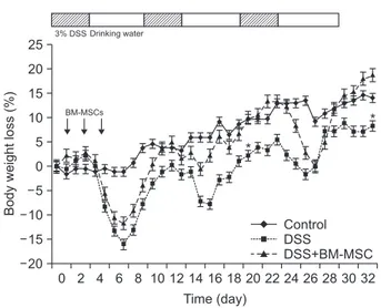
We first investigated the clinical therapeutic efficacy of BM-MSCs in a DSS-induced colitis model. Administration of 3% DSS in three cycles resulted in three peaks of colitis that were characterized by substantial weight loss and bloody diarrhea compared with the normal, untreated mice. However, DSS-treated mice injected with BM-MSCs (BM-MSC-DSS-treated group) showed less weight loss than mice in the DSS-treated only group. Furthermore, body weight restoration was significantly promoted in BM-MSC-treated mice compared with the DSS-treated only group. Significant differences were noted at days 20 and 33 of the recovery periods of each DSS administration cycle (Fig. 1). In particular, regarding the course of weight change, BM-MSC injection in the first cycle of DSS administra-tion showed a preventive effect on weight loss in the second cycle, and rapid restoration of weight loss rather than a preven-tive effect in the third cycle, compared with the untreated DSS-induced colitis group (Fig. 1).
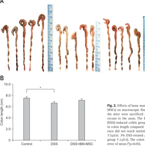
Macroscopic examination also revealed that entire colons from the DSS-treated mice were short, hyperemic, and edema-tous, indicating chronic inflammation in the colon. As shown in Fig. 2, BM-MSC treatment appeared to attenuate the colon shortening, though not to a statistically significant extent.
In summary, injection of BM-MSCs in the first cycle showed a sustained beneficial effect throughout the three cycles of DSS administration.
2. BM-MSCs injection showed sustained histologic im-provement in DSS-induced chronic colitis
We sacrificed mice on day 33 for further evaluation of colon inflammation and crypt damage and microscopic evaluation of the long-term, anti-inflammatory effect of BM-MSCs. Colon tissue samples were microscopically examined following H&E staining. DSS-treated mice that were not treated with BM-MSCs showed severe infiltration of inflammatory cells and disruption of crypt architecture. BM-MSCs treatment of mice with DSS-induced colitis exhibited a protective effect against DSS-DSS-induced histological damage, including fewer inflammatory infiltrates (less severe inflammation), a lower extent of inflammation, and less crypt structure damage, compared with untreated DSS-induced colitis mice. DSS-treated mice transplanted with BM-MSCs displayed significant reduction of total histological scores compared with scores for the DSS-treated only group (Fig. 3).
The IHC staining performed to identify transplanted BM-MSCs in the colon on day 33 revealed no definite GFP positive cell in the colons of BM-MSC-injected chronic colitis mice (data not shown). 0 2 4 6 8 20 25 20 15 10 5 0 5 10 15 Body weight loss (%) Time (day) 10 12 14 16 18 20 22 24 26 28 30 32 Control DSS DSS+BM-MSC BM-MSCs * * 3% DSS Drinking water
Fig. 1. Clinical effects of bone marrow-derived mesenchymal stem cells (BM-MSCs) in repeated dextran sulfate sodium (DSS)-induced chronic colitis. Mice (n=5/group) received 3% dextran sulfate sodium (DSS) in their drinking water in a cyclic manner consisting of three cycles of DSS for 4 days/cycle and 6 days of normal water. Green fluorescent protein-transgenic mouse BM-MSCs were injected intra-venously on days 1, 3, and 5 during the first cycle. DSS- and BM-MSCs-treated mice showed a significant increase in body weight compared with DSS-only-treated mice. The data are presented as the mean±standard error of mean. *p<0.05 vs 3% DSS+BM-MSCs.
3. BM-MSCs injection maintained anti-inflammatory cyto-kine response in DSS-induced chronic colitis
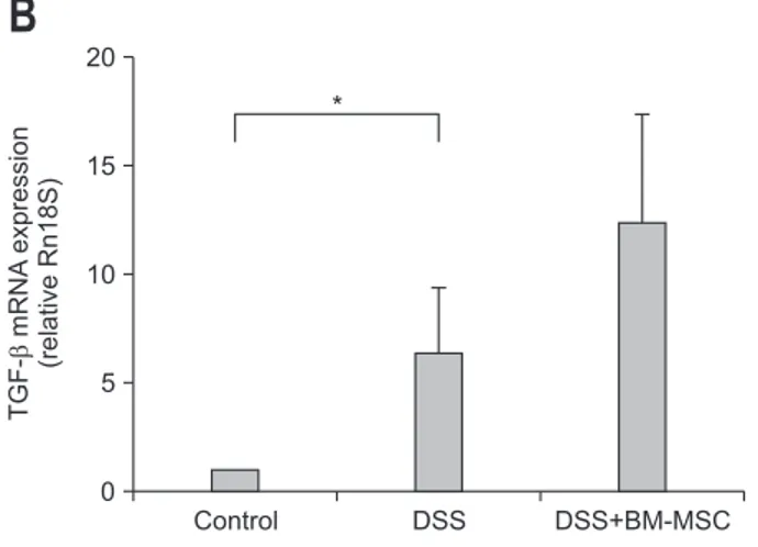
To identify the underlying mechanisms for the beneficial effect of BM-MSCs in DSS-induced chronic colitis, we inves-tigated the influence of BM-MSC transplantation on pro- and anti-inflammatory cytokine production. Real-time PCR analysis of colonic tissues showed that mRNA expression of TNF-α was significantly increased in DSS-treated only mice and decreased in BM-MSC-treated mice with DSS-induced colitis. In contrast, mRNA expression of IL-10 and TGF-β was increased in the in-flamed colons of BM-MSC-injected mice compared with that in colon samples of DSS-treated only mice. Notably, IL-10 produc-tion, which is known to act as an anti-inflammatory regulatory cytokine,23,24 was significantly upregulated by about 10-fold in
BM-MSC-treated mice, compared with DSS-treated only mice (Fig. 4).
DISCUSSION
In the present study, we identified the long-term beneficial role of BM-MSCs in the improvement of clinicopathologic ac-tivity in a DSS-induced chronic colitis mouse model.
Remark-ably, initial injection of BM-MSCs at disease onset manifested a sustained beneficial effect throughout three cycles of DSS administration. Immunological assays indicated that the ef-fect of BM-MSCs could be mediated by up-regulation of anti-inflammatory cytokine, IL-10.
Due to their immunoregulatory function and differentiation potential, BM-MSCs are increasingly being used to treat au-toimmune and systemic inflammatory disease. BM-MSCs are known to be immunoprivileged because of their low expression of class II major histocompatibility complex (MHC-II) and co-stimulatory molecules in their cell surface, which makes them invisible to the immune system.10
Previous studies report that co-culture with MSCs influences cytokine production of T-cell subsets, which results in suppression of T-cell effector func-tion (by decreasing levels of pro-inflammatory cytokines such as TNF-α, IFN-γ, IL-6, and IL-17) and enhancement of T-cell suppressor functions (by increasing IL-10 and TGF-β levels).25,26
Similarly, because IBD is related to immune dysregulation, recent studies have considered immunomodulatory cytokines as therapeutic targets4,27
and BM-MSCs are emerging as one of the promising therapies for IBD. However, though accumu-lating data have shown beneficial effects of BM-MSCs in an experimental colitis model,15,17,28,29 the mechanisms are yet to
A
Control 0 10.0 8.0 6.0 4.0 2.0 Colon length (cm)B
DSS DSS+BM-MSC *Fig. 2. Effects of bone marrow-derived mesenchymal stem cells (BM-MSCs) on macroscopic findings (A) and colon length (B). On day 33, the mice were sacrificed and their colons were removed from the cecum to the anus. The BM-MSC-treated, dextran-sulfate sodium (DSS)-induced colitis group showed a trend toward a lesser decrease in colon length compared with the DSS-treated group, but the differ-ence did not reach statistical significance (control, untreated group 7.5±0.6; 3% DSS-treated group 6.6±0.7; 3% DSS+BM-MSC-treated group 7.1±0.5). The colon length is presented as the mean±standard error of mean (*p<0.05).
A
B
C
D
E
F
G
Control 0 3.5 3.0 2.5 2.0 1.5 1.0 0.5 Inflammatory severity DSS DSS+BM-MSC Control 0 14 12 10 8 6 4 2 Histological score DSS DSS+BM-MSC Control 0 3.5 3.0 2.5 2.0 1.5 1.0 0.5 Inflammatory extent DSS DSS+BM-MSC Control 0 5 4 3 2 1 Crypt damage DSS DSS+BM-MSC * * * * * * *Fig. 3. Histological improvement in bone marrow-derived mesenchymal stem cells (BM-MSCs)-transplanted, dextran-sulfate sodium (DSS)-induced chronic colitis. (A-F) Histological analysis was performed with H&E staining of colon tissues. (G) In the BM-MSC-treated, DSS-induced colitis group, total histological scores were significantly reduced compared with the only-treated group (control, untreated group 4.0±1.9; 3% DSS-treated group 10.6±2.3; 3% DSS+BM-MSC-DSS-treated group 7.6±1.3; *p<0.05). The individual scores, including inflammatory severity, extent, and crypt damage, were also decreased with BM-MSC treatment compared with the scores of the DSS-only-treated group. The data are presented as the mean±standard error of mean. Representative histologic images are shown for the control group: A (×100) and D (×200); the 3% DSS-treated group: B (×100) and E (×200); and the 3% DSS+BM-MSC-treated group: C (×100) and F (×200).
be determined. Furthermore, most of the studies focused on the prevention and improvement of acute gut inflammation by BM-MSCs treatment.17,28,29 Therefore, in this study, we investigated
the long-term effect of BM-MSCs specifically in chronic colitis induced by repeated DSS administration, which is an established experimental model for human IBD.30
We found long-term beneficial effects of BM-MSCs on clini-copathologic colitis activity in our chronic colitis model. In ad-dition, with regard to the course of weight change, we found a greater preventive effect on weight loss in the second cycle of DSS administration and a more rapid restoring effect (without a preventive effect) in third cycle. These results suggest a decreas-ing beneficial effect of BM-MSC treatment with the progression of time.
In regard to the potential mechanisms for the therapeutic effect of BM-MSCs suggested by experimental models of IBD, BM-MSCs have been reported to downregulate the expression of Th1-type cytokines15,17,28,29 and upregulate the population
of FoxP3+ regulatory T cells, with a consequent increase in
IL-10 production.15,28,29,31 In agreement with previous studies, we
showed a significant increase in IL-10 production in BM-MSC-treated mice. IL-10, which is a signature cytokine for a subset of CD4 T cells that exhibit regulatory functions, induces Treg populations and attenuates experimental colitis.23,24
IL-10-se-creting Treg cells are known to play a critical role in maintain-ing intestinal homeostasis by suppressmaintain-ing immune response to resident commensal microbes.32 In addition, our results revealed
that TGF-β levels also were increased in DSS-induced colitis mice that were treated with BM-MSCs, suggesting the possibil-ity of FoxP3+ regulatory T cell induction23,24 and contribution to
the intestinal repair process.16
However, compared with previous studies that indicate BM-MSC treatment downregulates the Th1-driven inflammatory responses in DSS-induced colitis mice,15,17 our data did not show
a significant decrease in TNF-α levels upon BM-MSC trans-plantation. DSS administration in the present study, as in other recent studies,15,17,28,29 increased the proinflammatory cytokine of
TNF-α in inflamed mucosa, verifying the crucial role of TNF-α in the development of IBD.2,3 The lack of a definite inhibitory
ef-fect of Th1 response in BM-MSC-treated mice of our experiment might be attributable to the change of cytokine profiles accord-ing to the acute or chronic stage of colitis. DSS-induced colitis is known to switch from Th1-Th17-mediated acute inflamma-tion to a predominant Th2-mediated inflammatory response in the chronic state.33 In addition, because we used a chronic
DSS-induced colitis model to evaluate the long-term effect of BM-MSCs, the recoverable extent of TNF-α might be lower than that expected in acute colitis and, therefore, we could not observe
Control 0 80 60 40 20 IL-10 mRNA expression (relative Rn18S) DSS DSS+BM-MSC *
A
Control 0 20 15 10 5 TGF-mRNA expression (relative Rn18S) DSS DSS+BM-MSCB
* TNF-mRNA expression (relative Rn18S) Control 0 80 60 40 20 DSS DSS+BM-MSC *C
Fig. 4. Effect of bone marrow-derived mesenchymal stem cells (BM-MSCs) on the production of anti-inflammatory cytokines (IL-10 and TGF-β) (A, B) and pro-inflammatory cytokine, TNF-α (C). mRNA expression of inflammatory mediators was measured in colonic tis-sue samples by using real-time polymerase chain reaction. BM-MSC treatment reduced the mRNA expression of tumor necrosis factor α and increased the mRNA expression of interleukin-10 and transform-ing growth factor β. Similar results were obtained from three inde-pendent experiments. The data are presented as the mean±standard error of mean (*p<0.05).
significant suppressive effect of TNF-α expression after BM-MSCs treatment. No significant improvement in colon length in our experiment could be also explained by chronic repeated injury by three cycles of DSS administration, which could con-tribute to unrecoverable chronic injury like fibrosis.
The mechanism of the long-term effect of BM-MSCs is not likely due to long-term survival of injected BM-MSCs because we found no GFP positive cells in BM-MSCs-treated mice in our experiments and previous other studies demonstrate a rapid dis-appearance of injected MSCs.17,28,29 However, given the reported
long survival of T cells in peripheral tissues,34 long survival of
regulatory T cells induced by initial injections of BM-MSCs could contribute to the long-term beneficial effect of BM-MSCs. Additional detailed experiments regarding the fate of BM-MSC-induced regulated T cell population are needed to confirm this possibility.
In our study, we used chemically induced colitis model by DSS administration. Because acute, chronic, or relapsing model can be produced easily by changing the concentration of DSS, it is one of the most commonly used animal models for study-ing IBD.33,35 However, as other animal models of genetically
modified and adoptive transfer models, none of these models could represent the complexity of human disease. In addition, to date, there have been many experimental studies of the ef-fects of MSCs in colitis models. However, we need more data about quality control of MSCs, different effects by different cell sources, and effective dose, frequency, and route based on the duration of effect for clinical application. Our data could give some information on the effect of MSCs with the progression of time.
In conclusion, we demonstrated that BM-MSC transplantation could be an effective means to treat chronic colitis in mice. In particular, initial systemic infusion of BM-MSCs at disease on-set could exert preventive and rapid recovering effects through long-term immunosuppressive action in repeated DSS-treated chronic colitis.
CONFLICTS OF INTEREST
No potential conflict of interest relevant to this article was reported.
ACKNOWLEDGEMENTS
This study was supported by a grant of the Korea Healthcare technology R&D Project, Ministry for Health, Welfare & Family Affairs, Republic of Korea (A090898).
REFERENCES
1. Xavier RJ, Podolsky DK. Unravelling the pathogenesis of inflam-matory bowel disease. Nature 2007;448:427-434.
2. Baumgart DC, Carding SR. Inflammatory bowel disease: cause and immunobiology. Lancet 2007;369:1627-1640.
3. Bouma G, Strober W. The immunological and genetic basis of in-flammatory bowel disease. Nat Rev Immunol 2003;3:521-533. 4. Baumgart DC, Sandborn WJ. Inflammatory bowel disease:
clinical aspects and established and evolving therapies. Lancet 2007;369:1641-1657.
5. Torres J, Danese S, Colombel JF. New therapeutic avenues in ul-cerative colitis: thinking out of the box. Gut 2013;62:1642-1652. 6. Vermeire S, Ferrante M, Rutgeerts P. Recent advances:
person-alised use of current Crohn’s disease therapeutic options. Gut 2013;62:1511-1515.
7. Drakos PE, Nagler A, Or R. Case of Crohn’s disease in bone mar-row transplantation. Am J Hematol 1993;43:157-158.
8. Kim TI. Clinical trials with stem cells in digestive diseases and fu-ture perspectives. Korean J Gastroenterol 2011;58:139-143. 9. Phinney DG, Prockop DJ. Concise review: mesenchymal stem/
multipotent stromal cells. The state of transdifferentiation and modes of tissue repair: current views. Stem Cells 2007;25:2896-2902.
10. De Miguel MP, Fuentes-Julián S, Blázquez-Martínez A, et al. Im-munosuppressive properties of mesenchymal stem cells: advances and applications. Curr Mol Med 2012;12:574-591.
11. Song IH, Jang BI. Stem cells in inflammatory bowel disease: new potential therapeutic target. Intest Res 2013;11:79-84.
12. García-Bosch O, Ricart E, Panés J. Review article: stem cell thera-pies for inflammatory bowel disease: efficacy and safety. Aliment Pharmacol Ther 2010;32:939-952.
13. Le Blanc K, Rasmusson I, Sundberg B, et al. Treatment of severe acute graft-versus-host disease with third party haploidentical mesenchymal stem cells. Lancet 2004;363:1439-1441.
14. Fang B, Song YP, Liao LM, Han Q, Zhao RC. Treatment of severe therapy-resistant acute graft-versus-host disease with human adi-pose tissue-derived mesenchymal stem cells. Bone Marrow Trans-plant 2006;38:389-390.
15. Gonzalez-Rey E, Anderson P, González MA, Rico L, Büscher D, Delgado M. Human adult stem cells derived from adipose tissue protect against experimental colitis and sepsis. Gut 2009;58:929-939.
16. Tanaka H, Arimura Y, Yabana T, et al. Myogenic lineage differ-entiated mesenchymal stem cells enhance recovery from dextran sulfate sodium-induced colitis in the rat. J Gastroenterol 2011;46: 143-152.
17. He XW, He XS, Lian L, Wu XJ, Lan P. Systemic infusion of bone marrow-derived mesenchymal stem cells for treatment of experi-mental colitis in mice. Dig Dis Sci 2012;57:3136-3144.
18. Garcia-Olmo D, Herreros D, Pascual I, et al. Expanded adipose-derived stem cells for the treatment of complex perianal fistula: a phase II clinical trial. Dis Colon Rectum 2009;52:79-86.
19. Duijvestein M, Vos AC, Roelofs H, et al. Autologous bone marrow-derived mesenchymal stromal cell treatment for refractory luminal Crohn’s disease: results of a phase I study. Gut
2010;59:1662-1669.
20. Bruder SP, Kurth AA, Shea M, Hayes WC, Jaiswal N, Kadiyala S. Bone regeneration by implantation of purified, culture-expanded human mesenchymal stem cells. J Orthop Res 1998;16:155-162. 21. Whittem CG, Williams AD, Williams CS. Murine colitis modeling
using dextran sulfate sodium (DSS). J Vis Exp 2010;(35):e1652. 22. Williams KL, Fuller CR, Dieleman LA, et al. Enhanced survival
and mucosal repair after dextran sodium sulfate-induced colitis in transgenic mice that overexpress growth hormone. Gastroenterology 2001;120:925-937.
23. Groux H, O’Garra A, Bigler M, et al. A CD4+ T-cell subset inhib-its antigen-specific T-cell responses and prevents colitis. Nature 1997;389:737-742.
24. Boden EK, Snapper SB. Regulatory T cells in inflammatory bowel disease. Curr Opin Gastroenterol 2008;24:733-741.
25. Tse WT, Pendleton JD, Beyer WM, Egalka MC, Guinan EC. Sup-pression of allogeneic T-cell proliferation by human marrow stro-mal cells: implications in transplantation. Transplantation 2003; 75:389-397.
26. Prevosto C, Zancolli M, Canevali P, Zocchi MR, Poggi A. Genera-tion of CD4+ or CD8+ regulatory T cells upon mesenchymal stem cell-lymphocyte interaction. Haematologica 2007;92:881-888. 27. Strober W, Fuss IJ. Proinflammatory cytokines in the pathogenesis
of inflammatory bowel diseases. Gastroenterology 2011;140:1756-1767.
28. González MA, Gonzalez-Rey E, Rico L, Büscher D, Delgado M. Adipose-derived mesenchymal stem cells alleviate experimental colitis by inhibiting inflammatory and autoimmune responses. Gastroenterology 2009;136:978-989.
29. Castelo-Branco MT, Soares ID, Lopes DV, et al. Intraperitoneal but not intravenous cryopreserved mesenchymal stromal cells home to the inflamed colon and ameliorate experimental colitis. PLoS One 2012;7:e33360.
30. Strober W, Fuss IJ, Blumberg RS. The immunology of mucosal models of inflammation. Annu Rev Immunol 2002;20:495-549. 31. Parekkadan B, Upadhyay R, Dunham J, et al. Bone marrow
stro-mal cell transplants prevent experimental enterocolitis and require host CD11b+ splenocytes. Gastroenterology 2011;140:966-975. 32. Izcue A, Coombes JL, Powrie F. Regulatory T cells suppress
sys-temic and mucosal immune activation to control intestinal in-flammation. Immunol Rev 2006;212:256-271.
33. Alex P, Zachos NC, Nguyen T, et al. Distinct cytokine patterns identified from multiplex profiles of murine DSS and TNBS-induced colitis. Inflamm Bowel Dis 2009;15:341-352.
34. Nguyen VH, Zeiser R, Dasilva DL, et al. In vivo dynamics of regu-latory T-cell trafficking and survival predict effective strategies to control graft-versus-host disease following allogeneic transplanta-tion. Blood 2007;109:2649-2656.
35. Perše M, Cerar A. Dextran sodium sulphate colitis mouse model: traps and tricks. J Biomed Biotechnol 2012;2012:718617.