INTRODUCTION
Mesenchymal stem cells (MSCs) are capable of self-renew- al and differentiation into lineages of mesenchymal tissues, including bone, cartilage, fat, tendon, muscle, and hematopoi- etic supporting stroma (1, 2). While the most abundant sour- ce of MSCs is the bone marrow (BM), MSCs can be obtained from other tissues such as peripheral blood, periosteum, mus- cle, adipose tissue, and the connective tissue of human adults (3).
MSCs from cord blood (CB) and the placenta may serve as alternative sources to adult MSCs. These potential alter- native sources are important for a number of reasons, includ- ing the significant reduction in the number of MSCs with age and the high risk of viral contamination in the BM as well as the painful procedure required for the collection of BM MSCs. CB and placenta are potential alternative sources of MSCs; they are routinely discarded after delivery, and the ethical concerns and viral contamination issues are less of a problem. Several investigators have successfully isolated, ex- panded, and characterized the MSCs from CB and the pla- centa, and have evaluated their potential for differentiation into osteogenic, chondrogenic or adipogenic lineages (4-6).
In animal models, MSCs can be induced to circulate into
peripheral blood under certain conditions, such as hypoxia (7). The chemotactic signals that guide MSCs to appropriate microenvironments or induce their circulation have yet to be identified. In hematopoietic stem cells (HSC), the predomi- nant role of stromal derived factor-1 (SDF-1) and its receptor CXCR-4 is now well established (8). Some soluble factors have recently been reported to exert chemotactic effects on BM MSCs, including chemokines (9) and growth factors (10), however, their respective physiological relevance remains un- clear. An improved knowledge of the chemotactic factors that affect MSCs would be of clinical interest since modulation of their activity could affect not only engraftment efficiency at damaged sites but also their mobilization into peripheral blood. Ponte et al. (11) showed that, in contrast to what is known about HSCs, a wide range of soluble factors exert sig- nificant chemotactic activity on MSCs and some growth fac- tors are better chemoattractants than chemokines. Inflamma- tory cytokines such as tumor necrosis factor α(TNFα) can increase the sensitivity of MSCs to chemokines.
In this study, we characterized and compared the cytokine expression of BM-MSCs, CB-MSCs, and placental MSCs using a commercial human cytokine protein array assay to identi- fy the protein expression profiles of the MSCs from BM, CB and the placenta. This study focused on the cytokines associ-
547
Jong Ha Hwang1, Soung Shin Shim2, Oye Sun Seok3, Hang Young Lee4, Sang Kyu Woo4, Bong Hui Kim4, Hae Ryong Song5, Jae Kwan Lee1, and Yong Kyun Park1
Departments of Obstetrics and Gynecology1and Orthopedic Surgery5, Women’s Cancer Center Research Institute3, School of Medicine, Korea University, Seoul; Department of Obstetrics and Gynecology2, School of Medicine, Pochon CHA University, Seoul; Research Center4, RNL BIO CO., Ltd, Seoul, Korea
Address for correspondence Jae Kwan Lee, M.D.
Department of Obstetrics and Gynecology, Korea University Guro Hospital University College of Medicine, 80 Guro-dong, Guro-gu, Seoul 152-703, Korea Tel : +82.2-2626-2519, Fax : +82.2-838-1560 E-mail : jh36640@hanmail.net
DOI: 10.3346/jkms.2009.24.4.547
Comparison of Cytokine Expression in Mesenchymal Stem Cells from Human Placenta, Cord Blood, and Bone Marrow
Mesenchymal stem cells (MSCs) are capable of self-renewal and differentiation into lineages of mesenchymal tissues that are currently under investigation for a variety of therapeutic applications. The purpose of this study was to compare cytokine gene expression in MSCs from human placenta, cord blood (CB) and bone marrow (BM).
The cytokine expression profiles of MSCs from BM, CB and placenta (amnion, decidua) were compared by proteome profiler array analysis. The cytokines that were expre- ssed differently, in each type of MSC, were analyzed by real-time PCR. We evalu- ated 36 cytokines. Most types of MSCs had a common expression pattern includ- ing MIF (GIF, DER6), IL-8 (CXCL8), Serpin E1 (PAI-1), GROα(CXCL1), and IL-6.
MCP-1, however, was expressed in both the MSCs from the BM and the amnion.
sICAM-1 was expressed in both the amnion and decidua MSCs. SDF-1 was ex- pressed only in the BM MSCs. Real-time PCR demonstrated the expression of the cytokines in each of the MSCs. The MSCs from bone marrow, placenta (amnion and decidua) and cord blood expressed the cytokines differently. These results sug- gest that cytokine induction and signal transduction are different in MSCs from dif- ferent tissues.
Key Words : Mesenchymal Stem Cells; Placenta; Fetal Blood; Bone Marrow; Cytokines
Received : 17 March 2008 Accepted : 9 January 2009
ated with the monocyte chemotactic protein-1 (MCP-1), sol- uble intercellular adhesion molecule-1 (sICAM-1) and SDF-1.
MATERIALS AND METHODS Isolation of mesenchymal stem cells
MSCs from human BM, CB, and the amnion and decidua of the placenta were studied. The BM MSCs, in the first pas- sage, were obtained from the Department of Orthopedic Sur- gery. The CB MSCs, in the first passage, were obtained from MEDIPOST Co. (Seoul, Korea). The human placentas (from clinically normal pregnancies, gestational age, 34-41 weeks) were obtained after vaginal deliveries or caesarean section bir- ths. All tissues were obtained with the approval of the Korea University Medical Center Institutional Review Board. The amnion and decidua were mechanically peeled from the pla- centa, and washed with phosphate-buffered saline (PBS) sev- eral times to remove excessive blood. The tissues were incu- bated with 1 mg/mL type I collagenase for three hours after being cut into small pieces (1 cm3) with scissors. The mononu- clear cells in the collagenase were collected. After centrifuga- tion, the cells were washed with PBS and resuspended in an α-minimum essential medium (MEM, Gibco BRL, Life Tech- nologies, Merelbeke, Belgium) supplemented with 10% fetal bovine serum, and 10 ng/mL basic fibroblast growth factor (bFGF). The cells were seeded into a T75 flask. The cultures were maintained at 37℃in a humidified atmosphere with 5% CO2. Three to five days after initiating the incubation, the small digested residues were removed and the culture was continued. The medium was replaced one to two times every week, every third to fourth day.
Flow cytometry analysis
The specific surface antigens of MSCs in the cultures of pas- sage two to four were characterized by flow cytometry anal- ysis. The cells in culture were trypsinized and stained with fluorescein isothiocyanate (FITC)- or phycoerythrin (PE)- conjugated antibodies against CD29, CD31, CD34, CD44, CD45, HLA-DR, CD73, CD90, and CD105 (Immunotech, Marseille, MN, U.S.A.). Thereafter, the cells were analyzed using a Becton Dickinson flow cytometer (Becton Dickin- son, San Jose, CA, U.S.A.).
Adipogenic induction
The medium was replaced with adipocyte induction medi- um or control (stromal) medium, as previously described (12).
The induction medium contained a low glucose Dulbecco’s Modified Eagle’s Medium (LG-DMEM) with 10% FBS, 200 μM indomethacin, 1 μg/mL insulin, 1 mM dexamethasone, 0.5 mM isobutylmethylxanthine (IBMX), 100 U/mL peni- cillin, 100 mg/mL streptomycin, and 0.25 mg/mL ampho-
tericin B. After 3 days, the medium was changed to adipocyte maintenance medium that was identical to the induction me- dia but without IBMX. The cells were maintained in culture for 14 days, with 90% of the maintenance media replaced every 3 days.
Osteogenic induction
The medium was replaced with osteoblast induction medi- um or control (stromal) medium, as previously described (12).
Osteoblast induction medium contained DMEM (low glu- cose) with 10% fetal bovine serum, 10 mM β-glycerophos- phate, 0.15 mM ascorbate-2-phosphate, 10 nM 1,25-(OH) 2 vitamin D3, 10 nM dexamethasone, 100 U/mL penicillin, 100 mg/mL streptomycin, and 0.25 mg/mL amphotericin B.
The cells were maintained in culture for 21 to 28 days, with 90% of the media replaced every 3 days.
The assessment of cell viability and cell number in cultures
The cultured cells were detached from the culture dishes with 0.05% trypsin-EDTA (Gibco BRL, Life Technologies) at 72 hr of culture under different culture conditions. The cells were stained with trypan blue (Gibco BRL, Life Tech- nologies), and the viable cells that did not stain were count- ed on a hemocytometer.
The focused protein array
The culture media were collected at different incubation periods. All focused protein array analyses were performed according to the manufacturer’s instructions. Positive controls were located in the upper left-hand corner (two spots), lower left-hand corner (two spots) and lower right-hand corner (two spots) of each array kit. Each culture media was measured using the human cytokine array panel A (proteome profil- erTM) (R&D Systems, Minneapolis, MN, U.S.A.). Horseradish peroxidase substrate (Bio-Rad, Hercules, CA, U.S.A.) was used to detect protein expression and data were captured by exposure to Kodak BioMax Light film. Arrays were scanned into a computer and optical density measurements were ob- tained with the Image Pro Plus v 5.1 software (Media Cyber- netics, Silver Spring, MD, U.S.A.).
Total RNA isolation and reverse-transcriptase reaction
RNA extraction and purification were performed using an RNeasy mini kit as described in the manufacturer’s protocol (Qiagen, Valencia, CA, U.S.A.). The concentration of RNA was measured using a spectrophotometer (DU�530, Beck- man, Fullerton, CA, U.S.A.), and the RNA quality was con- firmed by agarose gels. A total RNA sample (2 μg/sample) was used with 20 μL of sample to generate cDNA using the SuperScriptTMIII First-Strand Synthesis System RT-PCR kit (Invitrogen, Milan, Italy). RNA was reverse-transcribed un-
der the following conditions: 25 mM MgCl2, 10 mM dNTP mix, 10×RT buffer, 0.1 M DTT, 200 U of SuperScriptTM III (Invitrogen), 40 U of RNaseOut, and 50 μM oligo d(T) primers in a final volume of 20 μL. The reaction was run at 65℃for 5 min and at 50℃for 50 min, and then the enzyme was heat inactivated at 85℃for 5 min. For the real-time PCR reaction, 4 μL of reaction product were used.
Real-time PCR analysis
The proteome profiler results showed that there was dif- ferent cytokine expression for each type of MSC including sICAM-1 (CD54), MCP-1 (CCL2), and SDF-1 (CXCL12). A real-time PCR analysis was used to quantify the sICAM-1 (CD54), MCP-1 (CCL2), and SDF-1 (CXCL12) transcripts.
Their expression was normalized using the GAPDH house- keeping gene product as an endogenous reference. The primers and probes were designed for human sICAM-1, MCP-1, and SDF-1 using Primer Express 2.0 (Applied Biosystems, Fos- ter City, CA, U.S.A.). sICAM-1 (CD54), MCP-1 (CCL2), and SDF-1 (CXCL12) mRNA levels were quantified using Taq- Man Real-Time PCR with an ABI 7700 system (Applied Biosystems). Gene-specific probes and primer pairs for ICAM- 1 (Assays-on-Demand, Hs00164932_m1; Applied Biosys- tems), MCP-1 (Assays-on-Demand, Hs00234140_m1; Ap- plied Biosystems), and SDF-1 (Assays-on-Demand, Hs00- 171022_m1; Applied Biosystems) were used. For each probe/
primer set, a standard curve was generated, which was con- firmed to increase linearly with increasing amounts of cDNA.
The amplification conditions were 2 min at 50℃, 10 min at 95℃, and a two-step cycle of 95℃for 15 sec, and 60℃ for 60 sec for a total of 45 cycles.
Western blotting
Protein lysates were obtained with a buffer containing 50 mM HEPES (pH 7.5), 150 mM NaCl, 1.5 mM MgCl2, 1 mM EDTA, 10% glycerol, 1% Triton X-100, and a mixture of
protease inhibitors (aprotinin, PMSF, and sodium orthovana- date). Equal amounts of total protein were resolved on a 12%
SDS-polyacrylamide gel. The proteins were transferred to a nitrocellulose membrane. After blocking (TBS, 0.1% Tween- 20) at 4℃overnight, the membranes were incubated with primary antibodies of anti-mouse SDF-1, anti-mouse MCP- 1, and anti-mouse sICAM-1 (R & D systems); all monoclonal antibodies were used at a dilution of 1:1,000 for 2 hr followed by incubation with secondary antibodies linked to HRP (Bio- Rad) and anti-rabbit GAPDH (dilution 1:2,000, Assay De- signs, Inc., Ann Arbor Michigan, U.S.A.). Immunoreactive proteins were visualized by chemiluminescence using Super- Signal West Dura Extended Duration Substrate (Pierce Che- mical Co., Rockford, IL, U.S.A.). Fujifilm Luminescent Image Analyzer LAS-3000, with a charged-coupled device camera (Science Imaging Scandinavia AB), was used for imaging.
Statistics
Data are presented as the mean±SD. Data were analyzed with SPSS statistical software, version 12.0 (SPSS Inc, Chica- go, IL, U.S.A.). The differences in cytokine concentrations among four groups of subjects were analyzed by the Kruskal- Wallis and Mann-Whitney tests. A probability of 0.05 was considered significant.
RESULTS
Isolation, culture, flow cytometry analysis and differentiation of placental MSCs
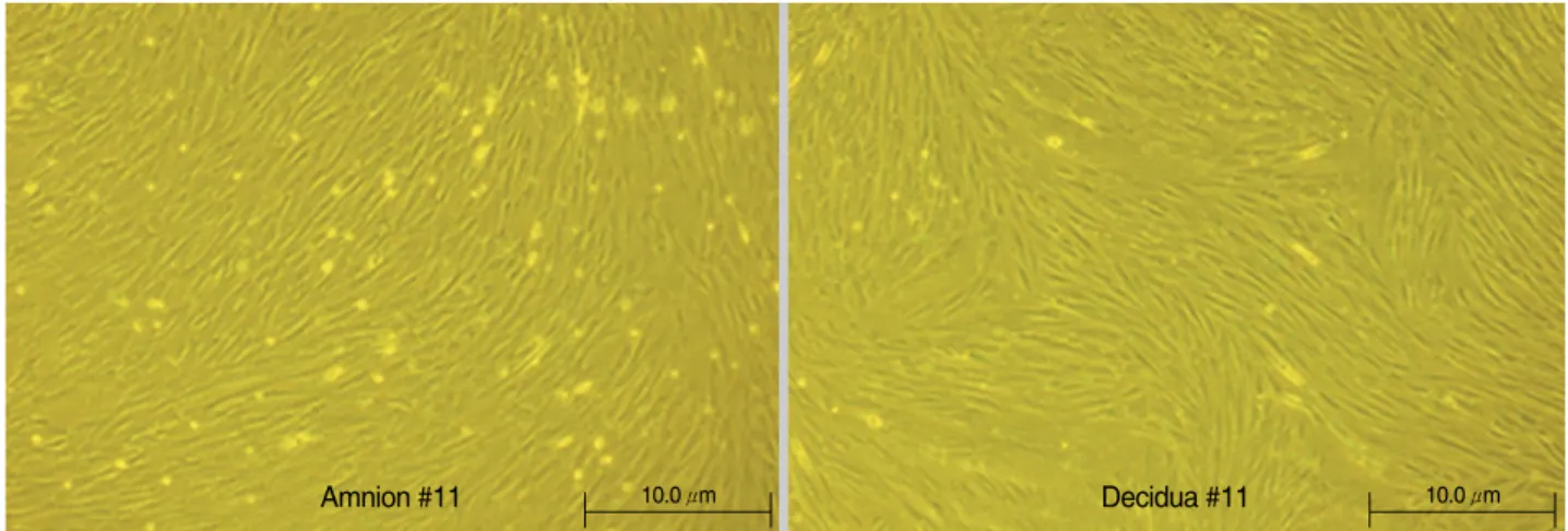
After enzyme digestion of the placental amnion and decid- ua, the cells were seeded into T-75 cell culture flasks at a con- fluence of 80%. The adherent cells with fibroblastic morphol- ogy were analyzed. The cells formed a monolayer of homoge- nous bipolar spindle-like cells with a whirlpool-like array in two to three days (Fig. 1), and these adherent cells could be
Fig. 1. Appearance and growth of fibroblastoid cells or placental mesenchymal stem cells at passage 1 on day 11.
Amnion #11 10.0 μm Decidua #11 10.0 μm
readily expanded in vitro by successive cycles of trypsinization, seeding and culture every three days for three passages with- out visible morphologic change. The MSCs from term placen- tal amnion and decidua were successfully isolated. We found no correlation between gestational age and the successful estab- lishment of MSC cultures from the amnion and decidua.
We examined the surface marker profile of the amnion and decidua derived cell lines using fluorescence activated cell sorting (FACS). The phenotype of the MSCs derived from amnion was similar to that of MSCs derived from the decid- ua. These cells were positive for CD29, CD44, and CD73 but were negative for CD31, CD34, CD45, and HLA-DR (Fig.
2). CD 105 was positive in amnion MSC whereas CD 90 was positive in the decidua.
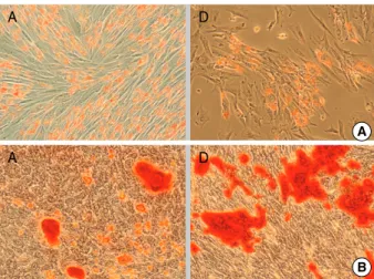
To estimate their potential to differentiate into several tis- sue lineages, the MSCs from the amnion and decidua were cultured in adipogenic, osteogenic, myogenic, and neurogenic medium. At the end of the induction period, the cells were differentiated into their respective tissues. The confirmation of differentiation was made by Oil Red O for adipogenic dif- ferentiation (Fig. 3A), Alizarin Red S staining for osteogenic differentiation (Fig. 3B), respectively. Culture expanded cells from the amnion and decidua were all able to differentiate into adipogenic and osteogenic lineages.
Cytokine array analysis
The conditioned medium, after 3-4 days of culture of the MSCs from CB, BM, and placenta (amnion, chorion), was assayed using the human cytokine array panel A (R & D sys- tems) according to the manufacturer’s instructions. We ana- lyzed 36 cytokines at a time. The 36 cytokines included: C5a, CD40 Ligand, G-CSF, GM-CSF, GROα, I-309, sICAM-1, IFN-γ, IL-1α, IL-1β, IL-1ra, IL-2, IL-4, IL-5, IL-6, IL-8, IL- 10, IL-12p70, IL-13, IL-16, IL-17, IL-17E, IL-23, IL-27, IL- 32α, IP-10, I-TAC, MCP-1, MIF, MIP-1α, MIP-1β, Serpin E1, RANTES, SDF-1, TNFα, and sTREM-1.
CB MSCs secreted: MIF (GIF, DER6), IL-8 (CXCL8), Ser- pin E1 (PAI-1), GROα(CXCL1), and IL-6. BM MSCs secret- ed: MIF (GIF, DER6), IL-8, Serpin E1 (PAI-1), GROα(CX- CL1), IL-6, MCP-1 (CCL2), and SDF-1 (CXCL12). Amnion MSCs secreted: GROα(CXCL1), sICAM-1 (CD54), IL-6, IL-8, MCP-1 (CCL2), MIF (GIF, DER6), and serpin E1. De-
cidua MSCs secreted: GROα(CXCL1), sICAM-1 (CD54), IL-6, IL-8, MIF (GIF, DER6), and Serpin E1.
Each of the MSCs expressed: MIF (GIF, DER6), IL-8 (CX- CL8), Serpin E1 (PAI-1), GROα(CXCL1), and IL-6. How- ever, MCP-1 (CCL2) was expressed only in the BM MSCs and amnion MSCs. sICAM-1 (CD54) was expressed in both the amnion and decidua MSCs. SDF-1 was expressed in only the BM MSCs (Fig. 4A). The relative expression level of the cytokines was calculated. The MCP-1 expression in the BM MSCs was higher than the expression in the amnion MSCs, whereas the expression of IL-6 in the CB MSCs was compar- atively lower. GROαexpression was higher in both the BM MSCs and amnion MSCs compared to the CB MSCs and the amnion MSCs (Fig. 4B).
The Detection of SDF-1 (CXCL12), MCP-1 (CCL2) and sICAM-1 (CD54) in each of the tissue-MSCs by RT-PCR
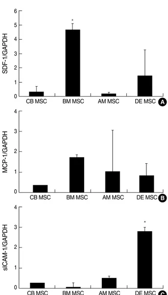
SDF-1 (CXCL12), MCP-1 (CCL2) and sICAM-1 (CD54) ex- pression of human MSCs derived from three different origins were determined at the level of gene expression (n=8). Real time RT-PCR demonstrated that the human MSCs derived from the three different origins express cytokines different- ly. The level of mRNA was quantified and compared with a
Fig. 2. Immunophenotypic results of passage 3 MSCs by FACS analysis. (A) MSC from amnion (B) MSC from decidua. Representative histogram (black line). The respective isotype control is shown as gray.
CD 29 CD 31 CD 34 CD 44 CD 45 CD 73 CD 90 CD 105 HLA DR
CD 29 CD 31 CD 34 CD 44 CD 45 CD 73 CD 90 CD 105 HLA DR B
A Fig. 3. Differentiation potential of MSCs obtained from the amnion and decidua. (A) Adipogenic differentiation of MSC shown by Oil red O staining of adipocytes (×200). (B) Osteogenic differentiation of MSC shown by Alizarin red S (×200). (A, amnion; D, decidua).
A D
B A
A D
housekeeping gene (GAPDH). The higher expression of SDF- 1 (CXCL 12) in BM MSCs was confirmed by RT-PCR (P=
0.006). However, the RT-PCR did not confirm differences for the gene encoding MCP-1 (CCL2) among the four MSCs (P=0.252) derived from different tissues. The gene encoding sICAM-1 was found to be expressed at a higher level in the decidua MSCs (P=0.002) (Fig. 5). SDF-1, MCP-1, and sICAM- 1 expression was confirmed by Western blot analysis in the amnion and decidua MSCs (n=2) (Fig. 6).
DISCUSSION
Mesenchymal stem cells are thought to have great thera-
peutic potential due to their capacity for self-renewal and multilineage differentiation (2, 13). They support hemato- poiesis and enhance the engraftment of hematopoietic stem cells after co-transplantation (14, 15). Experimental and clin- ical data have demonstrated an immunoregulatory function of BM-derived MSCs (BM MSC) that may contribute to the reduction of graft-versus-host disease following hematopoi- etic stem cell transplantation (16, 17). Currently, the BM rep- resents the major source of MSCs for cell therapy. However, aspiration of BM involves invasive procedures, and the fre- quency and differentiation potential of BM MSC decrease significantly with age (18). The search for alternative sources of MSCs, therefore, is of significant value. It has been report- ed that MSCs could be isolated from various tissues includ-
Fig. 4. (A) The cytokine expression in each of the MSCs using the proteome profiler. (B) Quantification of cytokine optical density. Measure- ments were obtained with the Image Pro Plus v 5.1 software (Media Cybernetics, Silver Spring, MD, U.S.A.).
CB, cord blood; BM, bone marrow; AM, amnion; DE, decidua MSCs; MSC, mesenchymal stem cells.
CB MSC GROα
GROα L6L8
MF Serpin E1
GROα
GROα
GROα MIF
SDF-1
MCP-1
MCP-1 MIF
MIF MIF
IL6
IL6
IL6
IL6
sICAM-1 sICAM-1
sICAM-1 sICAM-1
IL8
IL8
IL8
IL8 Serpin E1
Serpin E1
Serpin E1
Serpin E1 BM MSC
AM MSC
DE MSC
Pixel density
2,500 2,000 1,500 1,000 500 0
CB MSC
GROα L6
L8
MF Serpin E1
3DF-1 MCP-1
Pixel density
2,500 2,000 1,500 1,000 500
0 BM MSC
GROα L6
L8
MF Serpin E1
MCP-1
Pixel density
2,500 2,000 1,500 1,000 500 0
AM MSC
GROα
L6 L8
MF Serpin E1
Pixel density
2,500 2,000 1,500 1,000 500
0 DE MSC
A B
ing periosteum, trabecular bone, adipose tissue, synovium, skeletal muscle, deciduous teeth, fetal pancreas, lung, liver, amniotic fluid, CB, and placental tissues (19, 20). Among these sources, CB and the placenta may be ideal sources due to their accessibility, painless donor procurement, promising sources for autologous cell therapy, and lower risk of viral con- tamination.
We isolated MSCs from the amnion and decidua. After iso- lation and culture, under specified conditions, typical MSC- like cells similar to the cells from the amnion and decidua were identified. These cells were characterized by the same
shape and size, and the same monolayer appearance. In addi- tion, the cells expressed CD29, CD31, CD34, CD44, CD45, HLA-DR, CD73, CD90, and CD105 by cytofluorimetry.
Expression of the main marker genes of MSCs (CD29, CD44 and CD73) were positive and similar in all of the MSCs stud- ied. (Data for BM MSCs and CB MSCs: not shown). Expres- sion of CD31, CD34, CD45, and HLA-DR were negative.
CD90 was positive only in the decidua-derived MSCs, where- as CD105 was positive only in the amnion-derived MSCs.
The identity of MSCs from different sources has not been pre- viously proven. The results of this study of placenta-derived MSCs may provide an attractive and rich source of MSCs.
Initially, cytokine analysis was performed with the pro- teome profilerTMto determine which cyotokines were expre- ssed differently. Then, the cytokines were confirmed by PCR analysis. There was some discrepancy between the proteome profilerTMand the PCR analysis. For example, there was no significant difference in the expression of the gene encoding MCP-1 (CCL2) among the four differently derived MSCs by the PCR analysis. However, by the proteome profilerTManal- ysis, the amnion MSCs and BM MSCs were found to secrete MCP-1, whereas the CB MSCs and decidua MSCs did not secrete MCP-1. The gene encoding sICAM-1 was found to be expressed at a higher level in the decidua MSCs. The prote- ome profiler analysis, however, showed that the concentration of sICAM-1 was higher in the amnion MSCs compared to decid- ua MSCs. The manufacturing company recommended that the proteome profilerTMbe used as a screening tool. Therefore, we depended on the results of the PCR analysis and Western blot to confirm the expression of SDF-1, MCP-1, and sICAM-1 in the MSCs from the amnion and the decidua.
Potian et al. (21) recently analyzed the cytokine profile of BM MSCs using the protein array technique. IL-6, IL-8, MCP- 1, RANTES, GROα, INFγ, IL-1α, TGFβ, GM-CSF, angio- genin, and oncostatin M were constitutively expressed, and
SDF-1/GAPDH
6 5 4 3 2 1
0 CB MSC BM MSC AM MSC DE MSC
*
*
MCP-1/GAPDH
4
3
2
1
0
CB MSC BM MSC AM MSC DE MSC
sICAM-1/GAPDH
4
3
2
1
0
CB MSC BM MSC AM MSC DE MSC A
B
C
Fig. 5. The quantitative expression of SDF-1, MCP-1 and sICAM-1 in each of the mesenchymal stem cells. The mRNA levels were quantified using TaqMan Real-Time PCR with an ABI 7700 sys- tem (Applied Biosystems). The GAPDH housekeeping gene prod- uct was used as an endogenous reference. (A) SDF-1, (B) MCP- 1, (C) sICAM-1.
*Statistically significant difference (P<0.05).
SDF-1, stromal derived factor-1; MCP-1, monocyte chemotactic protein-1; sICAM-1, intracellular adhesion molecule.
AM MSC
SDF-1
8.0 KDa
MCP-1
8.7 KDa
sICAM-1 79 KDa
GAPDH 40 KDa
DE MSC
40 KDa
Fig. 6. SDF-1, MCP-1 and sICAM-1 expression profile by Western blot analysis in amnion-derived MSC and decidua-derived MSC.
AM MSC, amnion-derived MSC; DE MSC, decidua-derived MSC.
MIP-1α, IL-2, IL-4, IL-10, IL-12, and IL-13 were not ex- pressed by the BM MSCs. The results of this study showed that the cytokine profile of the BM MSCs was very similar to that of the CB MSCs, with the exception that the CB MSCs expressed IL-12 but not G-CSF under serum-free conditions.
Haynesworth et al. (22) reported that constitutively expressed cytokines in the growth phase include: IL-6, G-CSF, SCF, not detected in the growth medium of human BM derived MSCs.
Both MSCs from the BM and CB abundantly produced IL- 6, IL-8, and MCP-1.
The role that chemokines and their receptors play in the targeting of leukocytes to areas of inflammation, infection, or injury has been well characterized (23). As chemokine recep- tors are expressed on the cell surface of MSCs, and their stim- ulation has been shown to induce cell migration, it seems likely that they play a similar role in directing MSCs. MSCs have been shown to express a variety of chemokine receptors.
The reported chemokine receptors of MSCs, however, have been inconsistent under similar isolation and culture condi- tions. This might be due to the heterogeneous nature of a typical MSC population that obscures the detection of a dis- tinct receptor repertoire.
Our study findings showed that SDF-1 (CXCL12) was more highly expressed in the BM MSCs. SDF-1 is a small cytokine belonging to the chemokine family that is officially designat- ed CXCL12. CXCL12 is strongly chemotactic for lympho- cytes and is, therefore, an important hematopoietic growth factors for these cells (24-26). During embryogenesis it directs the migration of hematopoietic cells from the fetal liver to the BM. The receptor for this chemokine is CXCR4. This CXCL12-CXCR 4 interaction used to be considered exclu- sive, but recently it has been suggested that CXCL12 is also bound by the CXCR7 receptor (27, 28). SDF-1 could aug- ment the mobilization, migration, recruitment, and entrap- ment of MSCs. SDF-1-CXCR4 interactions mediate the hom- ing of MSCs. This suggests that SDF-1 plays an important role in the mobilization and homing of BM MSCs, although the signals required for this process have not been fully des- cribed.
The cytokine expression profile of placental MSCs remains poorly documented. We compared the cytokine expression of BM MSCs, CB MSCs, amnion MSCs, and decidua MSCs focusing on SDF-1 (CXCL12), MCP-1 (CCL2), and sICAM- 1 based on the protein array assay. In this study, sICAM-1 was more highly expressed in decidua MSCs compared to the BM MSCs, CB MSCs and amnion MSCs. sICAM-1 represents a cir- culating form of ICAM-1 that is constitutively expressed or is inducible on the cell surface of different cell lines (29). sICAM- 1 is a type of intercellular adhesion molecule continuously present in low concentrations in the membranes of leukocytes and endothelial cells. Upon cytokine stimulation, the concen- trations of this molecule greatly increase. ICAM-1 can be in- duced by interleukin-1 (IL-1) and TNFα, and is expressed by the vascular endothelium, macrophages and lymphocytes (30).
Although amnion MSCs and decidua MSCs are derived from the placenta, expression of sICAM-1 was found to be signif- icantly lower in the amnion MSCs. Cytokine induction and signal transduction may be different in the amnion MSCs and the decidua MSCs. sICAM-1 is thought to play a more important role in the decidua MSCs than in the amnion MSCs with regard to cell recruitment. There was no significant dif- ference in the expression of MCP-1 among the BM MSCs, CB MSCs, amnion MSCs, and the decidua MSCs.
In addition to their role in mediating cell migration, che- mokines may also play important autocrine and paracrine roles. CXCL12 promotes the growth, survival, and develop- ment of MSCs (31). MSCs are known to be able to synthesize this chemokine, which may act in an autocrine manner via CXCR4. Chemokines are also recognized as primary induc- ers of integrin up-regulation following their interaction with their cell surface receptors and various downstream signaling events.
Integrins are known to mediate the firm adhesion of leuko- cytes to endothelial cells and play an important role in their transendothelial migration. It is likely that they play a sim- ilar role for the MSCs. MSCs are known to express various integrin molecules and their roles have begun to be elucidat- ed. Pittenger et al. (2) reported the first study of integrin ex- pression of MSCs and noted the presence of α1, α2, α3 aα, αν, β1, β3, and β4 along with the other adhesion molecules IC- AM-1, ICAM-3, VCAM-1, ALCAM, and endoglin (CD105).
The results of the present study provide the characteriza- tion of cytokine expression of the MSCs from the BM, CB, amnion, and decidua. The cytokines play a role in migration of MSCs. The cytokine induction and signal transduction are important for migration of the MSCs. The characteristics of cytokine expression in MSCs derived from different tissues help with the understanding of the mechanisms of MSC migra- tion. Further studies are required to better understand the interactions of these cytokines.
REFERENCES
1. Deans RJ, Moseley AB. Mesenchymal stem cells: biology and poten- tial clinical uses. Exp Hematol 2000; 28: 875-84.
2. Pittenger MF, Mackay AM, Beck SC, Jaiswal RK, Douglas R, Mosca JD, Moorman MA, Simonetti DW, Craig S, Marshak DR. Multilin- eage potential of adult human mesenchymal stem cells. Science 1999;
284: 143-7.
3. In’t Anker PS, Scherjon SA, Kleijburg-van der Keur C, de Groot- Swings GM, Claas FH, Fibbe WE, Kanhai HH. Isolation of mes- enchymal stem cells of fetal or maternal origin from human placen- ta. Stem Cells 2004; 22: 1338-45.
4. Erices A, Conget P, Minguell JJ. Mesenchymal progenitor cells in human umbilical cord blood. Br J Haematol 2000; 109: 235-42.
5. Gutierrez-Rodriguez M, Reyes-Maldonado E, Mayani H. Character- ization of the adherent cells developed in Dexter-type long-term cul-
tures from human umbilical cord blood. Stem Cells 2000; 18: 46-52.
6. Miao Z, Jin J, Chen L, Zhu J, Huang W, Zhao J, Qian H, Zhang X.
Isolation of mesenchymal stem cells from human placenta: compar- ison with human bone marrow mesenchymal stem cells. Cell Biol Int 2006; 30: 681-7.
7. Rochefort GY, Delorme B, Lopez A, Herault O, Bonnet P, Charbord P, Eder V, Domenech J. Multipotential mesenchymal stem cells are mobilized into peripheral blood by hypoxia. Stem Cells 2006; 24:
2202-8.
8. Peled A, Petit I, Kollet O, Magid M, Ponomaryov T, Byk T, Nagler A, Ben-Hur H, Many A, Shultz L, Lider O, Alon R, Zipori D, Lapi- dot T. Dependence of human stem cell engraftment and repopulation of NOD/SCID mice on CXCR4. Science 1999; 283: 845-8.
9. Ji JF, He BP, Dheen ST, Tay SS. Interactions of chemokines and chemokine receptors mediate the migration of mesenchymal stem cells to the impaired site in the brain after hypoglossal nerve injury.
Stem Cells 2004; 22: 415-27.
10. Forte G, Minieri M, Cossa P, Antenucci D, Sala M, Gnocchi V, Fiac- cavento R, Carotenuto F, De Vito P, Baldini PM, Prat M, Di Nardo P. Hepatocyte growth factor effects on mesenchymal stem cells: pro- liferation, migration, and differentiation. Stem Cells 2006; 24: 23-33.
11. Ponte AL, Marais E, Gallay N, Langonne A, Delorme B, Herault O, Charbord P, Domenech J. The in vitro migration capacity of human bone marrow mesenchymal stem cells: comparison of chemokine and growth factor chemotactic activities. Stem Cells 2007; 25: 1737-45.
12. Gonzalez R, Maki CB, Pacchiarotti J, Csontos S, Pham JK, Slepko N, Patel A, Silva F. Pluripotent marker expression and differentia- tion of human second trimester mesenchymal stem cells. Biochem Biophys Res Commun 2007;362:491-7.
13. Jiang Y, Jahagirdar BN, Reinhardt RL, Schwartz RE, Keene CD, Ortiz-Gonzalez XR, Reyes M, Lenvik T, Lund T, Blackstad M, Du J, Aldrich S, Lisberg A, Low WC, Largaespada DA, Verfaillie CM.
Pluripotency of mesenchymal stem cells derived from adult marrow.
Nature 2002; 418: 41-9.
14. Angelopoulou M, Novelli E, Grove JE, Rinder HM, Civin C, Cheng L, Krause DS. Cotransplantation of human mesenchymal stem cells enhances human myelopoiesis and megakaryocytopoiesis in NOD/
SCID mice. Exp Hematol 2003; 31: 413-20.
15. Noort WA, Kruisselbrink AB, in’t Anker PS, Kruger M, van Bezooi- jen RL, de Paus RA, Heemskerk MH, Lowik CW, Falkenburg JH, Willemze R, Fibbe WE. Mesenchymal stem cells promote engraft- ment of human umbilical cord blood-derived CD34(+) cells in NOD/
SCID mice. Exp Hematol 2002; 30: 870-8.
16. Frank MH, Sayegh MH. Immunomodulatory functions of mesenchymal stem cells. Lancet 2004; 363: 1411-2.
17. Le Blanc K, Rasmusson I, Sundberg B, Gotherstrom C, Hassan M, Uzunel M, Ringden O. Treatment of severe acute graft-versus-host disease with third party haploidentical mesenchymal stem cells. Lancet 2004; 363: 1439-41.
18. Rao MS, Mattson MP. Stem cells and aging: expanding the possibil- ities. Mech Ageing Dev 2001; 122: 713-34.
19. Barry FP, Murphy JM. Mesenchymal stem cells: clinical applications and biological characterization. Int J Biochem Cell Biol 2004; 36:
568-84.
20. in’t Anker PS, Noort WA, Scherjon SA, Kleijburg-van der Keur C, Kruisselbrink AB, van Bezooijen RL, Beekhuizen W, Willemze R, Kanhai HH, Fibbe WE. Mesenchymal stem cells in human second- trimester bone marrow, liver, lung, and spleen exhibit a similar im- munophenotype but a heterogeneous multilineage differentiation potential. Haematologica 2003; 88: 845-52.
21. Potian JA, Aviv H, Ponzio NM, Harrison JS, Rameshwar P. Veto-like activity of mesenchymal stem cells: functional discrimination between cellular responses to alloantigens and recall antigens. J Immunol 2003; 171: 3426-34.
22. Haynesworth SE, Baber MA, Caplan AI. Cytokine expression by human marrow-derived mesenchymal progenitor cells in vitro: effects of dexamethasone and IL-1 alpha. J Cell Physiol 1996; 166: 585-92.
23. Miyasaka M, Tanaka T. Lymphocyte trafficking across high endothe- lial venules: dogmas and enigmas. Nat Rev Immunol 2004; 4: 360-70.
24. Ara T, Nakamura Y, Egawa T, Sugiyama T, Abe K, Kishimoto T, Matsui Y, Nagasawa T. Impaired colonization of the gonads by pri- mordial germ cells in mice lacking a chemokine, stromal cell-derived factor-1 (SDF-1). Proc Natl Acad Sci USA 2003; 100: 5319-23.
25. Askari AT, Unzek S, Popovic ZB, Goldman CK, Forudi F, Kiedrows- ki M, Rovner A, Ellis SG, Thomas JD, DiCorleto PE, Topol EJ, Penn MS. Effect of stromal-cell-derived factor 1 on stem-cell homing and tissue regeneration in ischaemic cardiomyopathy. Lancet 2003; 362:
697-703.
26. Ma Q, Jones D, Borghesani PR, Segal RA, Nagasawa T, Kishimoto T, Bronson RT, Springer TA. Impaired B-lymphopoiesis, myelopoiesis, and derailed cerebellar neuron migration in CXCR4- and SDF-1-defi- cient mice. Proc Natl Acad Sci USA 1998; 95: 9448-53.
27. Balabanian K, Lagane B, Infantino S, Chow KY, Harriague J, Moepps B, Arenzana-Seisdedos F, Thelen M, Bachelerie F. The chemokine SDF-1/CXCL12 binds to and signals through the orphan receptor RDC1 in T lymphocytes. J Biol Chem 2005; 280: 35760-6.
28. Burns JM, Summers BC, Wang Y, Melikian A, Berahovich R, Miao Z, Penfold ME, Sunshine MJ, Littman DR, Kuo CJ, Wei K, McMas- ter BE, Wright K, Howard MC, Schall TJ. A novel chemokine recep- tor for SDF-1 and I-TAC involved in cell survival, cell adhesion, and tumor development. J Exp Med 2006; 203: 2201-13.
29. Witkowska AM, Borawska MH. Soluble intracellular adhesion mole- cule-1 (sICAM-1) : an overview. Eur Cytokine Netw 2004; 15: 91-8.
30. Yang L, Froio RM, Sciuto TE, Dvorak AM, Alon R, Luscinskas FW.
ICAM-1 regulates neutrophil adhesion and transcelluar migration of TNF-alpha-activated vascular endothelium under flow. Blood 2005; 106: 584-92.
31. Kortesidis A, Zannettino A, Isenmann S, Shi S, Lapidot T, Gronthos S. Stromal-derived factor-1 promotes the growth, survival, and develop- ment of human bone marrow stromal stem cells. Blood 2005; 105:
3793-801.


