Abstract (J. Kor. Oral Maxillofac. Surg. 2008;34:419-427)
Ⅰ. 서 론
구강 및 악안면 영역의 외상, 종양, 선천적 질환에 의한 결손 은 저작, 발음과 같은 기능저하와 심미적인 부조화를 야기한 다. 악안면 영역의 골결손은 자가골이식, 동종골이식, 이종골 이식, 생체친화성 인공골을 이용하여 회복되어 왔고, 인공치 아의 발달로 인해서 구강악안면영역의 골이식재 사용은 더욱
증가하고 있다1,2)
.
그렇지만, 자가골은 부가적인 수술로 인한 공포감, 수술시간과 감염가능성의 증가, 제한적 골량 등의 문 제점이 대두되었다3).
동종골이나 이종골의 사용은 면역반응 과 관련된 항원성과 전염성질환에 대한 안전성이 완벽하게 검 증되지 않고 있다. 이에 생체친화성이 우수한 인공골들이 개 발 연구되고 임상에도 적용되고 있다4).
골형성에 있어서 생체친화적인 인공골의 역할은 골형성을 직접 유발하는 것이 아니라, 조골세포가 작용할 수 있는 환경 을 조성하는 지지체(scaffold)의 역할이 주된 기능이다. 이러한 인공골이 자가골과 유사한 골유도성(osteoinductivity)과 골전도 성 (osteoconductivity)을 동시에 가지기 위해서는 골형성(osteo-
genesis)을 유도할 수 있는 골형성 단백질과 골형성을 진행하는
세포의 존재가 필요하다5). 많은 연구들에서 인공골에 골유도
골수유래줄기세포에서 분화된 골유사세포에서 β -TCP와 rhBMP-2의 골형성 효과에 관한 연구
최용수1∙황경균1,2∙이재선2∙박창주1,2∙심광섭1
1한양대학교 의과대학 치과학교실 구강악안면외과, 2한양대학교 의과대학 의생명연구소
황 경 균
133-791
서울 성동구 행당동17
한양대학교 의과대학 치과학교실 구강악안면외과 Kyung-Gyun Hwang
Division of OMFS/Department of Dentistry, College of Medicine, Hanyang Univ.
#17 Haengdang-Dong, Seongdong-Ku, Seoul, 133-791, Korea Tel: +82-2-2290-8676 Fax: +82-2-2290-8673
E-mail: [email protected]
THE EFFECTS OF β -TCP/rhBMP-2 ON BONE FORMATION IN OSTEOBLAST-LIKE CELLS INDUCED FROM BONE MARROW-DERIVED MESENCHYMAL STEM CELLS
Yong-Soo Choi
1, Kyung-Gyun Hwang
1,2, Jae-Seon Lee
2, Chang-Joo Park
1,2, Kwang-Sup Shim
11
Division of Oral and Maxillofacial Surgery / Department of Dentistry, College of Medicine, Hanyang University
2
Institute of Biomedical Science, College of Medicine, Hanyang University
The present study aimed to investigate the osteogenic potentials of differentiated osteoblast-like cells (DOCs) induced from bone marrow-derived mesenchymal stem cells (MSCs) on β-tricalcium phosphate (β-TCP) with recombinant human bone morphogenetic protein (rhBMP-2) in vitro.
Osteoblast differentiation was induced in confluent cultures by adding 100 nM dexamethasone, 10 mM β-glycerophosphate, 50 mM L-ascorbic acid. The Alizarin red S staining and reverse transcriptase-polymerase chain reaction (RT-PCR) were perfomed to examine the mRNA expression of alkaline phosphatase (ALP), bone sialoprotein (BSP), osteocalcin (OCN), receptor activator for nuclear factor κB ligand (RANKL), runt-related tran- scription factor 2 (RUNX2), collagen-Ⅰ (COL-Ⅰ).
There were no significant differences in the osteogenic potentials of DOCs induced from MSCs on β-TCP(+/-). According to the incubation period, there were significant increasing of Alizadin red S staining in the induction 3 weeks. The mRNA expression of ALP, RUNX2, and RANKL were higher in DOCs/β-TCP(-) than DOCs/β-TCP(+). According to rhBMP-2 concentrations, the mRNA expression of BSP was significantly increased in DOCs/β-TCP(+) compared to that of DOCs/β-TCP(-) on rhBMP 10 ng/ml.
Our study presented the β-TCP will have the possibility that calcium phosphate directly affect the osteoblastic differentiation of the bone marrow- derived MSCs.
Key words: mesenchymal stem cell, rhBMP -2, β-TCP, differentiated osteoblast-like cell
※ 이 논문은 2006년도 정부재원(교육인적자원부 학술연구조성사업비)으로 한국학술진흥재단의 지원을 받아 연구되었음(KRF-
2006-331-E00330).
성을 부여하기 위해서 PRP(platelet-rich plasma), 인간재조합골형 성단백질(recombinant human bone morphorgenetic protein; rhBMP) 을 동시에 이식하기도 하고, 골형성을 향상시키기 위해 전기 자극 또는 맥동전자기장의 원리를 이용하기도 한다. 골수유래 줄기세포(bone marrow derived stem cell; BMSC)와 골형성유도물 질을 인공골과 같이 이식하는 조직공학적 골이식술도 최근 시 도되고 있다6-14)
.
골형성단백질은 transforming growth factor-β(TGF-β
) superfami- ly의 일종으로 7개의 subgroup이 발견되었다
15). 골형성단백질
은 중간엽줄기세포증식 (mesenchymal cell proliferation), 연골 형 성, 혈관형성, 골형성, 골의 재형성 (remodeling), 발생(embryonicdevelopment processes)등에 관여한다. 골형성단백질을 임상적
으로 이용하기 위해서는 bovine bone에서 추출을 하거나, cDNA 를 이용하여 클로닝하는 방법으로 rhBMP를 만든다. 골형성단 백질을 골결손부에 사용하기 위해서는 collagen sponge나 calci-um phosphate particle 등의 전달체(carrier material)가 필요하다. 최
근 β-tricalcium phosphate(β-TCP)와 같은 골전도성 인공골이BMP의 전달체로 임상에서 많이 이용되고 있다
16-18).
줄기세포(stem cell)는 미분화 상태로 존재하면서 다양한 세 포로 분화할 수 있는 능력을 가진 세포이다. 줄기세포는 기원 에 따라 배아줄기세포와 성체줄기세포로 나눌 수 있고, 성체 줄기세포는 배아줄기세포보다는 제한적인 분화능을 가지며, 신체의 특정 위치에서 성장하고 특정 조직 세포로의 증식과 분화를 준비하고 있다. 조혈과 골형성 대사가 항상 진행되는 골수는 중간엽줄기세포(mesenchymal stem cells) 및 조혈세포 생 성에 관여하는 세포외기질(extracellular matrix; ECM)로 구성된 복잡한 구조를 가지고 있어 골화세포 및 연골세포로의 분화가 가능한 줄기세포를 추출할 수 있고, 추출된 줄기세포의 골모 유사세포(osteoblast-like cell)로의 분화에 대한 연구도 많이 진행 되고 있다19-22)
.
최근 줄기세포의 추출, 확립 및 분화의 기술이 급속히 발달 함에 따라 조직공학적 생체재료에 대한 연구가 활발히 진행되 고 있으며, 골재생을 향상시키기 위한 줄기세포의 임상적 적 용도 조금씩 진행되고 있다. 이에 본 연구는 골수유래줄기세 포의 분화와 이에 따른 골형성능력을 확인하고, 서로 다른
rhBMP-2의 농도와 β -TCP 여부에 따른 골형성의 차이를 평가하
여 보다 적합한 골재생을 위한 환경을 찾음으로서 조직공학적 골재생의 임상적 적용에 기여하고자 한다.
Ⅱ. 연구 방법 1. 세포 배양(Cell culture)
사람의 중간엽줄기세포는 Cambrex (USA)에서 구입하여 사 용하였다. 세포는 같은 회사의 mesenchymal stem cell basal medi-
um과 mesenchymal stem cell growth supplements, 그리고 L-gluta-
2. 골모유사세포의 분화 실험
실험군으로 미분화상태의 중간엽줄기세포를 100 nM Dex
(dexamethasone, Sigma, USA), 50 mM β-GP (β-glycerophosphate, Fluka, Switzerland), 50 μ M L-ascorbic acid (Sigma, USA), 10% FBS (fetal bovine serum, Gibco, USA)가 혼합된 LG-DMEM (low-glu- cose Dulbecco's modified Eagle's medium, Gibco, USA) 배지에서 1
주부터 4주동안 배양하여 각각 조골세포로의 분화를 유도하 였다. 대조군으로 미분화상태의 중간엽줄기세포를 10% FBS 가 혼합된 LG-DMEM 기본 배양배지에서 4주동안 배양하였다.배양은 5% CO2
, 37.5℃ 배양기내에서 배양하였으며 10% FBS
가 포함된 LG-DMEM은 3~4일마다 교환하였다. 배양이 끝난 후, 각 세포의 배지를 걷어내고 4% paraformaldehyde 용액으로 4℃에서 30분간 고정시켰다. 이후에 phosphate buffered saline
(PBS)로 세척하고 증류수로 한번 더 세척한 다음 40 mM Alizarin red S (Usb, OH, USA) 용액(pH 4.2)을 1 ml 넣어 실온에서 10분간 염색하였다. 증류수로 한번 세척하여 여분의 용액을 제
거한 다음 염색 정도를 관찰하고, 10 mM sodium phosphate가 포 함된 10% cetylpyridinium chloride (Sigma, USA) 용액에 탈염(de-staining)하여 562 nm 파장에서 흡광도를 측정하여 염색정도를
정량화하였다.3. 인간재조합골생성 단백질(rhBMP)과 인공이식재(β- TCP)하에서 골모유사세포 배양 후 RT-PCR을 통한 mRNA발현 관찰
중간엽줄기세포가 분화하는 동시에 인공이식재(β-TCP)와
rhBMP-2 (R&D systems, USA)를 0, 1, 10ng/ml의 농도로 각각 배
지에 혼합하여 배양하였다.배양이 끝난 세포를 회수하여 배지를 걷어내고 Easy Blue
(Intron, Korea)를 처리하여 전체 mRNA를 추출하였다. 이를 정
제하기 위해서 200㎕의 chloroform을 넣고 잘 섞은 다음 13000rpm에서 15분간 원심분리하고, 상등액을 취해 동량의 iso- propanol을 넣어 잘 섞은 다음 mRNA를 침전시켜, 13000 rpm에
서 10분간 원심분리하여 pellet을 얻었다. Pellet을 75% ethanol로 세척한 다음 nuclease free water (Promega, USA)에 녹여 cDNA합 성에 사용하였다. 추출된 mRNA를 cDNA로 바꾸기 위해서, 0.5㎍ random nonamer (Genotech, Korea) 와 70 ℃에서 5분간 반응시 킨 후 4 ℃로 식혔다. mRNA-nonamer 중합체는 10 mM dNTP mix
(Intron, Korea) 1 ㎕, reverse transcriptase M-MuLV (MBI Fermentas,
USA) 1 unit, 5X M-MuLV dilution buffer (MBI, USA) 4 ㎕, 그리고
RNase inhibitor (MBI, USA) 0.5 ㎕와 섞고, nuclease-free water를 사
용하여 전체 volume을 20 ㎕로 맞추었다. 이를 25 ℃에서 10분,42 ℃에서 60분, 72 ℃에서 10분, 4 ℃ 5분간 차례로 반응시킨 다
음 PCR에 사용하였다. 1 ㎕의 cDNA와 2.5 mM dNTP mix (Intron,Korea) 2 ㎕, 10 × Taq buffer (Intron, Korea) 2 ㎕, Taq polymerase
Table 1.
Primer sequencestarget genes sequences conditions size
temp(℃) time cycle (bp)
GAPDH - FW GAA GTG GAA GGT CGG AGT C 94 3 min
94 30 sec 25
57 30 sec 313
GAPDH - RV GAA GAT GGT GAT GGG ATT TC 72 30 sec
72 7 min
4
∞ALP - FW ACG TGG CTA AGA ATG TCA TC 94 3 min
94 30 sec 35
55 30 sec 475
ALP - RV CTG GTA GGC GAT GTC CTT A 72 30 sec
72 7 min
4
∞OCN - FW GGC CAG GCA GGT GCG AAG C 94 3 min
94 30 sec 35
70 30 sec 255
OCN - RV GCC AGG CCA GCA GAG CGA CAC 72 30 sec
72 7 min
4
∞RUNX2 - FW ATG CTT CAT TCG CCT CAC AAA C 94 2 min
94 30 sec 35
50 30 sec 261
RUNX2 - RV CCA AAA GAA GTT TTG CTG ACA TGG 72 60 sec
72 7 min
4
∞RANKL - FW CAG GAG ACC TAG CTA CAG A 94 3 min
94 30 sec 29
55 30 sec 381
RANKL - RV CAA GGT CAA GAG CAT GGA 72 30 sec
72 7 min
4
∞OL-I - FW GGA CAC AAT GGA TTG CAA GG 94 3 min
94 60 sec 30
60 60 sec 461
OL-I - RV TAA CCA CTG CTC CAC TCT GG 72 60 sec
72 7 min
4
∞BSP - FW GCA TCG AAG AGT CAA AAT AG 94 3 min
94 60 sec 40
55 30 sec 534
BSP - RV TTC TTC TCC ATT GTC TC TC 72 40 sec
72 7 min
4
∞Abbreviation: GAPDH (glyceraldehydes-3-phosphate dehydrogenase), ALP (alkaline phosphatase), OCN (osteocalcin), RUNX2 (runt related transcription
factor 2), RANKL (receptor activator of nuclear factor kB ligand), COL-Ⅰ (collagen type I), BSP (bone sialoprotein), FW (forward), RV (reverse), Temp
(temperature).
line phosphatase (ALP), bone sialoprotein (BSP), osteocalcin (OCN), receptor activator for nuclear factor κ B ligand (RANKL), runt-related transcription factor 2 (RUNX-2), collagen- I (COL-Ⅰ) 를 증폭하였
다. 각 mRNA의 primer sequence는 Table 1과 같다.결과는 GAPDH의 band intensity에 대한 각 mRNA의 band inten-
sity를 각각 Gel-doc (Bio-Rad, USA)으로 측정, 비율을 산출하여
그래프로 나타내거나, BMP-2로 자극된 그룹의 intensity/자극하 지 않은 그룹의 intensity 값으로 표시하였다.4. 통계처리
Alizarin red-S 염색결과는 특별한 통계처리를 하지 않았고 배
양기간이나 rhBMP-2의 배양 농도에 따른 각 mRNA 발현의GAPDH에 대한 비율은 β -TCP가 없이 배양된 경우와 β -TCP와
함께 배양된 경우를 SigmaStat Version 8 (Systat software Inc, USA) 을 이용하여 Mann-Whitney U-test로 비교하였다. 유의수준이
.05보다 적게 나타나는 경우를 통계적으로 유의한 차이가 있는
것으로 검정하였다.Ⅲ. 연구결과 1. 세포 배양
골수로부터 분리된 중간엽줄기세포는(Cambrex, USA)는 10%
FBS (fetal bovine serum)가 포함된 기본배지인 LG-DMEM (low- glucose Dulbecco's modified Eagle's medium)에서 4회 계대 배양을
시킨 후 실험에 이용하였다(Fig. 1).2. 골모유사세포의 분화 실험
1) 분화기간에 따른 Alizarin red S staining 변화
줄기세포의 골모유사세포로의 분화를 확인하기 위해서
Alizarin red S staining을 시행한 결과 2주, 3주, 4주 시간이 지남에
따른 staining은 증가하는 양상이 보였고, β-TCP를 동시에 배양
하였을 경우에도 유사한 staining결과를 보이나, β-TCP가Alizarin red S를 흡수하는 양상을 보여 두 군 간의 시각적인 분
화 능력의 비교는 어려웠다. 하지만 Alizarin red S의 정량분석을 시행하여 각 군간의 결정화의 정도를 비교한 결과 β-TCP가 없
는 군에서는 3주부터 골결정화가 본격적으로 이루어지는 것 으로 보이는 반면 β-TCP가 첨가된 군에서는 2주부터 골결정화
가 본격적으로 진행되는 것으로 보였다(Fig. 2).2) rhBMP-2의 농도에 따른 Alizarin red S staining 변화
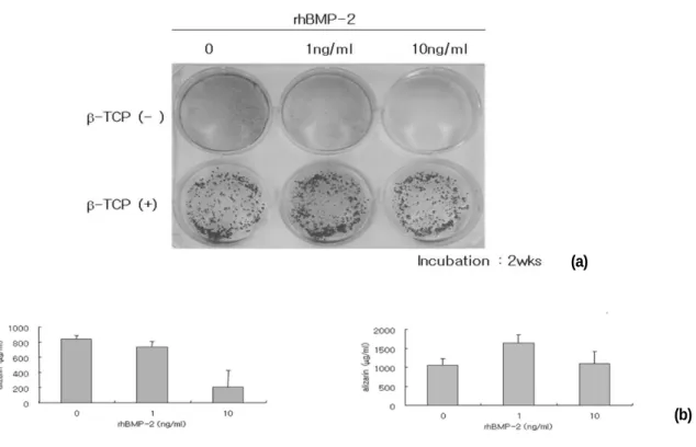
중간엽줄기세포에 Alizarin red S staining을 시행한 결과 β
-TCP
가 없는 군에서는 rhBMP-2의 농도가 높을수록 염색의 정도가 낮았으며 β-TCP가 첨가된 군에 있어서는 β -TCP에 흡수가 일어
나 시각적 판단이 어려웠다. Alizarin red 정량분석을 시행한 결 과 β-TCP가 없는 군에서는 rhBMP-2농도가 0과 1 ng/ml인 경우
에 결정화가 높게 나타났으나 β-TCP가 있는 군에서는 rhBMP-2
농도가 1 ng/ml인 군에서 가장 높게 나타났다(Fig. 3).3. β-TCP와 rhBMP-2하에서 골모유사세포 배양 후 mRNA 발현 양상
1) β -TCP여부와 배양기간에 따른 mRNA 발현양상
전반적으로 골유사세포로 분화된 골수유래줄기세포에서의
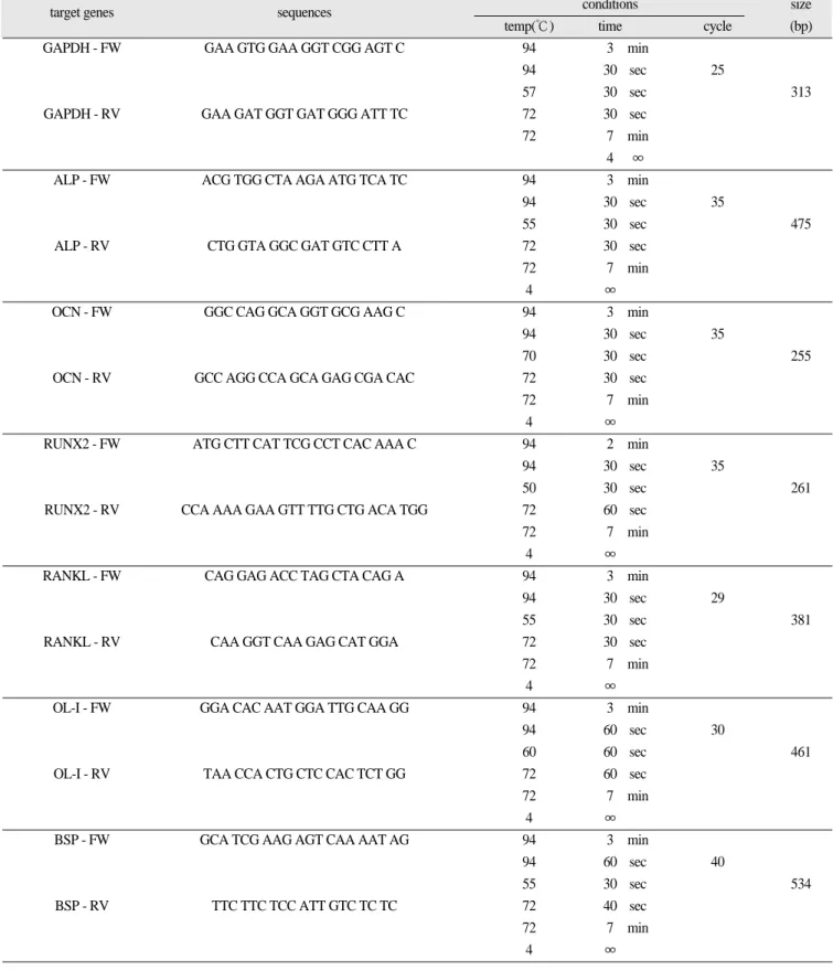
a. BMSCs b. Osteoblast-like cells
Fig. 1. Phase-contrast microphotographs of Bone marrow derived MSCs (BMSCs)
(a) osteoblast-like cells (b) at 1st week.
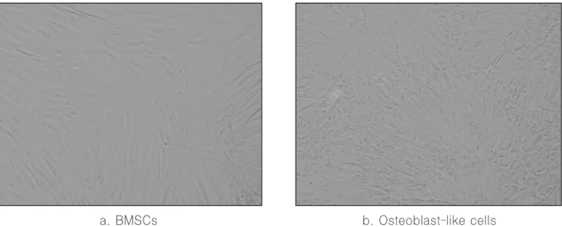
Fig. 2. Effect of osteogenic supplements and β-TCP on osteogenic differentiation. The 4th passage MSCs were seeded at 5×10
3cells/cm
2in each dish, grown in control medium or medium containing osteogenic supplements, fixed and stained by Alizarin red S staining.
Fig. 3 Pictures of alizarin red stained MSCs which were treated with different concentrations of rhBMP-2 (a). The effect of different concentrations of rhBMP-2 and β-TCP presence (b).
(a)
(b)
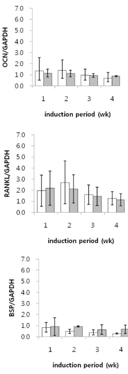
배양기간에 따른 mRNA발현은 다른 단백질에 비하여 RUNX2 와 RANKL의 발현이 두드려졌으며 이는 모두 배양 2주에서 최 고점을 이루고 이후 감소하는 양상을 보였다. 또한 β
-TCP가 없
이 배양된 경우, β-TCP와 함께 배양된 경우에 비하여 ALP,
RUNX2, 그리고 RANKL의 mRNA 발현이 배양 2주에서 통계적
으로 유의성 있게 높게 나타났다(Fig. 4).Fig. 4. mRNA expression of ALP, OCN, RUNX2, COL-Ⅰ, RANKL, and BSP after MSCs were cultured for 1, 2, 3, and 4 weeks
in osteogenic medium (data were expressed as mean ± SD; white bar = β-TCP(-) and dark bar = β-TCP(+)
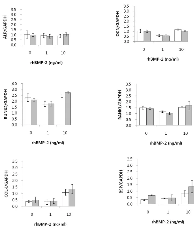
2) rhBMP-2농도에 따른 mRNA 발현양상
전반적으로 골유사세포로 분화된 골수유래줄기세포를
rhBMP-2의 각 농도 별로 4주간 배양하였을 때 각 단백질의 mRNA발현은 rhBMP-2의 농도에 따라 특별한 변화가 관찰되
지 않았으나 COL-I과 BSP가 rhBMP-2를 10 ng/ml의 농도로 배양하였을 때 증가하였다.
또한 β
-TCP와 함께 배양된 경우, β -TCP가 없이 배양된 경우
와 비교하여 BSP의 mRNA발현이 rhBMP-2 10 ng/ml으로 배양 하였을 때 통계적으로 유의성 있게 높게 나타났다(Fig. 5).
Fig. 5. mRNA expression of ALP, OCN, RUNX2, COL-Ⅰ, RANKL, and BSP after MSCs were cultured for 4 weeks in osteogenic medium with different concentrations of rhBMP-2. (data were expressed as mean ± SD; white bar = β-TCP(-) and dark bar
= β-TCP(+)
Ⅳ. 총괄 및 고찰
골전도성만을 가진 인공골 대체재료가 자가골과 유사한 기 능을 발휘하기 위해서는 골형성세포를 직접 유도하는 신호전 달 역할을 가진 단백질과 골형성세포의 동시이식이 필수적이 라고 볼 수 있다. 최근 이와 관련된 조직공학적 생체재료에 대 한 연구가 활발히 진행되고 있으며 골형성을 위한 줄기세포의 적용도 조금씩 진행되고 있다12-14)
. 골수의 간질에는 지방세포,
망상적혈구, 내피세포 및 섬유모세포 등을 포함한 불균일한 세포들이 혼합되어 있으며 골, 연골, 지방과 조혈간세포의 분 화를 돕는 결합조직으로 분화하는 세포들을 포함한다는 사실 이 이미 알려져 있었고, 이는 간엽줄기세포로 언급되었다19-23).
최근에는 골수간엽줄기세포를 dexamethasone, ascorbic acid, β- glycerophosphate가 첨가된 골형성배지에서 배양하여 골유사세
포로 유도분화가 가능함도 밝혀졌다24,25). 이렇게 분리배양된
골수간엽줄기세포는 일차 골재생세포로서 그 수가 아주 적지 만 필요한 양만큼의 증식이 가능하며 배아줄기세포와 같은 윤 리적 문제의 소지가 없어 임상적인 적용에 많은 기대를 걸고 있다. 하지만 계대배양의 수가 지나치게 늘어날 경우 세포노 화현상과 같은 한계를 보이며 분화능력이 떨어지는 결과를 보 이므로 이번 실험과 같이 주로 3-5회 계대배양된 초기의 세포 들을 이용하게 된다26).
골수줄기세포를 임상적으로 적용하기 위해서는 이식부위 내에서의 안정성을 위한 전달체가 필요하며 이 같은 전달체로 는 동종골이나 이종골, HA, β
-TCP 등과 같은 합성골이나 gelatin sponge 등이 있다. 특히 합성골은 감염의 문제나 비용적인 문제
들을 극복할 수 있어 활발하게 연구되고 있는데 생체 내에서 의 흡수가 거의 일어나지 않는 HA에 비하여 β-TCP 는 흡수를
통한 자가골 치환율이 높고 형태 유지를 위한 충분한 강도를 가지고 있어 이번 실험에 전달체로 선택하였다.줄기세포의 골세포유도를 촉진시키고 골재생을 활발히 이 루기 위해 BMP와 같은 골유도물질이 사용된다27-30)
. 특히 BMP- 2는 TGF-β 1 계열로 ALP활성의 증가와 더불어 OCN과 COL-I의
합성을 유도하여 광화 과정을 촉진시키며 골세포와 지방세포 로의 분화에 영향을 끼치는 것으로 보고되고 있다31). 그러나
이번 실험에서는 이런 연구와는 달리 BMP-2의 처리가 상기 보 고된 단백질의 발현으로 나타나지는 않았다. 이는 처리 농도 와 연관되는 것으로 사료되며 Lecanda 등15)은 인간 골수간엽줄 기세포에서 BMP-2는 증식은 억제하면서 분화를 유도하며, 골 세포분화와 골광화를 위해 100 ng/ml의 BMP-2농도가 필요하 다고 보고한 반면 Chaudhary 등29)은 골세포 분화를 위해 40ng/ml의 BMP-7 농도가 필요하다고 하였다. 이번 실험에서는 Alzarin red-S 염색 정도로 판단되는 골형성능이 β -TCP 없이 배
양하였을 경우보다 β-TCP와 함께 배양하였을 경우 10 ng/ml의
농도에서 가장 증가한 것으로 보아 전달체로 β-TCP를 사용할
경우 기존의 BMP농도보다는 적은 농도에서도 골형성능을 증골유사세포에서 β
-TCP 없이 배양한 경우에 β -TCP와 함께 배
양한 경우와 비교하여 배양 2주째 ALP, RUNX2, RANKL의 발 현이 보다 높은 이유는 보다 추가적인 연구가 필요하다고 생 각된다. 각 단백질의 기능을 간략히 살펴보면 ALP는 골형성의 지표로 알려져 있으며 조골세포 표면에서 골활성도가 증가할 때 농도가 증가하고32), RUNX2은 조골세포분화와 골격 형성에
필수적인 전사인자이다33). 특히 RANKL은 파골세포의 자극과
관계가 있어 골흡수에 관련하는 골대사에 중요한 성분이다.본 연구결과에서 β
-TCP(-)군에서 ALP의 mRNA 발현의 증가
가 배양 2주에 증가하다 감소하는 경향을 보였지만, β-TCP(+)
에서는 발현 증가를 관찰할 수 없는 것은 β-TCP에 의해서 ALP
의 발현이 억제되는 것으로 판단할 수 있다. 일반적으로 ALP 는 조골세포의 초기 분화에 관여하는 전사인자로 줄기세포 및 조골세포에서 2주에서 발현이 증가하는 것으로 보고되는 것 으로 판단할 때에 calcium phosphate에 의한 ALP의 발현에 관련 성은 더욱 추가적인 연구가 필요할 것으로 판단된다34,35).
RUNX2 또한 초기 조골전구세포 및 조골세포의 분화에 밀접
하게 관여하는 전사인자로 본 연구결과에서 1-3주경에 높은 발현을 나타내고 있다. 그리고, ALP와 유사하게 β-TCP(-)에서
β-TCP(+)보다 유의한 증가를 보여주고 있다. β -TCP(+)에서 발
현이 β-TCP(-)보다 낮은 것은 ALP와 비슷하게 calcium phosphate
의 영향으로 볼 수 있다.최근 calcium phosphate의 표면 자체가 골형성을 유도한다는 보고와36)
Alizarin red S staining결과를 가지고 판단한다면, 골형
성의 양은 차이가 없는데, ALP와 RUNX2의 발현 차이는 초기 줄기세포의 분화과정에서 calcium phosphate에 의한 직접적인 관여가 있는 것으로 판단할 수 있다.골유사세포를 BMP-2의 각 농도로 처리한 결과 높은 발현을 나타내는 RUNX2와 RANKL보다는 β
-TCP를 함께 배양한 경우
β
-TCP 없이 배양한 경우보다 BSP가 유의성있게 나타난 이유
는 β
-TCP와 함께 배양한 경우 초기광화가 보다 조속하게 일어
날 수 있는 가능성을 제시한다고 볼 수 있다. 특히 BSP는 골, 상 아질, 백악질과 석회화된 연골과 같은 광화된 조직의 구성성 분으로 골의 세포외기질에 중요한 요소이며 골과 백악질에서 발견되는 비교원성 단백질의 대략 8%를 구성하는 것으로 알 려져있다.37)골과 상아질의 BSP의 양은 대략 비슷하지만 광화 된 조직내에서의 기능은 명확히 알려져있지는 않으며 초기
apatite 결정의 형성에서 핵으로서 작용한다
38). 세포외기질에서
교원섬유를 따라 apatite 가 형성될 때 BSP가 방향을 결정하거 나 결정의 성장을 억제하기도 한다.
현재까지 중간엽줄기세포의 골세포분화를 명령하는 동적인 세포간의 상호관계에 대한 분자적 기전은 많이 알려져 있지 않다. 본 연구에서 이러한 상호관계에 대한 기반을 제시하며 추가적인 실험이 필요할 것으로 사료된다.
참고문헌