Fate of Transplanted Bone Marrow Derived Mesenchymal Stem Cells Following Spinal Cord Injury in Rats by Transplantation Routes
This research was performed to investigate the differences of the transplanted cells’ survival and differentiation, and its efficacy according to the delivery routes following spinal cord injury. Allogenic mesenchymal stem cells (MSCs) were transplanted intravenously (IV group) or intralesionally (IL group) at post-injury 1 day in rats. Behavioral improvement,
engraftment and differentiation of the transplanted cells and the expression of
neurotrophic factors of the transplanted groups were analyzed and compared with those of the control group. At 6 weeks post-injury, the mean BBB motor scales in the control, IV and IL groups were 6.5 ± 1.8, 11.1 ± 2.1, and 8.5 ± 2.8, respectively. Regardless of the delivery route, the MSCs transplantation following spinal cord injuries presented better behavioral improvement. The differentiations of the engrafted cells were different according to the delivery routes. The engrafted cells predominantly differentiated into astrocytes in the IV group and on the other hand, engrafted cells of the IL group demonstrated relatively even neural and glial differentiation. The expressions of neuronal growth factor were significantly higher in the IL group (mean relative optical density, 2.4 ± 0.15) than those in the control (2.16 ± 0.04) or IV group (1.7 ± 0.23).
Transplantation of MSCs in the early stage of spinal cord injury gives a significant clinical improvement. However, the fate of the transplanted MSCs and expression of neuronal growth factors are different along the transplantation route.
Key Words: Mesenchymal Stem Cells; Spinal Cord Injuries; Stem Cell Transplantation;
Neuronal Differentiation; Neuroprotection Eun-Sun Kang, Kee-Yong Ha,
and Young-Hoon Kim
Department of Orthopedic Surgery, Seoul St. Mary’s Hosptial, College of Medicine, The Catholic University of Korea, Seoul, Korea Received: 5 December 2011 Accepted: 13 March 2012 Address for Correspondence:
Young-Hoon Kim, MD
Institute of Catholic Integrative Medicine and Department of Orthopedic Surgery, Seoul St. Mary’s Hospital, The Catholic University of Korea, 222 Banpodae-ro, Seocho-gu, Seoul 137-701, Korea
Tel: +82.2-2258-6118, Fax: +82.2-505-9834 E-mail: [email protected]
The authors wish to acknowledge the financial support of the Catholic Medical Center Research Foundation made in the program year of 2011.
http://dx.doi.org/10.3346/jkms.2012.27.6.586 • J Korean Med Sci 2012; 27: 586-593
INTRODUCTION
Cell transplantation for the regeneration of an injured spinal cord would be one of the promising regenerative trials. Cumu- lative research has demonstrated its feasibility and various stem cells have been tried to protect against the secondary damage and to enhance the regeneration of a damaged spinal cord (1-6).
As one trial of this cell therapy, mesenchymal stem cells (MSCs) have been highlighted because they can not only be easily har- vested, expanded and transplanted, but they can also be direct- ly harvested and transplanted, which obviates the ethical and immune rejection problems (7-10). However, several issues need to be addressed in order to establish a successful cell therapeu- tic strategy. Selection of the ideal cell, transplantation method, dosage, and the timing of transplantation are still the questions that should be addressed. Moreover, knowledge about the sur- vival, migration, proliferation and differentiation of the trans- planted cells in the injured site is also essential for successful cell therapy for spinal cord injury (SCI).
MSCs are known to have a homing effect and to be neuropro- tective following SCI when they are injected in the early stage of SCI (8, 11-13). The suggested neuroprotective effects of MSCs for SCI are that they act as an inductor of neurotrophic factor, a modulator of inflammation. Moreover, they are suggested to be able to replace damaged cells through trans-differentiation (9, 14-16). However, there is little research regarding the fate of the transplanted cells and their neuroprotective effects in different transplantation conditions (17, 18). In the present study, MSCs were delivered via different transplantation routes in a contu- sive SCI animal model to investigate whether there are any dif- ferent in terms of the fate of transplanted MSCs and the altera- tion of microenvironment of the injured site.
MATERIALS AND METHODS Animal Model and group allocation
All of the surgical interventions and the pre-surgical and post- surgical animal care were provided in accordance with the Lab-
oratory Animal Welfare Act and the Guidelines and Policies for Rodent Survival Surgery, as provided by the Animal Studies Committee of the Catholic University of Korea (IACUC approv- al No.2011-0174-01). A total 36 of adult male Sprague-Dawley rats (body weight: 250-300 g each) were used in this study. They were kept under standardized conditions (4 rat/cage, 20-24°C, 45%-65% humidity, and a 12 hr of daily light) and given free ac- cess to food and water throughout the study. Rats were random- ly assigned to one of the following three groups before opera- tion; the control group (n = 12, SCI only), the intravenous (IV) group (n = 12, SCI + IV administration of MSCs), intralesional (IL) group (n = 12, SCI + IL administration of MSCs).
After random allocation, the rats were anesthetized with ket- amine (50 mg/kg) and xylazine (2 mg/kg, intraperitoneal). Spi- nal cord injury was made as described previously (19, 20). Brief- ly, their backs were shaved and then sterilized with antiseptic betadine. Lamincetomies were performed at T9 after exposure of the paravertebral muscles from T8-10. All the spinal contu- sions were induced by a 25 g-cm contusion using the MASCIS (Multicenter Animal Spinal Cord Injury Study) impactor (a rod weighing 10 g and dropped from a height of 2.5 cm). The 25 g-cm lesion was chosen to evaluate the neuroprotective effect of the experimental trials in severe SCI. Postoperatively, 5 mg genta- mycin was administrated intramuscularly. The postoperative care procedures involved manual emptying of the bladder twice a day during the experiment.
Preparation of allogenic mesenchymal stem cells
The femoral bone was used to obtain bone marrow. After anes- thesia, the femoral bone was harvested and both ends of the fem- oral bone were cut. Bone marrow was aspirated with an 18-guage needle and then, diluted to 25 mL with Dulbecco’ Eagles medi- um (DMEM) (Sigma, St. Louis, MO, USA) supplemented with 10% heat inactivated fetal bovine serum (FBS) (GibcoBRL, Grand island, NY, USA), 2 mM L-glutamate (Sigma), 100 U/mL peni- cillin and, 0.1 mg/mL streptomycin (Sigma). The bone marrow aspirates was plated and then incubated in a humidified atmo- sphere of 5% CO2 at 37°C. For selecting the MSCs, the nonad- herent cells were eliminated by replacing the medium 48 hr af- ter cell seeding. For each passage, the cells were plated at about 8,000 cells/cm2 and they were grown to confluency.
Phenotypic characterization of the MSC and transplantation
Flow cytometric analysis of the cultured MSC was performed.
Briefly, the cells were detached with trypsin-EDTA solution (0.05%, 1 min, Sigma) and washed twice with PBS that contain- ing 0.1% bovine serum albumin. For direct assays, aliquots of cells at a concentration of 1 × 10⁶ cells per milliliter were immu- nolabelled at 4°C for 30 min with the following antibodies: FITC- conjugated CD 45, PE-conjugated CD 29 and CD 73. MSCs are
known to have the immunophenotype of CD 29 and CD 73 and they lack the CD 45 hematopoietic immunophenotype. All the monoclonal antibodies were purchased from Pharmingen/Bec- ton Dickinson (Flanklin Lakes, NJ, USA). As an isotype-matched control, mouse immunoglobulin G1-FITC or mouse immuno- globulin G1-PE was used. The labeled cells were analyzed by a FACS Calibur flow cytometer (Becton Dickinson) with the use of CellQuest software.
Before transplantation, cells were labeled with fluorescent membrane-intercalating dye PKH 26 (red fluorescence, 10-3 M, Sigma). PKH 26 is known to have the longest in vivo half-life and it is ideal for in vivo cell tracking and cell proliferation studies.
For the IV transplantation, 1 × 10⁶ cells in a 0.5 ml total volume were injected through the tail vein 24 hr after SCI and PBS of same volume was injected through the tail vein for the control group. For the IL transplantation, at post-injury day one the in- jured sites were re-exposed and a concentration of 1 × 10⁶ cells in 10 µL was injected using a Hamilton needle. With the help of a microscope, even administration was done at the caudal and cephalad portions of the injured site. All rats received a daily in- jection of cyclosporine A (10 mg/kg) intraperitoneally for 5 days starting 2 days before surgery.
Immunohistochemical staining for identifying of the transplanted cells
To evaluate the survival, homing ability and proliferation of the PKH 26 labeled MSCs, 8 rats in each group were randomly as- signed to undergo tissue harvesting. At 6 weeks post-injury, specimens that include spinal cord and spleen were obtained after transcardiac perfusion. Spleen was histologically evaluat- ed to observe the transplanted cells’ entrapment by host im- mune system. To identify the cell type of the transplanted cells, double-labeling studies were performed with the use of prima- ry antibodies to neurons (NeuN, 1:100, Chemicon, Pemecula, CA, USA), oligodendrocytes (CC-1, 1:50, Chemicon) and astro- cytes (GFAP, 1:50, Chemicon). The details followed the previ- ously described procedures (20). To quantify the differentiation of the transplanted cells in each condition, the cells that were exactly co-localized by expressing DAPI, PKH 26 and the cell markers NeuN, CC-1, and GFAP were considered as differenti- ated transplanted MSCs. Two tissue samples from each subject were used and on each slide, six fields were randomly selected and the high-powered images (× 400) were obtained using con- focal microscope. The positive cells were counted and the mean numbers of each specimen were recorded and the mean num- bers of each group were compared. We also performed staining for type II collagen to investigate the unintended mesenchymal differentiation of the transplanted cells.
Growth factor analysis
Recent studies have demonstrated that stem cell therapy, includ-
ing MSCs for SCI could alleviate secondary damage. The pro- duction of growth factors by the transplanted cells is suggested as one of the possible mechanisms of this. Therefore, we evalu- ated the expression of neurotrophic growth factors. Four rats in each group were used to assess the secretion of growth factors.
At 1 week post-injury, the spinal tissues were dissected and stored at -80°C. Subsequently, the samples were homogenized on ice in RIPA buffer (150 mM NaCl, 50 mM Tris-HCl pH 7.4, 2 mM EDTA, 1% NP-40, 0.1% Triton X-100, 0.1% SDS, 1 mM Na3VO4, 1 mM sodium deoxycholate, 1 mM PMSF, 10 mg/mL aprotinin, and 5 mg/mL leupeptin). The lysate was centrifuged at 16,000 rpm for 10 min at 4°C. The proteins were separated by SDS–polyacrylamide gel electrophoresis and they were trans- ferred to a polyvinylidene difluoride membrane (Hybond-P, Amersham Pharmacia Biotech, Buckinghamshire, UK). The membrane was blocked with 5% fat-free dry milk for 1 hr in Tris- buffered saline (0.1% Tween-20, 20 mM Tris-HCl, 137 mM NaCl, pH 7.4) and then it was incubated overnight at 4°C with the pri- mary antibodies. The antibodies used for immunoblotting were as follows: brain-derived neurotrophic factor (BDNF; 1:500, Santa Cruz, Santa Cruz, CA, USA), and neuronal growth factor (NGF; 1:500; Santa Cruz). After the membranes were washed, they were incubated with secondary peroxidase-conjugated anti-rabbit or anti-mouse antibodies (Amersham Pharmacia Biotech) that were diluted 1:2,000 in Tris-buffered saline with 0.01% Tween 20. An antibody detection system (ECL, Amersham Pharmacia Biotech) was used and the membranes were ex- posed to X-ray film. The protein band intensities were quanti- fied with a VDS densitometer (Amersham Pharmacia Biotech).
BBB locomotor rating scale
The BBB locomotor rating scale was used to evaluate the neu- rological outcomes over the time course of 6 weeks after SCI (21).
The behavior of each animal in an open field was observed and recorded by two researchers. Scores ranging from 0 to 21 were recorded every week after injury.
Statistical analysis
All the values in the figures and text are expressed as means ± S.E.Ms. The results were analyzed by one-way ANOVA followed by a Bonferroni post-hoc test for multiple comparisons. A P val- ue less than 0.05 was considered to be statistically significant.
CD 45
CD 29
CD 73
FL2HFL2HFL2H FL2HFL2HFL2H Cell countsCell countsCell counts
Control
Control
Control
FITC
FITC
FITC
FL1-H
FL1-H
FL1-H 100 101 102 103 104
100 101 102 103 104
100 101 102 103 104
100 101 102 103 104
100 101 102 103 104
100 101 102 103 104
100 101 102 103 104
100 101 102 103 104
100 101 102 103 104 104
103 102 101 100 104 103 102 101 100 104 103 102 101 100
104 103 102 101 100 104 103 102 101 100 104 103 102 101 100
200 160 120 80 40 0 200 160 120 80 40 0
200 160 120 80 40 0
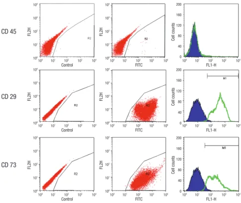
Fig. 1. Flow cytomertric analysis of cultured cells with CD 45, 73, and 29. The positive expression of CD 73 and CD 29, and negative expression of CD 45 indicate its mesenchymal stem cell lineage.
Fig. 2. Type II collagen expression of the transplanted cells were observed. In both transplanted groups, cells with a colocalization of PKH 26 and collagen II were not detected. (A) The IV transplanted MSCs do not express type II collagen. (B) Some type II collagen expression are noted in the IL group, however, no colocalization are found with PKH26 expression (collagen was tagged with green fluorescence, magnification
× 400, scale bar 20 µm).
A B
20 µm 20 µm
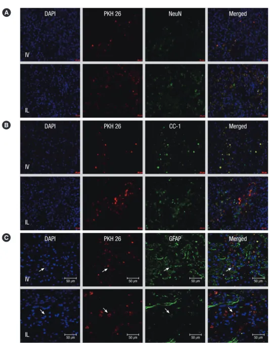
Fig. 3. Various expressions of neural and glial cell makers of engrafted MSCs. PKH26 positive cells were mainly found at the injured sites. (A) Neuronal differentiation of the transplanted MSCs (n = 4, two tissue samples and six fields in each sample). (B) Oligodendrocyte differentiation of the transplanted MSCs. (C) Astrocyte differentiation of the transplanted MSCs. Arrow indicates co-localization of PKH and GFAP expression. IV transplanted MSCs were mainly expressed the astrocyte differentiation. The proportion of neuronal and oligodendrocyte differentiation were lower than that of IL transplanted MSCs (magnification × 200, scale bar 50 µm).
A DAPI
IV
IL
NeuN
PKH 26 Merged
B DAPI PKH 26 CC-1 Merged
IV
IL C
50 µm 50 µm
50 µm 50 µm
50 µm
50 µm
50 µm
50 µm
DAPI PKH 26 GFAP Merged
IV
IL
RESULTS
Characterization of MSC
Before transplantation, the third passage cells were evaluated to confirm their phenotype as MSCs. Flow cytometry analysis
of the MSCs showed positive cell surface markers in CD 29 and CD 73. The cultured MSCs lacked the expression of CD 45 (Fig.
1). This represents that the cultured cells have characteristics of MSCs.
Table 1. The mean numbers of engrafted and differentiated cells by transplatation routes
Routes PKH 26 positive
( × 200)
Neuron differentiation (× 400, PKH26+NeuN)
Oligodendrocyte differentiation (× 400, PKH26+CC-1)
Astrocyte differentiation ( × 400, PKH 26+GFAP)
Other organ (spleen)*
IV 30 ± 6.8 9.4 ± 0.9 8.2 ± 1.1 27 ± 1.0 ++
IL 47 ± 7.3 28.4 ± 2.4 20.4 ± 1.3 20 ± 1.3 -
*PKH26 positive cell in spleen. × 200 and × 400 represent the magnification rates which were used for cell counting. IV, intravenous transplantation; IL, intralesional trans- plantation.
Identification of the transplanted cells in vivo
In both treated groups, the transplanted MSCs were found at the posterior portion (injured site) of the spinal cord and some scattered cells were also observed at the gray and white matter adjacent to the injured site. Under magnification (× 400), the cells expressing PKH staining coincident with DAPI were counted and quantified. In the control group, there were no cells express- ing PKH 26. The mean numbers of PKH 26 positive cells in the IV and IL groups were 30.4 ± 6.9 and, 47 ± 7.3, respectively (Table 1). The numbers of implanted MSCs were greater in the IL group than that in the IV group with statistical significance (P < 0.05).
Abundant MSCs were found in the spleen in the IV group but no MSCs were observed in the spleen of the IL group.
Aberrant differentiation of the transplanted cells
To identify an aberrant differentiation of the transplanted cells to mesenchymal lineage, type II collagen staining was performed.
Primary antibody to type II collagen (1:100, Lab Vision, Fre- mont, CA, USA) was used. Alexa Fluor 594 was used for immu- nofluorescence detection of collagen. Neither of the transplant- ed groups showed collagen expressing transplanted cells (Fig. 2).
Differentiation of the transplanted cells
To observe any differences in the differentiation of transplanted cells depending on the transplantation routes, double staining was performed and the numbers of cells that were colocalized
with DAPI, PHK 26 and other cell markers including NeuN, CC-1 and GFAP were counted. In the IV group, most of the PKH 26 positive cells were colocalized with GFAP labeling and the num- bers of cells showing immunofluorescence colocalization with NeuN and CC-1 markers were very low. In the IL group, the transplanted MSCs were evenly colocalized with GFAP, NeuN and CC-1. Table 1 shows the numbers of colocalized expres- sions of the transplanted cells (Fig. 3).
Neurotrophic factors expression in the SCI lesion
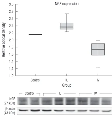
BDNF and NGF levels in the spinal cord tissue (n = 4, each group) were measured. BDNF level in the IL group (mean rela- tive optical density, 1.70 ± 0.2) were slightly increased com- pared to those in the control group (1.58 ± 0.22) and the IV group (1.39 ± 0.35). However, there was no statistical signifi- cance (Fig. 4). NGF level in the IL group (mean relative optical density, 2.4 ± 0.15) was significantly increased compared to the control (2.16 ± 0.04) and the IV group (1.7 ± 0.23) (P < 0.05) (Fig. 5).
Behavioral assessment
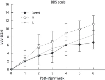
All the injured rats manifested complete hindlimb paraplegia immediately after the operation. In all the groups, the rats grad- ually recovered varying degrees of motor function over the time of observation (Fig. 6). At 6 weeks post-injury, the mean BBB motor scales in the control, IV and IL groups were 6.5 ± 1.8,
Fig. 4. Expression of BDNF. Post-injury 1 week BDNF levels in the spinal cord tissues were measured. The relative optical densities in the control, IV and IL groups are 1.58
± 0.22, 1.39 ± 0.35, 1.70 ± 0.2 respectively. The IL group shows slightly higher level compared to those in the control and IV groups. However, BDNF expression does not show any significant difference between the groups (n = 4, P > 0.05).
Relative optical density
Group BDNF expression
Control IL IV
2.2 2.0 1.8 1.6 1.4 1.2 1.0 0.8 0.6
BDNF (32 kDa)
β-actin (43 kDa)
Control IL IV
Fig. 5. Expression of NGF. Post-injury 1 week NGF levels in the spinal cord tissues were measured. The relative optical densities in the control, IV and IL groups are 2.16
± 0.04, 1.70 ± 0.23, 2.41 ± 0.15 respectively. The IL group shows significantly higher level compared to those in the control and IV groups. And there is also signifi- cant difference between the groups (n = 4, P < 0.05).
Relative optical density
Group NGF expression
Control IL IV
3.0 2.8 2.6 2.4 2.2 2.0 1.8 1.6 1.4 1.2 1.0
NGF (27 kDa)
β-actin (43 kDa)
Control IL IV
11.1 ± 2.1, and 8.5 ± 2.8, respectively. The functional recovery seen in the rats that underwent MSCs transplantation was sig- nificantly better than that in the control group (P < 0.05).
DISCUSSION
There is no doubt that cell based therapy, including stem cells, is an attractive and promising therapeutic strategy for many clinical conditions that currently lack efficacious treatment.
However, there are many issues and concerns to be addressed before its clinical translation. As one of the efforts to address these topics, we tried different transplantation routes of stem cells for SCI. The efficacy and fate of the transplanted cells were observed according to different transplantation routes. In the present study, we used allogenic MSCs. MSCs have been dem- onstrated to have an ability to “home” into the injured site and trans-differentiate into neural lineage cells (13, 22-24). We were also able to observe this homing and trans-differentiation abili- ty even when the MSCs were applied intravenously at the acute stage of SCI. Moreover, its results were connected to behavioral improvement in the both IV and IL groups compared to the control group.
Studies on applying MSCs application for various central nervous disorders have demonstrated that transplantation of MSCs alleviated further tissue damage and it yielded significant clinical improvement (7, 10, 12, 25, 26). These results were ex- plained by the possibilities of a neuroprotective function and a tissue repair by the transplanted cells. In most of the reported studies, the differentiation of transplanted cells could be ob- served and to a limited extent, neuron and GFAP positive dif- ferentiation were reported (10, 25, 27). However, these studies
did not present the differences depending on the transplanta- tion route. A few studies that focused on comparing the efficacy following MSCs transplantation for SCI demonstrated more efficient engrafting of transplanted cells into lesion site when grafting by the intralesional or lumbar puncture routes (14, 28, 29). These studies demonstrated this difference of efficacy only through examining the engrafting MSCs volume as counted by histological or radioisotope labeling examination. Clinical as- sessment or the differentiation of transplanted cells has not been addressed. As suggested in many studies, intravenous delivery has inherent concerns of its efficacy. Although the IV route has the advantages of easy and safe delivery, trapping of the transplanted cells in the other organs and the high chance of exposure to an immune reaction limits its clinical utility. In the present study, as predicted and suggested by other previous studies, IV delivery showed a lesser number of engrafted MSCs as compared to that of IL delivery. Many MSCs were observed in the spleen. However, IV delivery showed more effective clini- cal improvement as compared to that of the control group and the IL group. It is hard to conclude from our results that IV de- livery could result in better clinical improvement in the early stage of transplanting MSCs for SCI. However, as other studies have reported, IV delivery could be an effective delivery route for early MSCs transplantation following SCI. Homing of the MSCs to the disrupted blood-spinal cord barrier tissue and avoidance of additional injury that can be caused by intrale- sional delivery could account for these results (9, 12, 25).
Homing of MSC into the injured spinal cord is well known and stromal derived factor-1 (SDF-1)/CXCR-4 has recently been demonstrated to take part in the migration of MSCs (11, 30, 31).
In our results, even though the absolute number of engrafted cells in the IV group was lower than that of the IL group, the de- gree of behavioral improvement was better in the IV group. This phenomenon suggests that the neuroprotective effects of early transplanted MSCs do not merely depend on the absolute num- ber of the engrafted cells. Although it is controversial, some stud- ies have suggested the possibility of replacement of damaged tissue by the transplanted MSCs (32, 33). However, the secre- tion of neurotrophic factors (BDNF, NGF, and VEGF), the mod- ulation of inflammation and immune reactions and enhance- ment of axonal sprouting in the pathologic condition following SCI have been recently suggested as the primary effects of MSCs transplantation at the early stage, which is beyond their potential to differentiate to form glial and neural lineage cells (8, 27, 32).
And in terms of clinical results, our study has some limita- tions. IL transplantation needed the secondary surgery which might influence the final clinical results. Further research is needed to clarify the neuroprotective mechanism of MSCs trans- plantation for early stage of SCI.
In terms of the fate of the transplanted cells, numerous stud- ies have reported that MSCs have the capability to differentiate
BBS scale
Post-injury week BBS scale
0 1 2 3 4 5 6
16 14 12 10 8 6 4 2 0
Control IV IL
Fig. 6. Open field locomotor assessment using Basso-Beattie-Bresnahan scale tested at every week after SCI. All subjects show a gradual improvement in hindlimb func- tion during the 6 week observation period. Stastistical analysis indicated that BBB scales in the MSCs transplanted group were significantly higher than those in the control group (n = 8, P < 0.05). The rats in the IV group show the highest improve- ment at the last follow-up.
into neuronal cell lineages (5, 10, 24). Even though MSCs were intravenously transplanted, the transplanted MSCs expressed neuron or glial cell phenotypes. Our immunohistochemical data also demonstrated that the transplanted PKH26 positive cells were predominantly located at the damaged area. Surpris- ingly, the phenotype expressions of the transplanted cells are different according to the delivery routes. In the intralesionally transplanted group, the transplanted cells showed NeuN, CC-1, and GFAP positivity. It suggests that the transplanted cells ex- pressed all the neuronal lineage phenotype differentiation. In contrast, the intravenously transplanted MSCs mainly expressed GFAP positivity. NeuN and CC-1 expressing MSCs were rarely observed. With our results, it is hard to conclude that one deliv- ery route is superior to the other. However, the intravenous de- livery route also showed effective neuroprotective results and this is supported by the histologic findings and the clinical im- provement.
Preferential astrocytic differentiation of the transplanted MSCs has been reported several times (10, 25, 34). Some au- thors have suggested that differentiation into astrocytes is the default pathway for transplanted MSCs or neuronal stem cells through in vivo study (22, 35). However, of importance is that this predominant astrocyte differentiation of MSCs in models of central nervous system injuries has been associated with fa- vorable functional results. To interpret these results, most au- thors agree with the hypothesis that differentiation into astro- cytes is beneficial for early stage injury through the neuropro- tective functions of astrocytes (34, 35). However, astrocytes are known to have a wide variety of biological activities following central nervous system injury (20, 36, 37). In the acute phase of injury, astrocytes have a major role to maintain and restore ho- meostasis in the injured site and this is also important to pro- tect against further damage. On the other hand, in the chronic phase, reactive proliferation of astrocytes results in gliosis and this gliosis is a major obstacle to regeneration. Therefore, the long term effect of this predominant astrocyte differentiation remains to be established.
In terms of the neurotrophic factors expression, the IL group showed higher BDNF and NGF expression compared to those in the control and IV groups. As suggested by the previous stud- ies, this might be related to the absolute number of the engraft- ed MSCs. However, in this study, the clinical improvement was not correlated to the absolute number of the engrafted MSCs, and the expression of BDNF and NGF. These findings might be also related to the additional injury during the transplantation in the intralesional injections.
Before stem cell therapy for SCI can be a successful treatment, many issues need to be resolved including the optimal delivery route and the fate of the transplanted cells. According to our re- sults, we suggest that early delivery of allogenic MSCs following SCI provided favorable behavioral improvement compared to
the control group. And then the fate of the transplanted MSCs and expression of neuronal growth factors following MSCs trans- plantation are different along the transplantation route.
REFERENCES
1. Wright KT, El Masri W, Osman A, Chowghury J, Johnson WE. Concise review: Bone marrow for the treatment of spinal cord injury: mecha- nisms and clinical applications. Stem Cells 2011; 29: 169-78.
2. Nandoe Tewarie RS, Hurtado A, Bartels RH, Grotenhuis A, Oudega M.
Stem cell-based therapies for spinal cord injury. J Spinal Cord Med 2009;
32: 105-14.
3. Lindvall O, Kokaia Z. Stem cells in human neurodegenerative disorders:
time for clinical translation? J Clin Invest 2010; 120: 29-40.
4. Kim BG, Hwang DH, Lee SI, Kim EJ, Kim SU. Stem cell-based cell thera- py for spinal cord injury. Cell Transplant 2007; 16: 355-64.
5. Akiyama Y, Radtke C, Honmou O, Kocsis JD. Remyelination of the spi- nal cord following intravenous delivery of bone marrow cells. Glia 2002;
39: 229-36.
6. Yoon SH, Shim YS, Park YH, Chung JK, Nam JH, Kim MO, Park HC, Park SR, Min BH, Kim EY, et al. Complete spinal cord injury treatment using autologous bone marrow cell transplantation and bone marrow stimulation with granulocyte macrophage-colony stimulating factor:
Phase I/II clinical trial. Stem Cells 2007; 25: 2066-73.
7. Yoo SW, Kim SS, Lee SY, Lee HS, Kim HS, Lee YD, Suh-Kim H. Mesen- chymal stem cells promote proliferation of endogenous neural stem cells and survival of newborn cells in a rat stroke model. Exp Mol Med 2008;
40: 387-97.
8. Vaquero J, Zurita M. Bone marrow stromal cells for spinal cord repair: a challenge for contemporary neurobiology. Histol Histopathol 2009; 24:
107-16.
9. Caplan AI, Dennis JE. Mesenchymal stem cells as trophic mediators. J Cell Biochem 2006; 98: 1076-84.
10. Azizi SA, Stokes D, Augelli BJ, DiGirolamo C, Prockop DJ. Engraftment and migration of human bone marrow stromal cells implanted in the brains of albino rats: similarities to astrocyte grafts. Proc Natl Acad Sci U S A 1998; 95: 3908-13.
11. Son BR, Marquez-Curtis LA, Kucia M, Wysocaynski M, Turner AR, Ratajczak J, Ratajczak MZ, Janowska-Wieczorek A. Migration of bone marrow and cord blood mesenchymal stem cells in vitro is regulated by stromal-derived factor-1-CXCR4 and hepatocyte growth factor-c-met axes and involves matrix metalloproteinases. Stem Cells 2006; 24: 1254-64.
12. Nomura T, Honmou O, Harada K, Houkin K, Hamada H, Kocsis JD. I.V.
infusion of brain-derived neurotrophic factor gene-modified human mesenchymal stem cells protects against injury in a cerebral ischemia model in adult rat. Neuroscience 2005; 136: 161-9.
13. Woodbury D, Schwarz EJ, Prockop DJ, Black IB. Adult rat and human bone marrow stromal cells differentiate into neurons. J Neurosci Res 2000; 61: 364-70.
14. Swanger SA, Neuhuber B, Himes BT, Bakshi A, Fischer I. Analysis of al- logeneic and syngeneic bone marrow stromal cell graft survival in the spinal cord. Cell Transplant 2005; 14: 775-86.
15. Lu P, Jones LL, Tuszynski MH. BDNF-expressing marrow stromal cells support extensive axonal growth at sites of spinal cord injury. Exp Neu-
rol 2005; 191: 344-60.
16. Kim DH, Yoo KH, Yim YS, Choi J, Lee SH, Jung HL, Sung KW, Yang SE, Oh WI, Yang YS, et al. Cotransplanted bone marrow derived mesenchy- mal stem cells (MSC) enhanced engraftment of hematopoietic stem cells in a MSC-dose dependent manner in NOD/SCID mice. J Korean Med Sci 2006; 21: 1000-4.
17. Jung DI, Ha J, Kang BT, Kim JW, Quan FS, Lee JH, Woo EJ, Park HM. A comparison of autologous and allogenic bone marrow-derived mesen- chymal stem cell transplantation in canine spinal cord injury. J Neurol Sci 2009; 285: 67-77.
18. Parr AM, Kulbatski I, Wang XH, Keating A, Tator CH. Fate of transplant- ed adult neural stem/progenitor cells and bone marrow-derived mesen- chymal stromal cells in the injured adult rat spinal cord and impact on functional recovery. Surg Neurol 2008; 70: 600-7.
19. Ha KY, Kim YH. Neuroprotective effect of moderate epidural hypother- mia after spinal cord injury in rats. Spine (Phila Pa 1976) 2008; 33:
2059-65.
20. Ha KY, Kim YH, Rhyu KW, Kwon SE. Pregabalin as a neuroprotector af- ter spinal cord injury in rats. Eur Spine J 2008; 17: 864-72.
21. Basso DM, Beattie MS, Bresnahan JC. A sensitive and reliable locomo- tor rating scale for open field testing in rats. J Neurotrauma 1995; 12:
1-21.
22. Chu K, Kim M, Park KI, Jeong SW, Park HK, Jung KH, Lee ST, Kang L, Lee K, Park DK, et al. Human neural stem cells improve sensorimotor deficits in the adult rat brain with experimental focal ischemia. Brain Res 2004; 1016: 145-53.
23. Kamada T, Koda M, Dezawa M, Anahara R, Toyama Y, Yoshinaga K, Hashimoto M, Koshizuka S, Nishio Y, Mannoji C, et al. Transplantation of human bone marrow stromal cell-derived Schwann cells reduces cys- tic cavity and promotes functional recovery after contusion injury of adult rat spinal cord. Neuropathology 2011; 31: 48-58.
24. Song L, Tuan RS. Transdifferentiation potential of human mesenchymal stem cells derived from bone marrow. FASEB J 2004; 18: 980-2.
25. Osaka M, Honmou O, Murakami T, Nonaka T, Houkin K, Hamada H, Kocsis JD. Intravenous administration of mesenchymal stem cells de- rived from bone marrow after contusive spinal cord injury improves functional outcome. Brain Res 2010; 1343: 226-35.
26. Yoshihara T, Ohta M, Itokazu Y, Matsumoto N, Dezawa M, Suzuki Y, Taguchi A, Watanabe Y, Adachi Y, Ikehara S, et al. Neuroprotective effect
of bone marrow-derived mononuclear cells promoting functional recov- ery from spinal cord injury. J Neurotrauma 2007; 24: 1026-36.
27. Lee KH, Suh-Kim H, Choi JS, Jeun SS, Kim EJ, Kim SS, Yoon do H, Lee BH. Human mesenchymal stem cell transplantation promotes function- al recovery following acute spinal cord injury in rats. Acta Neurobiol Exp (Wars) 2007; 67: 13-22.
28. Paul C, Samdani AF, Betz RR, Fischer I, Neuhuber B. Grafting of human bone marrow stromal cells into spinal cord injury: a comparison of de- livery methods. Spine (Phila Pa 1976) 2009; 34: 328-34.
29. de Haro J, Zurita M, Ayllón L, Vaquero J. Detection of 111In-oxine-la- beled bone marrow stromal cells after intravenous or intralesional ad- ministration in chronic paraplegic rats. Neurosci Lett 2005; 377: 7-11.
30. Imitola J, Raddassi K, Park KI, Mueller FJ, Nieto M, Teng YD, Frenkel D, Li J, Sidman RL, Walsh CA, et al. Directed migration of neural stem cells to sites of CNS injury by the stromal cell-derived factor 1alpha/CXC chemo- kine receptor 4 pathway. Proc Natl Acad Sci U S A 2004; 101: 18117-22.
31. Takeuchi H, Natsume A, Wakabayashi T, Aoshima C, Shimato S, Ito M, Ishii J, Maeda Y, Hara M, Kim SU, Yoshida J. Intravenously transplanted human neural stem cells migrate to the injured spinal cord in adult mice in an SDF-1- and HGF-dependent manner. Neurosci Lett 2007; 426: 69-74.
32. Neuhuber B, Timothy-Himes B, Shumsky JS, Gallo G, Fischer I. Axon growth and recovery of function supported by human bone marrow stromal cells in the injured spinal cord exhibit donor variations. Brain Res 2005; 1035: 73-85.
33. Krampera M, Marconi S, Pasini A, Gallè M, Rigotti G, Mosna F, Tinelli M, Lovato L, Anqhileri E, Andreini A, Pizzolo G, Sbarbati A, Bonetti B. In- duction of neural-like differentiation in human mesenchymal stem cells derived from bone marrow, fat, spleen and thymus. Bone 2007; 40: 382-90.
34. Zurita M, Vaquero J. Functional recovery in chronic paraplegia after bone marrow stromal cells transplantation. Neuroreport 2004; 15: 1105-8.
35. Jeong SW, Chu K, Jung KH, Kim SU, Kim M, Roh JK. Human neural stem cell transplantation promotes functional recovery in rats with experimen- tal intracerebral hemorrhage. Stroke 2003; 34: 2258-63.
36. Wang DD, Bordey A. The astrocyte odyssey. Prog Neurobiol 2008; 86:
342-67.
37. Ha KY, Carragee E, Cheng I, Kwon SE, Kim YH. Pregabalin as a neuro- protector after spinal cord injury in rats: biochemical analysis and effect on glial cells. J Korean Med Sci 2011; 26: 404-11.