• 검색 결과가 없습니다.

The lower-extremity muscle co-activation of flat-footed subjects wearing high-heels while descending stairs.

N/A
N/A
Protected

Academic year: 2021

Share "The lower-extremity muscle co-activation of flat-footed subjects wearing high-heels while descending stairs."

Copied!
7
0
0

로드 중.... (전체 텍스트 보기)

전체 글

(1)

1. Introduction

Biomechanical functions of the foot are primarily propulsion, absorbing shock, and providing posture

stability during walking(Abboud, et al., 2002; Perry, 1994). foot problems are related to impaired mobility and postural stability, which have a detrimental impact on quality of life(P.Kaoulla, et al., 2011; K.J. Gorter, et

The lower-extremity muscle co-activation of flat-footed subjects wearing high-heels while descending stairs.

Na-Hee Kim 1 , Bo-ram Choi 2*

1

Graduate Student, Department of Physical Therapy, College of Health and Welfare, Silla University

2

Professor, Department of Physical Therapy, College of Health and Welfare, Silla University

평발 대상자가 하이힐을 신고 계단을 내려갈 때 하지의 근활성도 변화

김나희

1

, 최보람

2*

1신라대학교 보건복지대학 물리치료학과 석사과정, 2신라대학교 보건복지대학 물리치료학과 교수

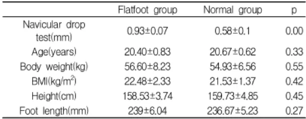
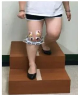
Abstract The purpose of this study was to examine the lower-extremity muscle activation of flat-footed and normal-footed subjects descending stairs while wearing high-heels, thereby identifying any imbalance between the medial and lateral muscles.Thirty female students volunteered to participate in this study. The navicular drop test (NDT) was applied to the selection criteria for the flat-footed group and the normal-footed group. Surface electromyographic data was collected from the medial and lateral quadriceps, hamstrings, and gastrocnemius.

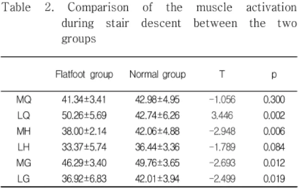
Activation of MG and LG was significantly lower in the flat-footed group than in the normal-footed group. Both groups showed significant increases in MQMH and MHMG, but the co-activation in the medial and lateral muscles was lower in the flat-footed group. The co-activation ratios showed a significantly greater MQMH/LQLH in the flat-footed group. Flat-footed subjects who wear high-heels are more likely to experience impaired knee joint alignment than normal-footed subjects. Therefore, flat-footed subjects should use caution when descending stairs while wearing high-heels.

Key Words : Flat-foot, descending stairs, high-heel, muscle co-activation, EMG

요 약 본 논문의 평발인 대상자가 하이힐을 신고 계단을 내려갈 때 하지의 근활성도 변화를 알아보고자 한다. 이 연구의 대상자는 30명의 여학생으로 구성되어 있다. NDT를 이용하여 평발군과 정상군을 구분하였으며 표전근전도를 사용해 측정 하였다. MG와 LG근활성도와 안, 가쪽 근공동활성도는 평발군에서 낮게 나타났고 근활성도 비율은 평발군이 안쪽에서 더 높게 나타났다. 그러므로 하이힐을 신은 평발군은 정상군보다 무릎관절 정렬에 더 많은 손상을 가질 가능성이 더 높으므로 계단을 내려갈 때 더 많은 주의가 필요할 것이다.

주제어 : 평발, 계단 내려가기, 하이힐, 근육공동활성화, 근전도

*Corresponding Author : Bo-ram Choi ([email protected]) Received August 22, 2018

Accepted November 20, 2018

Revised November 6, 2018 Published November 28, 2018

(2)

al, 2000; H.B. Menz, et al, 2006). Flat foot might be characterized as a mechanical imbalance by a decrease in the medial arch height and medial rotation of the talus. Flat foot is often accompanied by pain and frequently affects walking speed and balance, which increases the risk of falls(H.B. Menz, et al, 2006; J.M.

Frances, et al, 2015; C.A. Demetracopoulos, et al, 2015).

Literature showed that wearing high heels can produce injurious effects on musculoskeletal system(J.

Yu, et al, 2008; K.A. Opila-Correia, 1990). affecting structures from the toes to the pelvis. Wearing high heels leads to alterations in balance, regardless of the height of the heel (A. Mika, et al, 2016). Indeed, compared with barefoot, heels are responsible of difficulties in maintaining balance and enhanced risk of falls because of the changes of the center of mass and the decrease of the base of support (H.-L. Chien, et al, 2014; V. Dewi Hapsari, et al, 2016).

As can be seen, when an individual with flat feet, which cause changes in foot and ankle alignment, descends stairs wearing high-heels, it is likely to cause an imbalance in the muscles that maintain ankle joint stability. Therefore, the purpose of this study was to examine the lower-extremity muscle activation of flat-footed and normal-footed subjects descending stairs while wearing high-heels, thereby identifying any imbalance in the medial and lateral muscle co-activation.

2. Methods

2.1 Subjects

This study included 30 women in their 20s, none of whom had any medical history related to neurological problems. There were 15 flat-footed and 15 normal-footed women volunteering in the study. The navicular drop test (NDT) was applied to the selection criteria for the flat-footed group and the normal-footed group. Subjects with 0.8mm or above were assigned to the flat-footed group and those below 0.8mm were assigned to the normal-footed group. Table 1 This

study received ethics approval from the Human Research Ethics Committee of Silla University (1041449-201605-HR-001)

Flatfoot group Normal group p Navicular drop

test(mm) 0.93±0.07 0.58±0.1 0.00

Age(years) 20.40±0.83 20.67±0.62 0.33

Body weight(kg) 56.60±8.23 54.93±6.56 0.55 BMI(kg/m

2

) 22.48±2.33 21.53±1.37 0.42 Height(cm) 158.53±3.74 159.73±4.85 0.45 Foot length(mm) 239±6.04 236.67±5.23 0.27 Mean ± 1 Standard Deviation (SD)

Table 1. General characteristics of the subjects (Mean±SD) (N=30)

2.2 Instruments

A wireless surface electromyography (EMG) device (4D-SES, RELIVE, Korea) was used to measure the lower-extremity muscle activation during stair descent.

The sampling rate was 1000Hz, and the filter was a band-pass filter of 25-500Hz anda notch filter of 60Hz.

The EMG data was collected from the medial and lateral quadriceps (MQ, LQ), hamstrings (MH, LH), and gastrocnemius (MG, LG). The reference electrode was placed on the patella. The EMG data was analyzed using the RELIVE EMG software (4D-SES, RELIVE, Korea).

All 30 subjects used high-heels that were 7cm in height and 235mm or 245mm in length. A study reported that the most comfortableheel height during regular walking is in the range of 3 to 5cm (Chun and Choi, 2000), so in this regard this study selected a heel height of 7cm to increase the weight bearing on the feet. In this study, two flights of stairs without any railings were used. The first was 60cm wide with 20cm risers and 28cm treads, and the second was 60cm wide with 20cm risers and 90cm treads.

2.3 Procedure

The navicular drop test (NDT) used in this study to

identify flatfeet was introduced in a study by

Brody(Brody, 1982) and indicates changes in the

sagittal plane while loads are imposed. It has high

(3)

reliability in evaluating navicular height (Sell, et al., 1994). The NDT was performed after a mark was put on the navicular tuberosity. Two raters derived the NDT value of each individual by subtracting the value measured with weight bearing on a foot from the value measured without weight bearing on the same foot.

Each rater performed this step three times. The raters did not share the results of their measurement to increase reliability. In the NDT, the interclass correlation coefficient (ICC) as an estimate of intra-rater reliability was 0.973, and the ICC as an estimate of inter-rater reliability was 0.947. Both were statistically significant with a p-value of 0.000 for both.

Maximal voluntary isometric contraction (MVIC) was recorded to normalize the EMG data. MVIC was recorded for five seconds, and data was calculated from the middle threeseconds. For the quadriceps femoris, the subjects were instructed to maintain knee extension while sitting in a chair and then perform muscle contraction. For the hamstring, the subjects were instructed to maintain knee flexion in a prone position and then perform muscle contraction. For the gastrocnemius, the subjects were instructed to fix their body by holding a table in a standing position and then perform muscle contraction by pushing themselves up with their toes(Peterson-Kendall, et al., 2005).

Two or three times, dead skin cells were removed through rubbing the areas to which the electrodes would be attached, and this was done by using a disposable razor to minimize skin resistance to the surface EMG signals. Skin fat was then removed using cotton balls and rubbing alcohol, and then the electrodes were attached.

Muscle activation was measured during the stair descent task. Subjects stood on the second stair with both feet parallel and pelvic-width apart. Upon the first step, the measurement foot was descended tothe first floor, then the second step non-measurement foot was descended to the ground at a self-comfortable speed.

Fig. 1 We measured the muscle activation of the first step for both groups. This stair descent task was

repeated five times with a 30-second rest between tasks.

Fig. 1. measured during the stair descent

2.4 Data analysis

Muscle cocontraction was assessed between the MQ and MH, between LQ and LH, between MH and MG, between LH and LG. Cocontraction was determined

using the equation described by

EMGS/EMGL*(EMGS+EMGL), where EMGS was the level of activity in the less active muscle and EMGL was the level of activity in the more active muscle. The index was multiplied by the sum of the activity found in the 2 muscles. We calculated the ratio of lateral-to-medial co-contraction by dividing the lateral H:G co-contraction index by the Medial H:G co-contraction index(Palmieri-smith et al., 2009). The study compared the muscle activation, co-activation, and co-activation ratios during stair descent between the two groups. The ICC (2, 1) was measured to examine intra-rater reliability (Cote, et al., 2005;

Vicenzino, et al., 2005). The independent t-test was performed to compare differences in the lower-extremity muscle activation of the flat-footed group and normal-footed group during stair descent.

The statistical significance level was set at α=0.5, and the software package SPSS version 24.0 was used for statistical processing.

3. Results

In this study, the flat-footed group showed a

(4)

statistically significant lower level of muscle activation in the gastrocnemius than the normal-footed group (P<0.05). The lateral quadriceps showed a higher level of muscle activation in the flat-footed group. The medial hamstring showed a higher level of muscle activation in the normal-footed group (P<0.05). Table 2.

Flatfoot group Normal group T p

MQ 41.34±3.41 42.98±4.95 -1.056 0.300

LQ 50.26±5.69 42.74±6.26 3.446 0.002

MH 38.00±2.14 42.06±4.88 -2.948 0.006

LH 33.37±5.74 36.44±3.36 -1.789 0.084

MG 46.29±3.40 49.76±3.65 -2.693 0.012

LG 36.92±6.83 42.01±3.94 -2.499 0.019

Mean ± 1 Standard Deviation (SD) MQ: Medial Quadriceps,LQ: Lateral Quadriceps, MH: Medial Hamstring, LH: Lateral Hamstring MG: Medial Gastrocnemius, LG: Lateral Gastrocnemius

Table 2. Comparison of the muscle activation during stair descent between the two groups

Comparison of the muscle co-activation during stair descent between the two groups

In this study, both groups showed higher levels of muscle co-activation in the medial muscles in terms of the MQMH, LQLH, MHMG, and LHLG, while the flat-footed group showed lower levels of muscle co-activation in the medial and lateral muscles compared to the normal-footed group (P<0.05). Table 3.

Flatfoot group Normal group t p

MQMH 71.24±4.98 78.45±10.24 -2.453 0.021 LQLH 56.11±13.22 67.64±6.44 -3.036 0.005 MHMG 67.70±4.28 76.27±10.16 -3.012 0.005 LHLG 60.14±9.93 68.42±9.43 -2.341 0.027 Mean ± 1 Standard Deviation (SD)

MQMH: Medial Quadriceps Medial Hamstring, LQLH: Lateral Quadriceps Lateral Hamstring MHMG: Medial Hamstring Medial Gastrocnemius, LHLG: Lateral Hamstring Lateral Gastrocnemius

Table 3. Comparison of the muscle co-activation during stair descent between the two groups

Comparison of the muscle co-activation ratios during stair descent between the two groups

The flat-footed group showed lower LQLH/MQMH and LHLG/MHMG ratios compared to the normal-footed group. Fig. 2

Fig. 2. Comparison of the muscle co-activation ratios during stair descent between the two groups

4. Discussion

The comparison of the muscle activation during stair descent between the two groups showed that the flat-footed group had statistically significant lower levels of muscle activation in the gastrocnemius compared to the normal-footed group. The comparison of the two groups during stair descent showed that both groups had higher levels of muscle co-activation on the medial side in the Q:H co-activation and H: G co-activation. For the H: G co-activation, the flat-footed group exhibited a higher level of muscle co-activation on the medial side compared to the normal-footed group.

In this study, the flat-footed group showed an

overall lower level of muscle activation in the

gastrocnemius than the normal-footed group, and this

was statistically significant. The subjects wore 7cm

high-heels, creating a position that shortened the

length of the gastrocnemius (Peterson-Kendall, et al.,

2005). As a result, the length-tension relationship that

could enable the gastrocnemius to generate greater

force was not formed, and thus, muscle contraction was

reduced(K. H. Lee, et al., 1990). In addition, considering

that the flat-footed group showed lower muscle

activation than the normal-footed group, the lowered

(5)

medial longitudinal arch in a flat foot may have further increased plantar flexion, thereby further shortening the length of the gastrocnemius. moreover the pes planus foot will exhibit excessive dorsiflexion of the midfoot, especially late in stance (Franco, 1987; Olsen and Saidei, 1983). Therefore, muscle activity decreased because the gastrocnemius muscles relaxed by dorsifoxion of midfoot.

In this study, both groups showed higher levels of muscle co-activation in the medial muscles in terms of medial and lateral muscle co-activation of the MQMH, LQLH, MHMG, and LHLG, and the flat-footed group exhibited lower levels of medial and lateral muscle co-activation than the normal-footed group.

Muscular co-activation is a well-known mechanism for lower limb joint stabilization in both healthy and pathological individuals. This muscular feature appears particularly important for the knee joint(Alessandro Menarelli. et al., 2018). The flat-footed group exhibited an internal rotation of the tibia due to a loss of the medial longitudinal arch. This forms the movement of knee valgus that could change a normal kinematic mechanism (Neumann, 2010).

Therefore, during the stair descent of flat-footed subjects in high-heels, the medial knee muscles limited the movement of knee varus through their greater muscle activation to control ankle instability against pronation (Palastanga, et al., 2006). The level of experience is associated with perception which influences the level of coactivation (Marras et al., 2006).

Inexperienced people tend to be more rigid during task execution, which impedes upon the functional synergy needed for fluidic motion (Bernstein, 1967). Whereas, for experienced workers, motor patterns cooperate more optimally to execute the task (Magill, 2004).

Overall, coactivation is influenced by the level of experience(Peter Le et al., 2016). But in this study participated young women are not accustomed to wearing high-heeled. In addition Wearing High-heeled shoes puts the foot and ankle joints in an unstable position resulting in reduced shock absorption due to

the smaller supporting bases (Srivastava et al., 2012).

Therefore, the overall lower muscle activation shown in the flat-footed group may suggest that a flat foot experiences more inversion than the normal foot due to a loss of the medial longitudinal arch, and thus is limited in the movement of knee varus in regard to lower muscle activation.

Both groups recorded less than 1 in the LQLH/MQMH and LHLG/MHMG ratios. This means that the co-activation of the medial muscles was greater than that of the lateral muscles. In addition, the flat-footed group’s lower values indicate that the flat-footed group experienced larger co-activation in the medial muscles than the lateral ones. The co-contraction of the muscles around the knee joint helps maintain joint stability and balance through a balanced activation of the medial and lateral or anterior and posterior sides. However, muscle activation with imbalanced co-contraction can disturb knee joint alignment (Markolf, et al., 1995). Therefore, a flat foot with a larger imbalance between the left and right sides may involve a higher risk of disrupting knee joint alignment.

5. Conclusion

Both groups showed higher levels of muscle activation and co-activation in the medial muscles than the lateral ones while descending stairs while wearing high-heels, and the flat-footed group exhibited higher co-activation ratios in the medial muscles than in the lateral ones. Accordingly, when flat-footed subjects wear high-heels, they can suffer damage to their medial knee joints because of greater activation in the medial knee muscles.

REFERENCES

[1] Abboud P, Mansour G, Zejli A. (2002). Transient

anhydramnios after early amniocentesis complicated by

(6)

membrane rupture. Ultrasound in Obstetrics &

Gynecology, 20(5), 519-521.

DOI : 10.1046/j.1469-0705.2002.00849_3.x

[2] Perry J. (1994). Gait analysis: technology and the clinician. J. Rehabil. Res. Dev, 31(1), vii.

[3] P. Kaoulla, N. Frescos, H.B. Menz. (2011). A survey of foot problems in community-dwelling older Greek Australians. J Foot Ankle Res, 4 (2011), 23.

DOI: 10.1186/1757-1146-4-23

[4] K. J. Gorter, M. M. Kuyvenhoven & R. A. de Melker.

(2006). Nontraumatic foot complaints in older people A population-based survey of risk factors, mobility, and well-being. J Am Podiatr Med Assoc, 90, 397-402.

DOI: 10.7547/87507315-90-8-397

[5] H. B. Menz, A. Tiedemann, M. M. Kwan, K. Plumb &

S. R. Lord. (2006). Foot pain in community-dwelling older people: an evaluation of the Manchester foot pain and disability index. Rheumatology, 45, 863-867.

DOI: 10.1093/rheumatology/kel002

[6] H. B. Menz, M. E. Morris & S. R. Lord. (2006). Foot and ankle risk factors for falls in older people: a prospective study. J Gerontol A Biol Sci Med Sci, 61, 866-870.

DOI: 10.1093/gerona/61.8.866

[7] J. M. Frances & D. S. Feldman. (2015). Management of idiopathic and nonidiopathic flatfoot. Instr Course Lect, 64, 429-440.

[8] C. A. Demetracopoulos, P. Nair, A. Malzberg & J. T.

Deland. (2015). Outcomes of a stepcut lengthening calcaneal osteotomy for adult-acquired flatfoot deformity. Foot Ankle Int, 36, 749-755.

DOI: 10.1177/1071100715574933

[9] J. Yu, J. T.-M. Cheung, Y. Fan, Y. Zhang, A.K.-L.

Leung & M. Zhang. (2008). Development of a finite element model of female foot for high-heeled shoe design. Clin. Biomech, S31-S38.

DOI: 10.1016/j.clinbiomech.2007.09.005

[10] K. A. Opila-Correia. (1990). Kinematics of high-heeled gait with consideration for age and experience of wearers. Arch. Phys. Med. Rehabil. 71, 905-909 [11] A. Mika, Ł. Oleksy, R. Kielnar & M. Świerczek. (2016).

The influence of high- and low-heeled shoes on balance in young women. Acta Bioeng. Biomech, 18, 97-103.

DOI: 10.5277/ABB-00483-2015-02

[12] H.-L. Chien, T.-W. L &, M.-W. Liu. (2014). Effects of long-term wearing of high-heeled shoes on the control of the body’s center of mass motion in relation to the center of pressure during walking. Gait Posture, 39, 1045-1050.

DOI: 10.1016/j.gaitpost.2014.01.007

[13] V. Dewi Hapsari, S. Xiong. (2016). Effects of high heeled shoes wearing experience and heel height on human standing balance and functional mobility. Ergonomics, 59(2), 249-264.

DOI: 10.1080/00140139.2015.1068956

[14] Gefen A, Megido-Ravid M, Itzchak Y, Arcan M.(2002).

Analysis of muscular fatigue and foot stability during high-heeled gait. Gait Posture, 15(1), 56-63.

DOI: 10.1016/S0966-6362(01)00180-1

[15] Lee C, Jeong E, Freivalds A. (2001). Biomechanical effects of wearing high-heeled shoes. Int. J. Ind.

Ergonomics, 28(6), 321-326.

DOI: 10.1016/S0169-8141(01)00038-5

[16] Chun J, Choi S. (2000). A study on purchase and use of women’s dress shoes. Journal of the Korean Society of Clothing and Textiles, 24(2), 185-191

[17] Brody DM. (1982). Techniques in the evaluation and treatment of the injured runner. Orthop. Clin. North Am, 13, 541-558.

DOI: https://europepmc.org/abstract/med/6124922 [18] Sell KE, Verity TM, Worrell TW, Pease BJ,

Wigglesworth J. (1994). Two measurement techniques for assessing subtalar joint position: a reliability study.

Journal of Orthopaedic & Sports Physical Therapy, 19, 162-167.

DOI: 10.2519 / jospt.1994.19.3.162

[19] Peterson-Kendall F, Kendall-McCreary E, Geise-Provance P, McIntyre-Rodgers M, Romani W.

(2005). Muscles testing and function with posture and pain.

DOI:10.1093/ptj/86.2.304

[20] Cote KP, Brunet ME, Gansneder BM, Shultz SJ. (2005).

Effects of Pronated and Supinated Foot Postures on Static and Dynamic Postural Stability. J. Athl Trai, 40(1), 41-46.

DOI:www.ncbi.nlm.nih.gov/pmc/articles/PMC1088344/

[21] Vicenzino B, Franettovich M, McPoil T, Russell T, Skardoon G. (2005). Initial effects of anti-pronation tape on the medial longitudinal arch during walking and running. Br. J. Sports Me, 39(12), 939-43.

DOI: 10.1136/bjsm.2005.019158

[22] K. H. Lee, J. C. Shieh, A. Matteliano, T. Smiehorowski.

(1990). Electromyographic changes of leg muscles with heel lifts in women: therapeutic implications. Arch. Phys.

Med. Rehabil, 71(1), 31-33.

DOI: https://europepmc.org/abstract/med/2297307

[23] Pter Le, Thomas M. Best Safdar N. Khan, Ehud Mendel,

(7)

William S. Marras. (2017). A review of methods to assess coactivation in the spine. Journal of Electromyography and Kinesiology, 32, 51-60.

DOI: 10.1016/j.jelekin.2016.12.004

[24] Neumann DA. (2010). Kinesiology of the musculo skeletal system: foundations for rehabilitation.. St Louis, MO: Mosby. Elsevier.

[25] Palastanga N, Field D, Soames R. (2006). Anatomy and human movement: structure and function. Elsevier Health Sciences.

[26] Markolf KL, Burchfield DM, Shapiro MM, Shepard MF, Finerman GA, Slauterbeck JL. (1995). Combined knee loading states that generate high anterior cruciate ligament forces. Journal of Orthopaedic Research, 13(6), 930-935.

DOI: 10.1002/jor.1100130618

[27] Franco, A.H. (1987). Pes cavus and pes planus: analyses and treatment. Phys. Ther,. 67, 688–694.

DOI: 10.1093/ptj/67.5.688

[28] Alessandro Mengarelli, Andrea Gentili, Annachiara Strazza, Laura Burattini, Sandro Fioretti, Francesco Di Nardo. (2018). Co-activation patterns of gastrocnemius and quadriceps femoris in controlling the knee joint during walking. Journal of Electromyography and Kinesiology, 42, 117-122.

DOI: 10.1016/j.jelekin.2018.07.003

[29] W.S. Marras, J. Parakkat, A.M. Chany, G. Yang, D. Burr, S.A. Lavender. (2006). Spine loading as a function of lift frequency, exposure duration, and work experience. Clin.

Biomech., 21, 345-352.

DOI: 10.1016/j.clinbiomech.2005.10.004

[30] N. Bernstein. (1967). The Coordination and Regulation of Movements. Pergamon Press, Oxford, NY.

[31] R.A. Magill. (2004). Motor Learning and Control:

Concepts and Applications (seventh ed.). McGraw Hill, Boston, MA

[32] A. Srivastava, A. Mishra, R. Tewari. (2012).

Electromyography analysis of high heel walking. Int. J.

Electron. Comm. Technol., 3 (1), 166-169

김 나 희(Na-Hee Kim) [학생회원]

․2018년 2월 : 신라대학교 보건복 지대학 물리치료학 보건학사

․2018년 2월 ∼ : 신라대학교 보건 복지대학 물리치료학 석사과정

․관심분야 : 생역학, 근골격계 물리 치료

․E-Mail : [email protected]

최 보 람(Bo-ram Choi) [정회원]

․2013년 2월 : 연세대학교 물리치료 학과 물리치료학 박사

․2014년 3월 : 신라대학교 보건복지 대학 물리치료학과 조교수

․관심분야 : 생역학, 근골격계 물리 치료

․E-Mail : [email protected]

수치

Table  1.  General  characteristics  of  the  subjects  (Mean±SD)                                                (N=30)
Fig. 1 We measured the muscle activation of the first step for both groups. This stair descent task was
Table  3.  Comparison  of  the  muscle  co-activation  during  stair  descent  between  the  two  groups

참조

관련 문서

A Convergence Study on Immediate Effects of kinesio taping on upper extremity pain and muscle activation of lateral

The muscle activity of the vastus medial and lateral oblique muscles was significantly increased in all three directions when barefoot rather than when wearing shoes

Purpose: This study aimed to investigate lower extremity muscle activation to the variable weight shift on the affected side of patients with hemiplegia.. Methods:

PURPOSE: This study researched the effects of different types of high heels on the muscles surrounding the cervical spine, the thoracic spine, and the lumbar spine by analyzing

and 56°. Thus, the core muscles are maximally activated when the legs are above the ground at an angle between 45° and 66° for the scissors exercise. The maximum activation

Result : In this study, the activity of the lower extremity muscle and the ratio of the vastus medial and lateral muscles according to the direction of use of the