INTRODUCTION
The core muscles are the group of muscles that are connected through the center of the body where the sagittal, coronal, and trans- verse planes intersect. Thus, the core muscles include muscles located where the three planes intersect, including the muscles of the abdomen, hip, and waist, in addition to the muscles around the spinal column where the sagittal and coronal planes intersect (Brill, 2002; Kendall, McCreary, Provance, Rodgers & Romani, 2005). Therefore, the core muscles connect to the pelvis, which connects the limbs to the spinal column, as well as to the scapula, and stabilize the spinal column, pelvis, and shoulders.
The core muscles are divided into two muscle systems: the global or superficial muscle system and the local or deep muscle system. The superficial muscles are not directly attached to the vertebrae but gene-
rate torque and assist in stability. Deep muscles are attached directly to the vertebrae and are located deep inside the trunk, and provide segmental stability and coordinate fine movements. For this reason, deep core muscles are called major core muscles and superficial core muscles are called minor core muscles.
The deep core muscles include the 1) spinal extensors (multifidus muscles), 2) deep neck flexors, 3) musculus transversus abdominis, 4) diaphragm, and 5) pelvic floor. The superficial core muscles include the 1) external oblique, 2) rectus abdominis (upper/lower), 3) erector spinae, 4) latissimus dorsi, and 5) quadriceps femoris (Faries & Greenwood, 2007;
Miyake, Nakamura & Nakajima, 2014). Of the core muscle groups, the multifidus muscles and transversus abdominis muscles are the most important muscles in maintaining lumbar stability and proprioceptive senses (O'Sullivan, Phyty, Twomey & Allison, 1997).
The core muscles are used for respiration, continence, postural control,
KJSB
http://e-kjsb.org eISSN 2093-9752ORIGINAL
Angular Differences between the Lower Extremity and the Ground that Express Maximum Core Muscle Activation According to Core-strengthening Exercises
Nam Jeong Son1, Hyun Jeong Jun2, Kyung Ock Yi1
1Division of Kinesiology & Sport Studies, Ewha Womans University, Seoul, South Korea
2Department of Physical Education, Graduate School of Ewha Womans University, Seoul, South Korea
Received : 16 November 2017 Revised : 23 December 2017 Accepted : 26 December 2017
Corresponding Author Kyung Ock Yi
Division of Kinesiology & Sport Studies, Ewha Womans University, Seoul 03760, South Korea
Tel : +82-10-8940-1215 Fax : +82-02-3277-2846 Email : [email protected]
Objective: The purpose of this study was to investigate the maximum core muscle activation angle according to core-strengthening exercises.
Method: Twenty-six young female football players (age: 17.84 ± 0.80 years, height: 163.08 ± 5.25 cm, weight:
54.96 ± 7.41 kg) registered in the Korea Football Association from D High School located in Seoul were the subjects of this research. An electromyogram (Noraxon, USA) was used for monitoring the maximum core muscle contraction activity.
Results: The angle for the maximum core muscle strength per core exercise and muscle was the smallest for the upper rectus abdominis in the windshield wiper exercise. The angle of the vastus medialis was significantly the largest. The range of angles at which the maximum strength was observed for each core exercise were as follows: 1) abdominal flutter kicks (11~40°), 2) leg raises (21~34°), 3) scissors (45~66°), 4) knee to elbow sit-ups (42~64°), 5) reverse crunches (9~40°), 6) butt-ups (24~32°), 7) V sit-ups (5~24°), 8) windshield wipers (11~20°), 9) bird dog (11~18°), and 10) raised leg plank (38~50°).
Conclusion: Four kinds of motion could be classified according to the range of angles at which the core muscles were maximally activated. The first group involves the range of motion that gives the maximum muscle strength when the lower extremity and ground angle was between 5° and 24°, such as the V sit-ups, windshield wipers, and bird dog. The second group comprised the flutter kicks and reverse crunches at an angle between 9° and 40°. The third group comprised the leg raise and butt-up exercises at an angle between 21° and 34°. The fourth group included the scissors, knee to elbow sit-ups, and raised leg plank at an angle between 38° and 66°. These results may be useful as basic data for core movement and core muscle training according to the purpose of exercise.
Keywords: Core-strengthening exercises, Core muscle strength, EMG
joint and segmental stabilization, movement generation, and energy transfer (Kiesel, Burton & Cook, 2004). For efficient movements, balance between deep muscles and superficial muscles is important. Develop- ment of only the superficial muscles is not enough to improve stability.
This is because the deep core muscles must work before the superficial muscles in order to enable deep stability not only for dynamic move- ments but also for static postures.
These core muscles contract around the spinous processes of the spine toward the center of the gravity line, and the loss of muscular strength and flexibility of the trunk may result in injury. The core muscles are the source of the strength that generates most body movements and work to stabilize the limb movements, playing a central role in the balance of the body. This stability comes from the continuous control of muscle tension by the central nervous system (Richardson, Jull, Toppenberg & Comerford, 1992).
Soccer is one of the most popular and fastest growing sports in Korea. However, as the soccer playing population grows, the injuries from playing this sport are also increasing. Moreover, soccer is a sport characterized by rapid acceleration or deceleration, and collision between players is frequent from tackling or failed sudden deceleration, making it one of the sports with a high frequency of injuries. Weak core muscles of players may reduce performance and result in a predisposition to injuries (Nesser & Lee, 2009). Of the core muscles, the hip muscles are especially important in maintaining balance while walking and running, and in preventing falling (Nadler, 2002). Moreover, the core muscles pull and push on the musculoskeletal structure to maintain balance, protecting important muscles and bones from injury (Willson, Dougherty, Ireland & Davis, 2005). For these reasons, the core muscles stabilize the upper and lower body movements and generate effective and power- ful movements. Thus, core muscle strength is especially important for athletes playing sports that require dynamic balance, such as soccer players. Core training for soccer players has been shown to have a positive effect on stability during rapid deceleration, preventing injury (Kim & Lee, 2010). It also positively affects physical fitness, including agility, balance, and muscular strength (Yun et al., 2013).
Generally, many people have imbalance in the abdominal and lumbar areas for various reasons. This imbalance may cause instability, pain, and injury when exercising or training. Increase in core stability provides movement stability and results in successful training (Kim & Yi, 2015).
The importance of the core muscles has become widely known in recent years, and many athletes as well as the general public are per- forming a number of exercises for core muscular development. Weight- bearing exercises for strengthening the core muscles generally involve working one's own body weight against gravity. This type of exercise enables individuals to adjust the exercise intensity according to their physical fitness (Brown, McCarney & Sale, 1990). In addition, weight- bearing exercises can be practiced anywhere, and as the exercise only involves supporting one's own body weight, exercise-related injuries are reduced. This type of exercise also positively affects muscle streng- thening (Munsat, McNeal & Waters, 1976), as it increases the number of proteins that regulate muscle contraction, resulting in muscle con- traction with a greater force (Kim, 2007). Moreover, it is the type of exercise mostly practiced by the general population, as it is not econo-
mically burdensome.
Weight-bearing core-strengthening exercises comprise various moves that focus on the abdomen, hips, waist, and spine. However, the acti- vation of core muscles varies according to the angles of the upper and lower body, angle of the knees, and angle of the hip joints even when performing the same exercise (Choi, 2015; Shinn & Yi, 2015). Thus, in order to enable maximal activation of the target muscle, it is necessary to identify the angles at which each core muscle is maximally activated.
The purpose of this study was to investigate the angles at which the core muscles are effectively activated by analyzing the muscle acti- vation of each core muscle for weight-bearing core training exercises.
The results obtained from this study may be useful as basic data for efficient core muscle training.
METHODS
1. ParticipantsA total of 26 young female football players (age: 17.84 ± 0.80 years, height: 163.08 ± 5.25 cm, weight: 54.96 ± 7.41 kg) registered in the Korea Football Association from D High School located in Seoul were the subjects of this research. All subjects were active soccer players and had extensive core-strengthening training experience. Soccer players were selected as the subjects of the study because it is difficult for women from the general population to perform the core exercises precisely for the muscle activity analysis, and surface electromyography (EMG) signals are less reliable with increased body fat. Subjects with a history of musculoskeletal surgery or musculoskeletal disease, and those who showed signs of musculoskeletal diseases were excluded from the study. Voluntary consent from all subjects was obtained before their participation in the study.
2. Measurements
Of the core-strengthening exercises required for soccer players, we selected 10 exercises that are generally performed by female players.
An eight-channel electromyogram (Myotrace 400, Noraxon, USA) was used to determine the angle at which each of the selected core muscle was maximally activated. SEED EMG single electrodes (SEEDTECH, Korea) were used. The upper rectus abdominis, lower rectus abdominis, external oblique, internal oblique, rectus femoris, vastus lateralis, and vastus medialis were selected for core muscle activation assessment. As some of the 10 core-strengthening exercises required the subjects to lie down on their back, the erector spinae was excluded from the assessment although it is a major core muscle. The selected seven superficial core muscles (Faries & Greenwood, 2007; Miyake et al., 2014) were located at sites where the female subjects felt comfortable to allow electrode placement.
The SENIAM guideline (Hermens & Freriks, 1999) was used as a re- ference for locating each muscle and for placing electrodes. The skin was cleaned with alcohol pads before placing the electrodes in order to eliminate noise that may affect the EMG signal results. Each exercise was repeated three times for 3 sec, and the mean value was used for
the analysis. In order to minimize muscle fatigue that may result from repeating the exercise for data collection, the subjects were given a
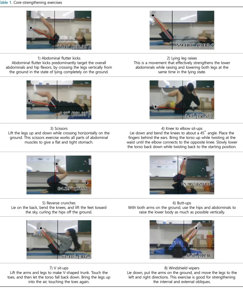
3-min break after each exercise. The 10 core-strengthening exercises selected for the study are shown in (Table 1).
Table 1. Core-strengthening exercises
1) Abdominal flutter kicks
Abdominal flutter kicks predominantly target the overall abdominals and hip flexors, by crossing the legs vertically from
the ground in the state of lying completely on the ground.
2) Lying leg raises
This is a movement that effectively strengthens the lower abdominals while raising and lowering both legs at the
same time in the lying state.
3) Scissors
Lift the legs up and down while crossing horizontally on the ground. This scissors exercise works all parts of abdominal
muscles to give a flat and tight stomach.
4) Knee to elbow sit-ups
Lie down and bend the knees to about a 45° angle. Place the fingers behind the ears. Bring the torso up while twisting at the waist until the elbow connects to the opposite knee. Slowly lower
the torso back down while twisting back to the starting position.
5) Reverse crunches
Lie on the back, bend the knees, and lift the feet toward the sky, curling the hips off the ground.
6) Butt-ups
With both arms on the ground, use the hips and abdominals to raise the lower body as much as possible vertically.
7) V sit-ups
Lift the arms and legs to make V-shaped trunk. Touch the toes, and then let the torso fall back down. Bring the legs up
into the air, touching the toes again.
8) Windshield wipers
Lie down, put the arms on the ground, and move the legs to the left and right directions. This exercise is good for strengthening
the internal and external obliques.
3. Data processing
During the isometric maximum contraction according to the core movement, the angle between the lower extremity and ground was measured when the activity of surface EMG RMS (a.u./sec) per second was the maximum. Because RMS shows a linear increase pattern with increasing muscle strength, it is used as an important index of linear muscle strength increase of the data collected while maintaining maximal voluntary isometric contraction (MVIC) for each exercise movement, the lower extremity and ground angle was measured at the peak activation on the surface electromyogram (root mean square [RMS]; a.u./sec). The RMS is used as an important index for the increase of static muscle strength because it shows a static increase pattern with increasing muscle strength (Gerdle, Karlsson, Crenshaw & Friden, 1997). Each ex- ercise movement was held for 3 sec, and each movement was limited to 120 bpm of the metronom. In order to compare data between subjects, the surface EMG data were processed with a recursive digital filter (Matlab Elliptic filter; 80-Hz low pass, 250-Hz high pass) and rectified using a full-wave rectification method. The rectified signals were then converted to RMS values, normalized to MVIC, and expressed as a percentage (%MVIC). Video recordings of the subjects performing each core-strengthening exercise were collected, and images in which the maximal muscle activation for each core muscle was expressed were captured. The angle between the two lines, the horizontal line crossing the femoral head of the limb and the line connecting the femoral head of the limb to the medial or lateral malleolus, was defined as the lower extremity and ground angle. The captured images were used to measure the lower extremity and ground angles, and the measured values were used for the analysis (Table 1).
4. Statistical analysis
All data were analyzed using Windows SPSS (version 20.0). One-way analysis of variance was used for the analysis of the lower extremity and ground angles at which each of the seven core muscles were maximally activated during the 10 core-strengthening exercises. A post- hoc least square difference test was used to identify differences among groups for each independent variable, and the significance level was
set at α = .05.
RESULTS
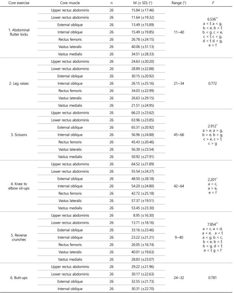
1. Angle of maximum core muscle strength per core exercise and muscle
The lower extremity and ground angles of maximum activation of the seven core muscles (upper rectus abdominis, lower rectus abdominis, external oblique, internal oblique, rectus femoris, vastus lateralis, and vastus medialis) for the 10 core-strengthening exercises are shown in (Table 2). The angles at which each core muscle was maximally activated were significantly different for the flutter kicks (F = 6.536**), scissors (F = 2.912*), knee to elbow sit-ups (F = 2.201*), reverse crunches (F = 7.854**), V sit-ups (F = 5.889**), and windshield wipers (F = 2.366*).
There were no significant differences in the angles for the leg raise, butt-up, bird dog, and raised leg plank exercises.
1) Flutter kicks
The rectus abdominis, internal oblique, and external oblique were maximally activated when the lower extremity and ground angle was between 11° and 15°. The rectus femoris, vastus lateralis, and vastus medialis were maximally activated at 27°, 40°, and 34°, respectively. For the flutter kick exercise, the range of angles at which the abdominal and femoral muscles were maximally activated was 11~40°. The abdominal muscles were mainly activated when the lower extremity and ground angle was between 11° and 15°, whereas the femoral muscles were mainly activated when the angle was between 27° and 40°. For the flutter kick exercise, the angle at which the vastus lateralis was maximally activated was the largest among the femoral muscles (rectus femoris and vastus medialis).
2) Lying leg raises
There were no significant differences among the angles at which each core muscle was maximally activated. Thus, both of the muscle groups (abdominal and femoral muscles) were maximally activated at Table 1. Core-strengthening exercises (Continued)
9) Bird dog
Get down on the hands and knees with the palms flat on the floor and shoulder-width apart. Raise the right arm and left leg
until they are in line with the body.
10) Raised leg plank
Alternately raise each leg in a flank posture.
Table 2. Angle of maximum core muscle strength per core exercise and muscle
Core exercise Core muscle n M (± SD) () Range () F
1. Abdominal flutter kicks
Upper rectus abdominis 26 15.84 (±17.46)
11~40
6.536**
a < f, a < g, b < e, b < f, b < g, c < e, c < f, c < g, d < f, d < g,
e < f Lower rectus abdominis 26 11.64 (±19.32)
External oblique 26 13.49 (±15.89)
Internal oblique 26 15.49 (±19.85)
Rectus femoris 26 26.78 (±24.15)
Vastus lateralis 26 40.06 (±31.13)
Vastus medialis 26 34.51 (±28.33)
2. Leg raises
Upper rectus abdominis 26 24.63 (±20.20)
21~34 0.772
Lower rectus abdominis 26 28.89 (±22.88)
External oblique 26 30.15 (±20.92)
Internal oblique 26 26.15 (±25.16)
Rectus femoris 26 34.03 (±22.99)
Vastus lateralis 26 26.63 (±29.15)
Vastus medialis 26 21.51 (±24.95)
3. Scissors
Upper rectus abdominis 26 66.23 (±23.62)
45~66
2.912* a > e, a > g, b > e, b > g, c > e, c > f,
c > g Lower rectus abdominis 26 63.96 (±23.85)
External oblique 26 65.31 (±20.92)
Internal oblique 26 56.96 (±24.88)
Rectus femoris 26 45.43 (±20.46)
Vastus lateralis 26 56.30 (±23.54)
Vastus medialis 26 50.92 (±27.91)
4. Knee to elbow sit-ups
Upper rectus abdominis 26 64.52 (±21.89)
42~64
2.201* a > c, a > e, e < f Lower rectus abdominis 26 55.54 (±24.27)
External oblique 26 48.50 (±28.18)
Internal oblique 26 54.20 (±24.80)
Rectus femoris 26 42.72 (±25.18)
Vastus lateralis 26 57.37 (±19.51)
Vastus medialis 26 53.45 (±23.30)
5. Reverse crunches
Upper rectus abdominis 26 8.95 (±16.30)
9~40
7.854**
a < c, a < d, a < e, a < f, a < g, b < c, b < e, b < f, b < g, d < f, e < f, g < f Lower rectus abdominis 26 13.71 (±18.16)
External oblique 26 33.16 (±23.46)
Internal oblique 26 23.22 (±21.21)
Rectus femoris 26 26.05 (±16.74)
Vastus lateralis 26 40.01 (±19.63)
Vastus medialis 26 28.83 (±23.07)
6. Butt-ups
Upper rectus abdominis 26 29.22 (±21.96)
24~32 0.781
Lower rectus abdominis 26 30.17 (±22.63)
External oblique 26 32.55 (±21.73)
Internal oblique 26 30.31 (±22.70)
an angle of 21~34° in the lying leg raise exercise.
3) Scissors
The rectus abdominis, internal oblique, and external oblique were maximally activated when the lower extremity and ground angle was
between 56° and 66°. The rectus femoris, vastus lateralis, and vastus medialis were maximally activated when the angle was between 45°
and 56°. Thus, the core muscles are maximally activated when the legs are above the ground at an angle between 45° and 66° for the scissors exercise. The maximum activation angle of the upper rectus abdominis was significantly greater than the maximum activation angles of the Table 2. Angle of maximum core muscle strength per core exercise and muscle (Continued)
Core exercise Core muscle n M (± SD) () Range () F
6. Butt-ups
Rectus femoris 26 23.75 (±11.09)
24~32 0.781
Vastus lateralis 26 26.22 (±15.00)
Vastus medialis 26 24.17 (±14.73)
7. V sit-ups
Upper rectus abdominis 26 8.52 (±17.10)
5~24
5.889**
a < b, a < e, a < f, a < g, c < b, b < e, c < e, c < g, e < d,
f < e, g < e Lower rectus abdominis 26 16.53 (±14.78)
External oblique 26 5.71 (±5.57)
Internal oblique 26 10.11 (±11.87)
Rectus femoris 26 24.74 (±12.32)
Vastus lateralis 26 10.70 (±14.10)
Vastus medialis 26 16.25 (±17.02)
8. Windshield wipers
Upper rectus abdominis 26 16.13 (±20.03)
11~20
2.366* b > e, b > f, b > g, c > e, c > f, d > e Lower rectus abdominis 26 20.03 (±11.52)
External oblique 26 20.25 (±10.64)
Internal oblique 26 17.43 (±11.76)
Rectus femoris 26 11.60 (±7.68)
Vastus lateralis 26 13.68 (±7.97)
Vastus medialis 26 15.53 (±9.21)
9. Bird dog
Upper rectus abdominis 26 14.07 (±11.37)
11~18 0.770
Lower rectus abdominis 26 11.30 (±7.75)
External oblique 26 17.90 (±18.15)
Internal oblique 26 11.49 (±7.62)
Rectus femoris 26 11.98 (±12.33)
Vastus lateralis 26 13.76 (±15.29)
Vastus medialis 26 12.75 (±16.41)
10. Raised leg plank
Upper rectus abdominis 26 42.02 (±22.39)
38~50 0.772
Lower rectus abdominis 26 44.63 (±22.09)
External oblique 26 45.25 (±24.61)
Internal oblique 26 38.24 (±28.70)
Rectus femoris 26 47.27 (±25.38)
Vastus lateralis 26 49.67 (±27.18)
Vastus medialis 26 50.92 (±27.15)
*p < .05, **p < .01.
SD, standard deviation; a, upper rectus abdominis; b; lower rectus abdominis; c, external oblique; d, internal oblique; e, rectus femoris; f, vastus lateralis; g, vastus medialis
vastus lateralis and vastus medialis. Likewise, the maximum activation angle of the lower rectus abdominis was significantly greater than the maximum activation angles of the vastus lateralis and vastus medialis.
The angle at which the upper and lower abdominal muscles were acti- vated per scissors exercise was relatively greater. The maximum activation angle of the internal oblique was greater than those of femoral muscles.
The angles of maximum activation for the abdominal and femoral muscles were different in the scissors exercise.
4) Knee to elbow sit-ups
The rectus abdominis, internal oblique, and external oblique were maximally activated when the lower extremity and ground angles were between 48° and 64°. The rectus femoris, vastus lateralis, and vastus medialis were maximally activated at 42°, 57°, and 53°, respectively.
Thus, the knee to elbow sit-up exercise is most effective when the lower extremity and ground angle is between 42° and 64°.
The maximum activation angle of the rectus abdominis was signifi- cantly greater than the maximum activation angles of the external oblique and rectus femoris. The maximum activation angle of the rectus femoris was significantly smaller than that of the vastus lateralis.
5) Reverse crunches
The upper rectus abdominis was maximally activated when the lower extremity and ground angle was at 9° whereas the lower rectus abdo- minis was maximally activated at an angle of 13°. The external oblique and internal oblique were maximally activated at an angle between 23° and 33°. The rectus femoris, vastus lateralis, and vastus medialis were maximally activated at 26°, 40°, and 28°, respectively.
The maximum activation angle of the upper rectus abdominis was significantly smaller than the maximum activation angles of the internal oblique, external oblique, rectus femoris, vastus lateralis, and vastus medialis. The maximum activation angle of the lower rectus abdominis was significantly smaller than the maximum activation angles of the external oblique, rectus femoris, vastus lateralis, and vastus medialis.
The maximum activation angles of the internal oblique, rectus femoris, and vastus medialis were significantly smaller than that of the vastus lateralis. Thus, the internal oblique, external oblique, and femoral muscles were all activated at an angle between 23° and 40° in the reverse crunch exercise.
6) Butt-ups
There were no differences among the angles at which each core muscle was maximally activated in the butt-up exercise. Thus, both of the muscle groups (abdominal and femoral muscles) were activated at an angle between 24° and 32° in the butt-up exercise.
7) V sit-ups
The upper rectus abdominis was maximally activated when the lower extremity and ground angle was at 8°. The lower rectus abdominis
was maximally activated when the angle was 16°. The external oblique and internal oblique were maximally activated at 5° and 10°, respec- tively. The rectus femoris, vastus lateralis, and vastus medialis were maximally activated at 24°, 10°, and 16°, respectively.
Thus, the V sit-up exercise was most effective when the motion is at the lower extremity and ground angle range of 5~24°, which is relatively low. The angles at which the abdominal and femoral muscles were activated were different in the V sit-up exercise.
8) Windshield wipers
The rectus abdominis muscles, internal oblique, and external oblique were maximally activated when the ground and lower extremity angle was between 16° and 20°. The femoral muscles (rectus femoris, vastus lateralis, and vastus medialis) were maximally activated at an angle between 11° and 15°. Thus, the femoral muscles were activated at smaller angles than the abdominal muscles in the windshield wiper exercise.
9) Bird dog
There were no differences among the angles at which each core muscle was maximally activated. Thus, both of the muscle groups (abdominal and femoral muscles) were maximally activated at an angle between 11° and 17° in the bird dog exercise.
10) Raised leg plank
There were no differences among the angles at which each core muscle was maximally activated. Thus, both of the muscle groups (abdominal and femoral muscles) were maximally activated at the range of angles of 38~50° in the raised leg plank exercise.
To sum up, the 10 core-strengthening exercises could be classified into four kinds of motion according to the range of angle at which each of the seven core muscles was maximally activated. The first group included V sit-ups, windshield wipers, and bird dog, which had the maximum muscle activation at a lower extremity and ground angle between 5° and 24°. The second group included flutter kicks and reverse crunches, which had the maximum muscle activation at a lower extremity and ground angle between 9° and 40°. The third group included leg raises and butt-ups, which had the maximum muscle activation at a lower extremity and ground angle between 21° and 34°.
The fourth group included scissors, knee to elbow sit-ups, and raised leg plank, which had the maximum muscle activation at a lower extremity and ground angle between 38° and 66°.
DISCUSSION
Core muscle strength relies on muscles located deep inside the trunk and plays an important role in joint stability, which contributes to the stabilization of movements. Core muscle training was reported to have positively affected the stability and balance of gymnasts during floor exercises (Yoon, Yoo & Yoon, 2016), and improved the physical fitness
and driver performance of male golfers (Beak & Park, 2013). Moreover, complex core stability exercises were effective in improving the physical fitness of stroke patients when walking (Park, 2008). Although there have been several researches on the effect of core muscle strength training in exercise performance or physical fitness improvement, studies investigating which muscle groups and at what angles such muscles are maximally activated for such training are insufficient. One study that investigated the relationship between angles and core muscles examined the effect of trunk stabilization exercises, with varying angles of the shoulder by using a sling, on the thickness of the core muscles in young adults (Park, 2015). However, this study examined only the thickness of the core muscles according to the shoulder joint angle, which was limited to the upper body. Thus, our study is different in that we examined both the upper and lower core muscles.
In the present study, we selected 10 core-strengthening exercises that are often employed in core training, and measured the muscle activation of four upper body muscles and three lower body muscles for each core-strengthening exercise. The results of our study are signifi- cant in that we analyzed the muscle activation values and identified the optimal angular difference between the ground and lower extremity for each core-strengthening exercise.
Movements that mainly engage the abdomen minimize the move- ment of the spine and provide stability in the sagittal, coronal, and trans- verse planes (Houglum, 2005; Kisner & Colby, 2002; Prentice, 2004). Thus, this type of training is the first phase of core-strengthening exercises.
Muscles that mainly use the femur (e.g., during walking in all directions) generate continuous movement and contribute to postural stability (Houglum, 2005; Kisner & Colby, 2002). This stability improves the control on the neuromuscular system, protecting the spine from injury and recurrent injuries (Hodges & Moseley, 2003). Core training that mainly utilizes the femoral muscles is the second phase of core-strengthening exercises. Movements that mainly engage the femoral muscles stabilize the pelvic floor muscles, which help maintain continence and support the organs within the pelvis (Ashton, Howard & Delancey, 2001; Constantinou
& Govan, 1982; Peschers, Gingelmaier, Jundt, Leib & Dimpfl, 2001;
Sapsford and Hodges, 2001). Lastly, core training that engages both the femoral and abdominal muscles includes multi-segmented exercises that involve movements in multiple planes, and this type of training is the third phase of core-strengthening exercises. Thus, selecting the type of exercise according to the subject's fitness level and the purpose of the exercise will result in effective core training.
CONCLUSION
Angular differences between the lower extremity and the ground that express the maximum activation of seven core muscles for each of the 10 core-strengthening exercises were measured and classified into four groups: V sit-ups, windshield wipers, and bird dog (5~24°); flutter kicks and reverse crunches (9~40°); leg raises and butt-ups (21~34°); and scissors, knee to elbow sit-ups, and raised leg plank (38~66°). These findings may be useful in selecting the type of exercise and the angle between the lower extremity and the ground according to the trainee's fitness level and goal. Moreover, these results may be useful as basic
data for determining which core muscles of the trainee are weak.
ACKNOWLEDGEMENTS
This work was supported by the "Convergence Female Talent Edu- cation Project for Next Generation Industries" through the Ministry of Science, ICT and Future Planning and the National Research Foundation of Korea (2015H1C3A1064579).
REFERENCES
Ashton, M. J., Howard, D. & Delancey, J. O. L. (2001). The functional anatomy of the female pelvic floor and stress continence control system. Scandinavian Journal of Urology and Nephrology, 35(207), 1-7.
Beak, S. K. & Park, J. Y. (2013). Effects of core Training on Physical Fitness and Driver Performance in Male Professional Golfers. The Korean Society Of Sports Science, 22(1), 1053-1066.
Brill, P. W. (2002). The Core Program. 1st ed. New York Bantam Books.
Brown, A. B., McCarney, N. & Sale, D. G. (1990). Positiveadaptations to weight-lifting training in the elderly. Journal of Applied Physiology, 69(5), 1725-1733.
Choi, W. I. (2015). An Analysis of Core Muscle Activity Differences by Knee Joint Angles in Squat Exercise on Unstable Surfaces, Un- published Master's Thesis. Graduate School, Korea National Sport University.
Constantinou, C. E. & Govan, D. E. (1982). Spatial distribution and timing of transmitted and reflexly generated urethral pressures in healthy women. The Journal of Urology, 127(5), 964-969.
Faries, M. D. & Greenwood, M. (2007). Core training: stabilizing the con- fusion. Strength and Conditioning Journal, 29(2), 10-25.
Gerdle, B., Karlsson, S., Crenshaw, A. G. & Friden, J. (1997). The relation- ships between EMG and muscle morphology throughout sustained static knee extension at two submaximal force levels. Acta Phy- siologica, 160(4), 341-351.
Hermens, H. J. & Freriks, B. (1999). SENIAM: European Recommendations for Surface Electromyography. Ressingh Research and Development.
Hodges, P. W. & Moseley, G. L. (2003). Pain and motor control of the lumbopelvic region: effect and possible mechanisms. Journal of Electromyography and Kinesiology, 13(4), 361-370.
Houglum, P. (2005). Therapeutic Exercise for Musculoskeletal Injuries, 2nd ed.
Kim, J. H. (2012). Effect of core training on physical strength and stroke ability in female tennis players. Un-published Master's Thesis.
Graduate School of Education Chonnam National University.
Kim, K. H. & Lee, S. C. (2010). Dynamic Stability Effect of Applicable Core and Neuromuscular Training for 12 Weeks. Korean Journal of Sport Biomechanics, 20(1), 101-108.
Kim, N. H. & Yi, K. O. (2015). The Effects of Corrective Hip Joint Ex- ercises and Foot Orthotics on RCSP, Ankle's Range of Motion, and Core Muscle Strength for Middle School Students with Pes Planus.
Korean Journal of Sport Biomechanics, 25(4), 401-412.
Kim, S. Y. (2007). The imoacts of weight-bearing exercise and aquatic
exercise on middle-aged women's body composition and isokinetic musle strength and bone mineral density. Un-published Master's Thesis. Rraduate School of Education Han-Yang University.
Kendall, F. P., McCreary, E. K., Provance, P. G., Rodgers, M. M. & Romani, W. A. (2005). Muscles testing and function with posture and pain.
5th ed. Lippincott/ Williams & wikins.
Kiesel, K., Burton, L. & Cook, G. (2004). Mobility screening for the core.
Athletic Therapy Today, 9(5), 38-41.
Kisner, C. & Colby, L. A. (2002). Therapeutic Exercise: Foundations &
Techniques, 4th ed.
Miyake, Y., Nakamura, S. & Nakajima, M. (2014). The effect of trunk coordination exercise on dynamic postural control using a Core Noodle. Journal of Bodywork and Movement Therapies, 18(4), 519 -525.
Munsat, T. L., McNeal, D. & Waters, R. (1976). Effect of nerve stimu- lation on human muscle. Archneural, 33, 608-617.
Nadler, R. B. (2002). Bladder training biofeedback and pelvic floor myalgia. Urology, 60(6l), 2-3.
Nesser, T. W. & Lee, W. L. (2009). The relationship between core strength and preformance in division I female soccer players. Journal of Exercise Physiology Online, 12(2).
O'sullivan, P. B., Phyty, G. D. M., Twomey, L. T. & Allison, G. T. (1997).
Evaluation of specific stabilizing exercise in the treatment of chronic low back pain with radiologic diagnosis of spondylolysis or spondy- lolisthesis. Spine, 22(24), 2959-2967.
Park, E. K. (2008). The Effects of Core Stability Exercises with Aero- Equipment on Fitness, Gait Development, EMG, FMRI in Stroke Patients. Un-published Doctor's Dissertation. Graduate School of Ewha Womans University.
Park, M. H. (2015). Effect of core muscle thickness and static or dynamic
balance on prone bridge exercise with sling according to shoulder joint angle in healthy adults. Un-published Master's Thesis. Graduate School of Sunmoon University.
Peschers, U. M., Gingelmaier, A., Jundt, K., Leib, B. & Dimpfl, T. (2001).
Evaluation of pelvic floor muscle strength using four different techniques. International Urogynecology Journal, 12(1), 27-30.
Prentice, W. E. (2004). Rehabilitation Techniques for Sports Medicine &
Athletic Training, 4th ed.
Richardson, C., Jull, G., Toppenberg, R. & Comerford, M. (1992). Tech- niques for active lumbar stabilisation for spinal protection: a pilot study. Australian Journal of Physiotherapy, 38(2), 105-112.
Sapsford, R. R. & Hodges, P. W. (2001). Contraction of the pelvic floor muscles during abdominal maneuvers. Archives of Physical Medicine and Rehabilitation, 82(8), 1081-1088.
Shinn, Y. K. & Yi, K. O. (2015). Difference in core stability and muscle balance of the pilates Teaser motion according to kinds of the ground and skill. Korean Society of Sports Biomechanics, 25(1), 65 -76.
Willson, J. D., Dougherty, C. P., Ireland, M. L. & Davis, I. M. (2005). Core stability and its relationship to lower extremity function and injury.
The Journal of the American Academy of Orthopaedic Surgeons, 13(5), 16-25.
Yoon, C. S., Yoo, S. H. & Yoon, S. H. (2016). Effects of Core Muscles Exercise on the Balance and Stability of V-sit in the Floor Exercise of Gymnastics. The Korean Journal of Physical Education, 55(5), 719 -727.
Yun, K. S., Jun, I. S., Kwak, H. M., Kim, J. H., Jeon, C. B., Kim, G. K. & Lee, H. J. (2013). The Effect of 12-weeks Core Stability Exercise Program on Physical Fitness and Soccer Techniques in Middle School Soccer Players. Journal of Coaching Development, 15(3), 205-213.