Funneling venoplasty for anomalous graft left hepatic vein in living donor liver transplantation using a split left lateral section graft for an infant patient
Jung-Man Namgoong
1, Shin Hwang
1, Tae-Yong Ha
1, Young-In Yoon
1, Yong Jae Kwon
1, Hyunhee Kwon
1, Kyung Mo Kim
2, Seak Hee Oh
21
Department of Surgery, Asan Medical Center, University of Ulsan College of Medicine, Seoul, Korea,
2
Department of Pediatrics, Asan Medical Center, University of Ulsan College of Medicine, Seoul, Korea
Case Report
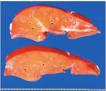
The left lateral section (LLS) can have an unusual variant left hepatic vein (LHV) anatomy. We present a case of customized funneling venoplasty of the graft LHV in a 22-month-old girl diagnosed with ornithine transcarbamylase deficiency undergoing deceased donor liver transplantation (LT) using a split LLS graft. The split LLS graft weighed 350 g, yielding a graft-to-recipient weight ratio of 3.2%.
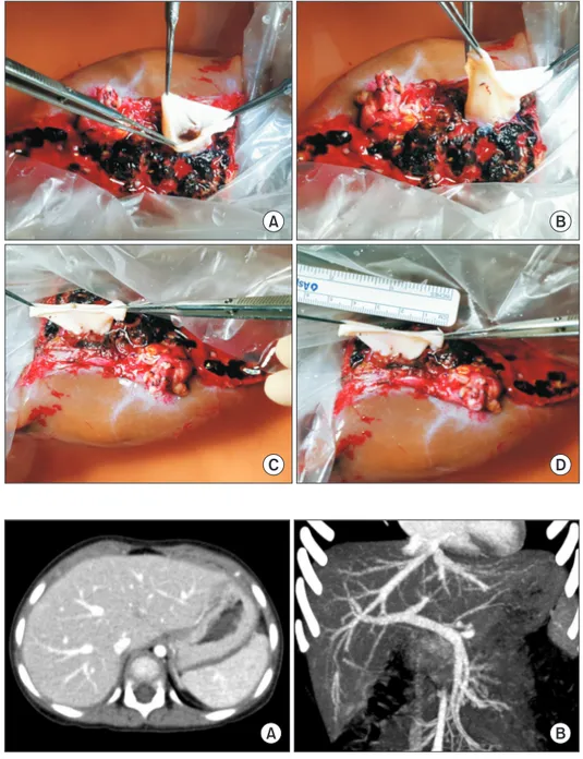
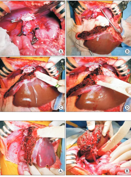
Notably, the graft LHV opening was located at the graft liver cut surface, which was only 1 cm in size and 2 cm away from the cepha- lad apex of the LLS graft. Since such a variant location of the small LHV opening was unsuitable for direct anastomosis, we performed a funneling venoplasty using an inferior vena cava fragment homograft obtained from the same donor. The graft implantation was performed according to standard procedures of infant split LT. Follow-up imaging studies showed no vascular complications. The patient recovered uneventfully from the LT operation. She had normal blood test findings, including normal ammonia level. She has been doing well for 6 months after the transplantation. In conclusion, our surgical technique using a funneling venoplasty enabled successful reconstruction of the anomalous graft LHV. Our results suggest that individualized reconstruction techniques should be applied to infant patients undergoing LT using a LLS graft with variant types of graft LHV anatomy.
Key Words: Left hepatic vein; Anatomical variation; Venoplasty; Interposition; Left lateral section graft
INTRODUCTION
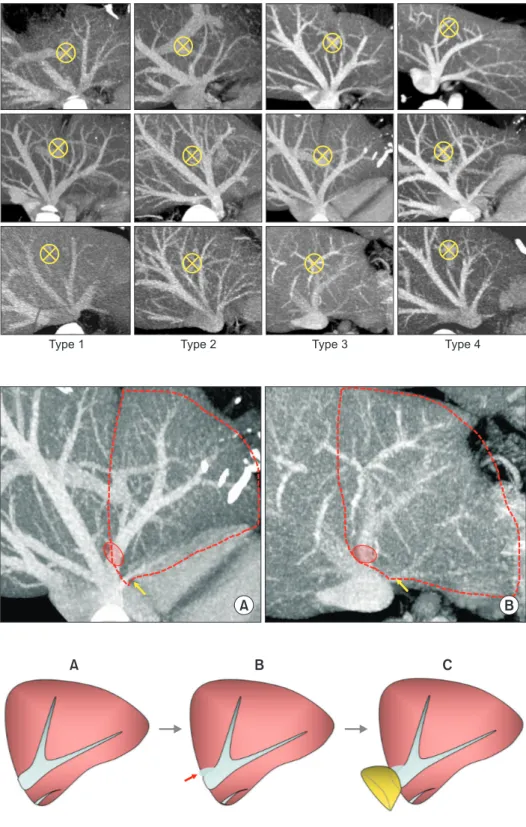
A left lateral section (LLS) graft is often used for liver trans- plantation (LT) in infant patients in the form of living donor liver transplantation (LDLT) or split LT. Although the anatomy of the left hepatic vein (LHV) is diverse [1-4], the majority of graft LHVs are suitable for direct anastomosis to the recipient hepatic vein stumps or directly to the inferior vena cava (IVC).
However, in rare instances, the LLS outflow drains through both the LHV and the middle hepatic vein (MHV) or the
LHV drains directly through the MHV trunk [1]. In donors with such unusual LHV anatomy, it is necessary to preserve the MHV trunk for the safety of the living donor or for the security of the split extended right liver graft. If the graft LHV opening is located at the cut surface of the LLS graft instead of at the cephalic apex, a customized venoplasty technique is nec- essary to make it suitable for graft hepatic vein reconstruction, especially for an infant recipient with a small IVC. We present a case of customized venoplasty of the graft LHV in an infant patient undergoing deceased donor LT using a split LLS graft.
CASE
The recipient was a 22-month-old girl who was diagnosed with ornithine transcarbamylase (OTC) deficiency. The pa- tient was born through a full-term cesarean-section delivery.
She showed irritability and decreased activity from one month after birth. At that time, laboratory studies showed hyper- ammonemia and metabolic acidosis with high levels of liver enzymes. Gene studies revealed OTC NM_000531.5:c.626C>T.
Received: January 15, 2021, Revised: January 21, 2021, Accepted: January 23, 2021
Corresponding author: Shin Hwang
Department of Surgery, Asan Medical Center, University of Ulsan College of Medicine, 88 Olympic-ro 43-gil, Songpa-gu, Seoul 05505, Korea
Tel: +82-2-3010-3930, Fax: +82-2-3010-6701, E-mail: [email protected] ORCID: https://orcid.org/0000-0002-9045-2531
Copyright Ⓒ The Korean Association of Hepato-Biliary-Pancreatic Surgery
This is an Open Access article distributed under the terms of the Creative Commons Attri- bution Non-Commercial License (http://creativecommons.org/licenses/by-nc/4.0) which permits unrestricted non-commercial use, distribution, and reproduction in any medium, provided the original work is properly cited.