대한외과학회지:제 74 권 제 4 호
□ 증 례 □
Vol. 74, No. 4, April, 2008
316
책임저자:유희철, 전북 전주시 덕진구 금암동 634-18
561-712, 전북대학교병원 외과 Tel: 063-250-1576, Fax: 063-271-6197 E-mail: [email protected]
접수일:2007년 11월 14일, 게재승인일:2007년 12월 31일 본 연구는 보건복지부 암정복추진연구개발사업 자원으로 이루어 진것임: 0620220
중심 단어: 문맥, 이상, 간이식
좌측 문맥 수평 부분의 선천적 결여에 대한 재고
전북대학교 의학전문대학원 1외과학교실, 2영상의학교실 및 3임상의학연구소
구본용1ㆍ유희철1,3ㆍ김광옥1,3ㆍ황홍필1ㆍ김영곤2ㆍ곽효성2ㆍ조백환1,3
Congenital Absence of the Horizontal Segment of the Left Portal Vein
Bon Yong Koo, M.D.1, Hee Chul Yu, M.D.1,3, Guang Yu Jin, M.D.1,3, Hong Pil Hwang, M.D.1, Young Kon Kim, M.D.2, Hyo Sung Kwak, M.D.2 and Baik Hwan Cho, M.D.1,3
Departments of 1Surgery and 2Diagnostic Radiology, 3Re- search Institute of Clinical Medicine, Chonbuk National University Medical School, Jeonju, Korea
As living-donor liver transplant techniques develop, variations in the portal vein are seen in approximately 20% of the population. However, congenital absence of the horizontal segment of the left portal vein is very rare and has not been reported in Korea. We present a case with a congenital ab- sence of the horizontal segment of the left portal vein that was detected during an evaluation for a living donor liver transplantation, with a review of the relevant literature. (J Korean Surg Soc 2008;74:316-318)
Key Words: Portal vein, Abnormalities, Liver transplantation
서 론
오늘날 간이식은 말기 간질환에 대한 치료방법으로 확고 히 정착되어 활발히 시행되고 있지만, 세계적으로 간이식 대기자수에 비해 상대적으로 뇌사자 간 공여가 부족한 상 황이다. 특히 우리나라는 뇌사자 장기기증에 대한 제도와 사회적 인식이 부족하여 장기의 수요와 공급의 불균형이 심각한 상황으로, 이러한 장기 부족 현상을 해결하기 위한
대안으로서 생체 간이식이 날로 증가하고 있다.
생체 간이식 수술 시 공여자에 대하여 혈액검사 및 영상 검사를 포함하는 자세한 검사를 시행함으로써 간공여자로 서의 적합성과 안정성을 평가하게 된다. 그중 영상 검사의 중요한 목적 중의 하나는 간혈관의 해부학적 변이를 파악 하는 것인데, 이는 공여자 간절제 가능 여부를 결정하는 중 요한 요소가 된다.
저자들은 생체 간이식을 위한 공여자 영상 검사 중 우연 히 좌측 문맥의 수평 부분(horizontal segment)이 선천적으로 결여되어 있는 1예를 발견하였고, 문헌 검색 결과 매우 드 물고 현재까지 국내에서 발표된 증례가 없어 문헌 고찰과 함께 보고하는 바이다.
증 례
20세 여자로 생체 간이식 공여자를 위하여 검사를 시행 하였다. 복강 내 수술이나 감염, 간담도 질환의 과거력은 없 었고 간기능과 혈액응고검사를 포함한 혈액검사도 정상범 위였다. 그러나 컴퓨터단층촬영상 주문맥은 좌측 문맥의 수평 부분이 결여된 채 우측 문맥으로 연속되며 우전 구역 및 우후 구역으로 분지를 내는 소견이 관찰되었다. 우전구 역으로의 문맥 분지는 크고 비정상적인 형태를 보이는 한 편, 좌엽의 내측 구역에서 횡방향으로 주행한 후 문맥의 제 대 부위(umbilical portion)를 형성하며 좌엽을 공급하는 문 맥 분지들을 내었다(Fig. 1). 이에 부분 간절제 시 수술 위험 도가 높을 것으로 판단되어 공여자에서 제외되었다.
고 찰
정상적으로는 문맥은 간문에서 우측과 좌측 문맥으로 나 뉘게 된다. 우측 문맥은 우엽의 전구역과 후구역을 공급하 는 두 개의 분지로 나뉘게 되고, 좌측 문맥은 근위부에 수평 부분이 존재하고 이 수평 부분이 간원인대 수준에서 앞쪽 으로 돌아 제대 부위, 즉 수직 부분(vertical segment)을 형성 하게 되는데, 이 수직 부분에서 좌엽의 내측과 외측 분절을 공급하는 문맥 분지들이 나오게 된다.(1)
문맥 변이는 인구의 약 20%에서 존재한다고 알려져 있 다.(2,3) 그중에서도 본 증례와 같이 좌측 문맥의 수평 부분
Bon Yong Koo, et al:Congenital Absence of the Horizontal Segment of the Left Portal Vein 317
Fig. 1. CT portograms in a 20-year-old woman with absence of the horizontal segment of the left portal vein. (A) The primary branch of the portal vein continued to the right branch of the portal vein without the horizontal segment of the left portal vein. (B, C) The right anterior sectional portal branch shows a large and aberrant shape and runs transversely in the medial section of the left hemiliver while forming the umbilical portion of portal vein. (D) The 3-D reconstruction demonstrates the entire portal vein and its branches.
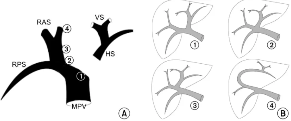
Fig. 2. Primary portal ramification patterns at the hilum are determined by the “sliding” characteristics of the reunion point of the left portal vein. (A) Each number indicates the point to which the left portal vein can reunite. Numbers ①, ②, and ③ were suggested by Ryu and Cho, and number ④ by this paper's authors. MPV = main portal vein; RPS = right posterior section; RAS = right anterior section; VS = vertical segment; HS = horizontal segment. (B) Each figure represents the results of where the left portal vein reunited to the four points of the right portal vein. ① bifurcation of the main portal vein, ② trifurcation of the main portal vein, ③ origin of the right posterior sectional branch from the main portal vein, and ④ absence of the horizontal segment of the left portal vein.
318 J Korean Surg Soc. Vol. 74, No. 4
이 선천적으로 결여되어 있는 변이 유형의 빈도는 0.03∼
0.9%로 보고되었다.(2-4)
좌측 문맥의 수평 부분이 결여되어 있는 변이 유형은 간 의 발생학으로 설명될 수 있다. 성인의 문맥과 간정맥은 태 아에서의 난황정맥(vitelline vein)과 제대정맥(umbilical vein) 에서 기원한 것이다.(1) 발생 초기에 난황정맥과 제대정맥 사이에 많은 연결 통로(anastomotic channel)가 존재하게 되 는데, 태아가 성숙함에 따라 일부 통로는 퇴화하고 다른 통 로는 두드러지게 된다. 그중 우측 난황정맥의 꼬리쪽 부분 과 좌측 제대 정맥을 연결하는 통로는 발생 5주째에 나타나 게 되고 성인에서 좌측 문맥의 수평부분을 형성하게 된 다.(5) 만약 이러한 연결이 정상적으로 발생하지 않는다면 또다른 정맥 통로가 우측 난황정맥과 좌측 제대정맥의 연 결을 유지시키게 되고, 임상적으로 이러한 일이 좌측 문맥 수평 부분의 결여와 좌엽을 공급하는 비정상적인 형태의 혈관의 발달로 나타나게 된다고 알려져 있다.
한편 Ryu와 Cho(6)는 문맥 변이의 발생학적 원인에 대해 보다 논리적으로 기술하였는데, 태생 3주가 되면 좌난황장 간막정맥(left vitellomesenteric vein)이 사라지고, 이후 제대 정맥에서 기원한 좌문맥과 우난황정맥에서 기원한 우문맥 과의 재결합 부위(reunion point)에 따라 간문부의 문맥 분지 형태가 결정된다고 하였다. 이는 좌문맥의 재결합 부위가 우문맥에서 마치 미끌어지듯이 이동하는(sliding) 특성을 가 리킨 것이다. 즉, 좌문맥이 우문맥의 본간(main trunk)과 결 합하였을 때는 전형적인 문맥 분지 형태가 되고 전구역문 맥과 후구역문맥의 분지 부위에 결합했을 때는 삼분지 (trifurcation) 형태가 되며 마지막으로 전구역문맥의 중간 부 분과 결합했을 때는 우후구역문맥이 직접 주문맥에서 분지 되는 형태가 되는 것을 말한다. 그런데 Ryu와 Cho가 주장한 이론으로는 본 증례와 같이 좌측 문맥의 수평 부분이 결여 되어 있는 변이를 설명할 수 없다. 그렇지만 만약 좌문맥이 우전구역문맥의 끝부분에 재결합되는 경우를 가정한다면 우전구역문맥에서 좌문맥으로 연속되는 본 증례의 경우가 설명될 수 있을 것이다(Fig. 2).
Lerut 등(7)은 본 증례와 같은 문맥 변이를 가진 성인 남 자의 간 좌엽을 구득하여 소아에 성공적으로 이식하였다는 보고를 하였다. 그러나 Duran 등(8)의 증례에서는 수혜자가 성인이기 때문에 공여자 간의 좌엽으로는 수혜자의 대사요 구량을 충족시킬 수 없을 뿐만 아니라 우엽 절제를 한다면
남아있는 좌엽의 문맥 혈류를 유지하기 위하여 이식편을 이용한 문맥 재건이 이루어져야 하고 이는 결국 공여자의 안전을 심각하게 위협할 수 있기 때문에 공여자로 이용하 지 못하였다고 기술하였다. 이와 같은 이유로 인하여 본 증 례도 간 공여자군에서 제외되었다.
컴퓨터 기술의 발달로 인해 문맥의 삼차원 재구성이 가 능하게 되었고, 이는 부분 간이식을 위한 간절제술을 계획 하는 데 있어서 아주 유용하게 되었다.(9) 따라서 문맥 변이 가 존재하는 경우에는 간절제 시 치명적인 결과가 발생할 수 있으므로 술 전 검사 시 발달된 기술을 이용한 적극적인 해부학적 평가가 이루어져야 할 것이다.
REFERENCES
1) Sherlock S, Dooley J. Anatomy and function. In: Sherlock S, Dooley J, editors. Diseases of the Liver and Biliary System.
Oxford: Blackwell; 1997. p.1-16.
2) Couinaud C. The hepatic pedicle. I. The intrahepatic portal vein. In: Couinaud C, editor. The Liver: Anatomic and Surgical studies. Paris: Masson; 1957. p.71-118.
3) Atri M, Bret PM, Fraser-Hill MA. Intrahepatic portal venous variations: prevalence with US. Radiology 1992;184:157-8.
4) Fraser-Hill MA, Atri M, Bret PM, Aldis AE, Illescas FF, Herschorn SD. Intrahepatic portal venous system: variations demonstrated with duplex and color Doppler US. Radiology 1990;177:523-6.
5) Elias H, Sherrick JC. Development of the human liver. In:
Morphology of the Liver. New York: Academic Press; 1969.
p.233-61.
6) Ryu M, Cho A. Surgical Anatomy of the Liver. Tokyo:
Igakushoin; 2004.
7) Lerut J, Ciccarelli O, Danse E, Goffette P, Van Beers B, Reding R, et al. Left lobe living related liver transplantation in the absence of an extrahepatic left portal vein. Transplanta- tion 2002;74:278-9.
8) Duran C, Taner B, Dayangac M, Akin B, Balci D, Kurt Z, et al. Why we did not use this donor. Liver Transpl 2007;
13:1199-200.
9) Lee HK, Nam YS, Park HK, Paik HK, Lee WJ, Lim HK, et al. 3D-reconstruction of right portal vein. Korean J Hepatobili- ary Pancreat Surg 1997;1:33-40.